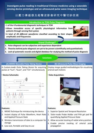
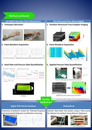
Overview on the project's aim, methodologies and results, potential applications include telemedicine as well as automated diagnosis via machine learning
Nuclear physics course instructor Dr. Waseem Ahmad presented on the medical use of radiation for imaging. There are several techniques that produce diagnostic images, including using an external source of radiation placed in front of the patient, like x-rays, or an internal radioactive source injected into the patient. Computed tomography (CT) uses a combination of x-rays and a computer to create detailed cross-sectional images of the inside of the body, showing more detail than regular x-rays. CT scans are commonly used to detect abnormalities in the brain, body, and organs.
Radiology uses medical imaging to diagnose and sometimes treat diseases. Modalities include X-ray, ultrasound, CT, nuclear medicine including PET, and MRI. Interventional radiology uses imaging to guide minimally invasive procedures. Plain radiography is commonly used for initial assessment but has lower sensitivity than newer modalities. It is useful for visualizing bone tumors, fractures, and arthritis. Ultrasound uses sound waves to image soft tissues in real time without radiation. CT obtains 3D images but has disadvantages of cost and radiation exposure. MRI provides high soft tissue contrast images in multiple planes but has contraindications for patients with metallic implants. Nuclear medicine involves radioactive tracers that accumulate in tissues to evaluate physiological function.
Ultrasound uses high frequency sound waves to produce images of the inside of the body. There are different modes of ultrasound including A-mode which displays amplitude over time, B-mode which produces two-dimensional images, and M-mode which depicts motion over time. Modern ultrasound systems can produce real-time 2D and 3D images using piezoelectric crystals and array transducers. Doppler ultrasound measures the frequency shift of reflected sound to analyze blood flow velocity.
This document provides an overview of biomedical instrumentation. It discusses how instrumentation is used to monitor and control process variables for measurement and control. Biomedical instrumentation specifically creates instruments to measure, record, and transmit data to and from the body. Some key types of biomedical instrumentation systems are direct/indirect, invasive/noninvasive, contact/remote for sensing and actuating in real-time or statically. Several important instruments are discussed in detail, including X-rays, electrocardiography, magnetic resonance imaging, ultrasound, and computed tomography. The document outlines the basic workings, advantages, and disadvantages of these key biomedical instruments.
Radiation diagnostics diseases of the brain and spinal cord ShieKh Aabid
Radiology plays an important role in diagnosing brain and spinal cord pathologies in children through various imaging techniques. Early diagnosis is key to treating issues in children's brains. There are three main neuroimaging methods - neurosonography, computed tomography, and magnetic resonance imaging. Sonography is a non-invasive way to evaluate the brain through fontanelles in infants, but has limitations. CT scans are also non-invasive and useful for detecting aneurysms and calcifications. MRI provides the best anatomical images and differentiation of structures, and can also perform spectroscopy to examine biochemical changes in tumors. Various contrast-based techniques like angiography can also evaluate the brain's vasculature in children.
Neuroimaging or brain imaging is the use of various techniques to either directly or indirectly image the structure, function, or pharmacology of the nervous system. It is a relatively new discipline within medicine, neuroscience, and psychology
SBRT is a precise form of radiation therapy that delivers very high ablative doses of radiation to tumors in a small number of fractions. It has become the standard of care for early stage non-small cell lung cancer (NSC LC) that is not surgically resectable. Key aspects of SBRT planning and delivery include delineating targets and organs at risk on imaging, determining appropriate dose and fractionation based on tumor location, using motion management strategies to account for tumor motion, precise daily image guidance, and ensuring dose constraints are met to minimize risks to critical structures like the spinal cord. SBRT provides superior local tumor control compared to conventional fractionation for early stage NSCLC with a favorable toxicity profile.
This document provides an introduction and overview of medical instrumentation. It begins by outlining the expectations and learning objectives of the course. It then discusses related classes, assessment weighting, and textbook references. The rest of the document defines key terms related to medical instrumentation and biomedical engineering. It provides classifications of medical equipment and describes examples within each classification, including diagnostic, therapeutic, surgical, and research devices. Feedback loops and the scientific method in relation to instrumentation are also summarized.
MRI has advanced significantly in recent years through improvements to hardware, software, and applications. Hardware advances include higher field strength scanners, new coil technologies, and wider bore sizes. Software now allows generating multiple contrasts from a single scan and uses techniques like SMS to reduce scan times. Applications have expanded MRI's use for imaging tissues like lungs and evaluating implants. Overall, MRI is providing more detailed images faster and for an increasing number of clinical uses.
This document provides an overview of biomedical instrumentation. It discusses key topics such as:
- The development of biomedical instrumentation from early devices like the electrocardiograph to modern advances enabled by surplus electronics after WWII.
- Key considerations for designing medical instrumentation systems, including range, sensitivity, linearity, and frequency response.
- Components of the man-instrument system including the subject, stimuli, transducers, signal conditioning equipment, and displays.
- Objectives of instrumentation systems like information gathering, diagnosis, evaluation, monitoring and control.
- Biometrics as the measurement of physiological variables and parameters that biomedical instrumentation provides tools to measure.
This document provides an introduction to magnetic resonance imaging (MRI). It discusses the history, instrumentation, working principles, and applications of MRI. MRI uses strong magnetic fields and radio waves to generate detailed images of organs and tissues in the body. Felix Bloch, Edward Purcell, and Raymond Damadian contributed to early discoveries around MRI. Key MRI machine components include the main magnet, gradient coils, and radiofrequency coils. MRI works by aligning hydrogen protons in tissues when a magnetic field is applied, and using radio waves to elicit signals to form images. MRI has many medical applications including neuroimaging, cardiology, musculoskeletal imaging, and angiography.
Nuclear medicine uses small amounts of radioactive tracers and imaging technology to diagnose and treat diseases. There are two main types of nuclear imaging - positron emission tomography (PET) and single photon emission computed tomography (SPECT). PET provides higher resolution images with short scan times, while SPECT is more widely used and less expensive, with longer-lasting radioactive tracers. Nuclear imaging offers unique functional and molecular information not available through other techniques like X-ray and CT scans, enabling early disease detection. It can evaluate organ function and metabolism to identify issues.
Recent advances in MRI technology include faster scans enabled by new software, simplified cardiac imaging workflows, and the ability to image lungs. New MRI systems have also been introduced, including the first 7T system approved for clinical use in the US. Additional software improvements have reduced scan times for brain exams and simplified scans for patients with implants.
This document provides an overview of magnetic resonance imaging (MRI). It begins by defining MRI as a medical imaging technique that uses magnetic fields and radio waves to produce detailed images of the body. The document then covers the history, components, principles, and types of MRI images. It discusses how MRI works to detect tissue properties using relaxation times and how varying pulse sequences produces different contrasts. The document concludes by outlining the clinical applications and benefits of MRI, such as its ability to clearly image soft tissues without radiation, as well as some limitations like expense and the enclosed scanner.
Radiology uses various imaging techniques like X-rays, CT scans, MRIs, ultrasounds, and fluoroscopy to diagnose and treat diseases. Radiologists interpret the images produced to make a diagnosis and report their findings to the ordering physician. Different techniques use different methods like radiation, magnetic fields, or sound waves to produce images of the body's internal structures. Radiation exposure can damage tissues over time through cell death, reduced blood cell counts, and increased cancer risks depending on the amount and area exposed.
Advanced Nuclear Medicine Through ResearchJMFitness
Nuclear medicine is an evolving field that uses radiopharmaceuticals and imaging techniques to diagnose and treat diseases. Research has led to advancements like improved diagnostic testing, new radiopharmaceuticals, and better treatment therapies. Current areas of focus include developing new molecular imaging agents and radiopharmaceuticals for diagnosing diseases with less radiation exposure. Research is also investigating therapeutic radiopharmaceuticals for treating cancers and reducing bone pain from metastases. Clinical trials are helping to establish the safety and efficacy of emerging nuclear medicine techniques and therapies.
Application of instrumentation in medical worldkanhaiya jha
This document discusses various medical instrumentation technologies and their applications. It begins by discussing digital thermometers and how they use thermoresistors to precisely measure body temperature. It then describes magnetic resonance imaging (MRI) in detail, including its history, working procedure, required magnetic field strengths, diagnostic capabilities, advantages over CT scans, and potential disadvantages like noise and motion restrictions. The document also discusses glucometers, which allow home testing of blood glucose levels, and continuous glucose monitors which can track glucose trends. Overall, the document outlines how instrumentation has improved medical diagnostics and patient monitoring.
This document discusses various types of medical imaging technologies. It describes radiologic/x-ray technology, ultrasound technology, CT scans, MRI scans, and nuclear imaging including PET and SPECT. The goal of medical imaging is to non-invasively examine the inside of the body to diagnose health problems and guide treatment. Each technology has advantages for certain applications based on the type of information and depth of imaging it provides. Together these modalities provide physicians a variety of tools to accurately diagnose and monitor patient health issues.
This document discusses respiratory motion management in radiotherapy. It notes that respiratory motion can cause artifacts during image acquisition and limitations in treatment planning and delivery. It describes several methods to account for respiratory motion, including motion encompassing methods like slow CT, inhale/exhale breath-hold CT, and 4D CT. It also discusses respiratory gating techniques using external markers or internal fiducials, noting that gating involves administering radiation within a particular portion of the breathing cycle. Respiratory gating systems synchronize radiation with the patient's breathing pattern to reduce motion effects.
Nuclear medicine uses small amounts of radioactive tracers and imaging equipment to produce images of internal organs and functions. Radiotracers are injected, inhaled, or swallowed and accumulate in tissues and organs, emitting gamma rays that are detected by a gamma camera and computer to create 2D and 3D images. Common radiotracers include technetium-99, which is administered depending on the organ or system being examined. Precautions are taken to minimize radiation exposure to patients and staff through proper handling and administration of radiotracers, use of shielding, monitoring of doses, and specialized safety training for the nuclear medicine team.
Single-photon emission computed tomography is a nuclear medicine tomographic imaging technique using gamma rays. It is very similar to conventional nuclear medicine planar imaging using a gamma camera. but is able to provide true 3D information
Nuclear medicine uses small amounts of radioactive material and imaging equipment like gamma cameras to diagnose and treat diseases. It can visualize the structure and function of organs and systems. Common uses in adults include evaluating bones, lungs, heart, and brain. The gamma camera detects radioactive emissions and converts them into images, composed of detector heads in a box shape attached to a circular gantry. Patients are given radioactive tracers orally or intravenously and imaged as the tracer accumulates in organs.
4D-CBCT (Symmetry) - a useful tool to verify and treat traditional ITV withou...Dr. Malhar Patel
4D-CBCT is latest software gadget in field of radiation oncology. It will calculate breathing movement during treatment of lung cancer and help in delineate the target better.
This presentation will convince you that even if you do not have 4D-CT simulation, you can confidently use 4D-CBCT at optimal level.
Radio imaging techniques are the noninvasive imaging of various organs,
tissues using radioisotopes for the purpose of formulation development,
improvement of dosage form or diagnosis and treatment of disease.
Using technology in the biomedical industryGraham Wilson
Biomedical engineering applies engineering principles and design concepts to medicine and biology for healthcare purposes. It combines engineering skills with medical and biological sciences to advance healthcare treatment, including diagnosis, monitoring, and therapy.
The document provides examples of current biomedical engineering applications: 1) A handheld dermatological scanner that analyzes tissue morphology to help doctors determine if a biopsy is needed; 2) An implantable nerve stimulating device for patients with migraines; 3) Non-invasive continuous glucose monitors and insulin pumps; and 4) Medical robots that can autonomously monitor patients and manage their charts and vital signs.
1. Dr. Sheetal R Kashid presented on the use of IGRT for head and neck cancers and central nervous system tumors at TMH.
2. IGRT uses image guidance to precisely position patients and correct for setup errors, allowing for accurate radiation delivery while minimizing dose to surrounding healthy tissues.
3. At TMH, IGRT is performed using CBCT, EPID, and offline protocols to correct for systematic and random errors in head and neck and neuro-oncology patients.
This slide introduces the research topic of HCI Lab, Gachon University, Korea (Professor Ahyoung Choi)
For more information, please visit our research web site.
https://sites.google.com/view/hcilab/home
medical instrumentation system for biomedical engineerskeerthikrishna41
Bioinstrumentation or biomedical instrumentation is engineering concerned with devices and mechanics used to measure, evaluate, and treat biological systems. It focuses on using multiple sensors to monitor the physiological characteristics of a human or an animal.In biomedical instrumentation we may have the sensing system measuring a physiological parameter directly, such as the average blood flow in an artery or indirect measurement where a parameter related to the physiologic parameter of interest such as ECG recording at the body surface which is related to propagation of the action potential in the heart but isn’t a measurement of the propagation waveform.Biomedical instrumentation involves the design, development, and application of various instruments and devices used in healthcare settings. These instruments are crucial in diagnosing diseases, monitoring patient vital signs, delivering therapies, and conducting research. They bridge the gap between medicine and technology, enabling healthcare professionals to make accurate diagnoses, provide effective treatments, and enhance patient care.Biomedical instruments such as X-ray machines, computed tomography (CT) scanners, magnetic resonance imaging (MRI) systems, and ultrasound devices have revolutionized the way diseases are diagnosed. These instruments provide detailed images of internal body structures, aiding in the early detection of various conditions.Instruments like electrocardiography (ECG) machines, pulse oximeters, and blood pressure monitors enable continuous monitoring of patients' vital signs. This real-time data helps healthcare professionals detect abnormalities, make informed decisions, and provide timely interventions.Biomedical instrumentation also includes devices used for therapy and treatment, such as infusion pumps, ventilators, and surgical instruments. These instruments ensure precise and accurate delivery of medications, gases, and surgical interventions, improving patient outcomes.Biomedical instrumentation is a dynamic field that has immense potential in both India and abroad. The advancements in this field are transforming healthcare delivery, enabling precise diagnostics, continuous monitoring, and effective therapies. As technology continues to evolve, the opportunities for innovation and growth in biomedical instrumentation are boundless. By embracing this field and fostering collaboration between healthcare professionals, engineers, and researchers, we can shape a future where advanced medical technologies improve the quality of healthcare worldwide.India has a rich pool of scientific talent and a thriving research community. Biomedical instrumentation plays a pivotal role in advancing research in areas such as genomics, personalized medicine, regenerative therapies, and nanotechnology, opening doors for groundbreaking discoveries and innovations.The popularity of wearable devices for health monitoring, fitness tracking, and disease management is on the rise.
This document discusses the use of computers in veterinary surgery and medicine. It outlines the history of computers from Charles Babbage's concept in the 1830s to their use in veterinary science in the 1980s. Computers can be used as virtual labs to model drug effects, as simulators for surgical and medical training, and for data management in veterinary hospitals. They also assist with diagnosis, developing treatment plans, education, and various imaging and surgical techniques like digital radiography, ultrasound, CT scans, and MRI.
Computers play an important role in veterinary surgery by assisting with diagnosis, surgical planning and guidance, patient management, and other applications. They allow virtual simulations that can replace animal testing and provide educational opportunities. Computers also help manage patient data and records in veterinary hospitals. Advanced imaging technologies like CT scans, MRIs, ultrasounds, and digital radiography integrate computer processing and allow veterinarians to non-invasively visualize internal structures. New computer-assisted techniques like laparoscopic surgery, robotics, and natural orifice procedures further aid veterinary specialists.
Role Of Computers IN VETERINARY SURGERY,DRF.MUDASIR BASHIRguestafb98a0
Computers play an important role in veterinary surgery by assisting with diagnosis, surgical planning and guidance, patient management, and other applications. They allow virtual simulations that can replace animal testing. Computers also help manage patient data and records in veterinary hospitals. Advanced computer-assisted technologies are used in diagnostic imaging like digital radiography, ultrasound, CT scans, MRI, and endoscopy. New techniques like robotics, laparoscopic surgery, and NOTES (natural orifice transluminal endoscopic surgery) also utilize computer technologies.
The document proposes a method for classifying electrocardiogram (ECG) arrhythmias using a 2D convolutional neural network (CNN). ECG beats are transformed into grayscale images as inputs for the CNN. The CNN achieves 99.05% average accuracy and 97.85% average sensitivity in classifying arrhythmias from the MIT-BIH database, demonstrating it can accurately classify arrhythmias without manual preprocessing of ECG signals. The method represents an improvement over previous 1D CNN approaches by leveraging the 2D structure of the ECG images.
This document describes a wearable bioimpedance monitoring system developed to enable continuous, context-aware clinical monitoring. The system measures bioimpedance using electrodes and analyzes accelerometer data to detect posture changes. Testing on volunteers found high correlation between measurements from the wearable device and a commercial device. The wearable device achieved context awareness with over 95% accuracy in posture detection. Its low power consumption allows continuous multi-day use for informed clinical decision making.
Virtual Navigator Real-Time Ultrasound Fusion Imaging with Positron Emission ...rosopeplaton
Enzo Di Mauro, Marco Solbiati, Stefano De Beni, Leonardo Forzoni, Sara D’Onofrio, Luigi Solbiati
Real-time fusion imaging technologies are
increasingly being used among interventional radiologists,
mostly Computed Tomography (CT) or Magnetic Resonance
Imaging (MRI) dataset, fused with Ultrasound (US) imaging. In
addition, fusion of Positron Emission Tomography (PET) and
CT is increasingly diffused in clinical practice, due to the wide
availability of PET scanners and the capability to make either a
direct (acquisitions performed within the same system) or an
indirect (procedure performed on an external workstation,
merging the two different sets of acquired data) fusion with CT
data. The present work describes the feasibility of real-time
fusion imaging directly between PET data and US imaging,
with CT scans being used only for PET-US fusion registration.
Data on multimodality registration precision and clinical
applications are presented as well.
Ultrasound Machine-A Revolution In Medical ImagingRAVI KANT
What is medical imaging?
Why ultrasound imaging is required?
History of ultrasound
What is ultrasound
Physical definition
Medical definition
Ultrasound production
The Returning echo
Doppler effect
What is Doppler ultrasound
Principles of instrumentation in ultrasonography
Transmitter and receiver circuits of ultrasound
Mechanical assembly of ultrasound machine
Manufacturing companies of USG
Sonoscape S40 color Doppler ultrasound system
Clinical applications of ultrasound
Future of ultraso
This research task develops a mobile healthcare analysis system (PHAS) which combines both easy ECG signal measurement and reliable analysis of heart rate variability for home care purpose. The PHAS is composed by a health care platform (HCP) and a data system analysis (DSA) module. The HCP consists of a self-developed two pole electrocardiography (ECG) measuring device and the DSA a data processing unit for detection and analysis of heart rate variability. For the DSA module, the adaptive R Peak detection algorithm is proposed to reliably detect the R peak of ECG for HRV analysis. A number of features are extracted from ECG signals. A data mining method is employed for HRV analysis to exploit the correlation between HRV and these features. Experiments are conducted by establishing a database of ECG signals measured from 29 subjects under rest and exercise condition. The results show the PHAS’s significant potential in mobile applications of personal daily health care.
This document summarizes principles and techniques of intracranial pressure (ICP) measurement and waveform interpretation. It discusses the history of ICP monitoring, indications for monitoring, invasive and non-invasive monitoring techniques, optimal sensor locations, ICP waveform analysis in both time and frequency domains, and guidelines for ICP monitoring in traumatic brain injury. The key points covered include different invasive sensor types, complications of external ventricular drainage, interpreting mean ICP and waveform trends, and using indices like pressure reactivity and variability for management.
This document provides an overview of CT simulation components and processes. It discusses the key elements of a CT simulator, including the CT scanner components like bore size and image quality, virtual simulation software features like contouring and image display, and other essentials like laser positioning and DICOM connectivity. CT simulation has advanced radiation therapy planning by providing detailed volumetric patient images to design customized treatment plans while reducing dose to healthy tissues.
This document discusses various techniques for optimizing radiation dose in thoracic computed tomography (CT) scans. It begins with an introduction to the growth of CT technology and increasing use of CT exams. It then covers conventional techniques like using indication-specific protocols, limiting scan passes and length, optimizing patient positioning, and adjusting tube current, potential, and rotation time. Contemporary techniques discussed include iterative reconstruction, high pitch scanning, automatic tube potential selection, and organ-based dose modulation. The document emphasizes that chest CT is important but doses should be optimized to get necessary information while keeping radiation exposure as low as reasonably possible.
1. A new handheld ECG device has been developed to allow users to easily record their electrocardiogram (ECG) data anywhere and anytime without the help of medical technicians.
2. Clinical studies show the handheld ECG provides accurate ECG measurements and reliable wireless data transmission capabilities.
3. The handheld ECG can be used for clinical screening and health monitoring as part of a new telemedicine system, allowing physicians to remotely monitor patients' heart function.
The development of a wireless LCP-based intracranial pressure sensor for trau...IJECEIAES
Raised intracranial pressure (ICP) in traumatic brain injury (TBI) patients can lead to death. ICP measurement is required to monitor the condition of a patient and to inform TBI treatment. This work presents a new wireless liquid crystal polymer (LCP) based ICP sensor. The sensor is designed with the purpose of measuring ICP and wirelessly transmitting the signal to an external monitoring unit. The sensor is minimally invasive and biocompatible due to the mechanical design and the use of LCP. A prototype sensor and associated wireless module are fabricated and tested to demonstrate the functionality and performance of the wireless LCP-based ICP sensor. Experimental results show that the wireless LCP-based ICP sensor can operate in the pressure range of 0 - 60.12 mmHg. Based on repeated measurements, the sensitivity of the sensor is found to be 25.62 µVmmHg-1, with a standard deviation of ± 1.16 µVmmHg-1. This work represents a significant step towards achieving a wireless, implantable, minimally invasive ICP monitoring strategy for TBI patients.
4D flow MRI is an advanced MRI technique that allows for the acquisition of three-directional blood flow data throughout the entire cardiac cycle. It provides a time-resolved 3D velocity field that offers improved characterization of cardiovascular disease compared to standard 2D phase contrast MRI. The 4D flow MRI data undergoes preprocessing to correct for errors from factors like eddy currents before blood flow visualization and quantification. Clinical applications of 4D flow MRI include assessing congenital heart disease, such as evaluating the severity of pulmonary regurgitation after tetralogy of Fallot repair. It also has potential benefits for predicting complications earlier and aiding surgical planning.
Imaging Informatics refers to improving the efficiency, accuracy, and reliability of medical imaging services. It involves studying how medical image information is retrieved, analyzed, enhanced, and exchanged within radiology and other areas of medicine. Key areas include PACS, RIS, image processing, 3D visualization, and standards like DICOM that allow integration of imaging technologies. Open source software tools like ImageJ, ITK, and GemIdent provide platforms for medical image analysis.
This document describes the design of a radial pulse detector device. It aims to help medical practitioners diagnose diseases by capturing pulse signals from the radial artery using various sensors. The device is designed according to principles of Ayurvedic pulse diagnosis. It analyzes pulse parameters like rate, volume, force and rhythm which change under different diseases. The proposed system uses pressure sensors on the wrist to pick up the three pulse signals (vata, pitta, kapha). It includes components like amplifiers, filters, AD/DA converters and a signal processor to analyze the digital pulse waveforms. This is intended to provide an objective measurement of pulses that can help diagnose diseases, as pulse characteristics vary in different conditions.
A summary of recent innovations in radiation oncology focussing on the priniciples of different techniques and their application. An overview of clinical results has also been given
Adhd Medication Shortage Uk - trinexpharmacy.comreignlana06
The UK is currently facing a Adhd Medication Shortage Uk, which has left many patients and their families grappling with uncertainty and frustration. ADHD, or Attention Deficit Hyperactivity Disorder, is a chronic condition that requires consistent medication to manage effectively. This shortage has highlighted the critical role these medications play in the daily lives of those affected by ADHD. Contact : +1 (747) 209 – 3649 E-mail : sales@trinexpharmacy.com
Muktapishti is a traditional Ayurvedic preparation made from Shoditha Mukta (Purified Pearl), is believed to help regulate thyroid function and reduce symptoms of hyperthyroidism due to its cooling and balancing properties. Clinical evidence on its efficacy remains limited, necessitating further research to validate its therapeutic benefits.
Clinic ^%[+27633867063*Abortion Pills For Sale In Tembisa Central19various
Clinic ^%[+27633867063*Abortion Pills For Sale In Tembisa Central Clinic ^%[+27633867063*Abortion Pills For Sale In Tembisa CentralClinic ^%[+27633867063*Abortion Pills For Sale In Tembisa CentralClinic ^%[+27633867063*Abortion Pills For Sale In Tembisa CentralClinic ^%[+27633867063*Abortion Pills For Sale In Tembisa Central
share - Lions, tigers, AI and health misinformation, oh my!.pptxTina Purnat
• Pitfalls and pivots needed to use AI effectively in public health
• Evidence-based strategies to address health misinformation effectively
• Building trust with communities online and offline
• Equipping health professionals to address questions, concerns and health misinformation
• Assessing risk and mitigating harm from adverse health narratives in communities, health workforce and health system
TEST BANK For Basic and Clinical Pharmacology, 14th Edition by Bertram G. Kat...rightmanforbloodline
TEST BANK For Basic and Clinical Pharmacology, 14th Edition by Bertram G. Katzung, Verified Chapters 1 - 66, Complete Newest Version.
These lecture slides, by Dr Sidra Arshad, offer a quick overview of the physiological basis of a normal electrocardiogram.
Learning objectives:
1. Define an electrocardiogram (ECG) and electrocardiography
2. Describe how dipoles generated by the heart produce the waveforms of the ECG
3. Describe the components of a normal electrocardiogram of a typical bipolar lead (limb II)
4. Differentiate between intervals and segments
5. Enlist some common indications for obtaining an ECG
6. Describe the flow of current around the heart during the cardiac cycle
7. Discuss the placement and polarity of the leads of electrocardiograph
8. Describe the normal electrocardiograms recorded from the limb leads and explain the physiological basis of the different records that are obtained
9. Define mean electrical vector (axis) of the heart and give the normal range
10. Define the mean QRS vector
11. Describe the axes of leads (hexagonal reference system)
12. Comprehend the vectorial analysis of the normal ECG
13. Determine the mean electrical axis of the ventricular QRS and appreciate the mean axis deviation
14. Explain the concepts of current of injury, J point, and their significance
Study Resources:
1. Chapter 11, Guyton and Hall Textbook of Medical Physiology, 14th edition
2. Chapter 9, Human Physiology - From Cells to Systems, Lauralee Sherwood, 9th edition
3. Chapter 29, Ganong’s Review of Medical Physiology, 26th edition
4. Electrocardiogram, StatPearls - https://www.ncbi.nlm.nih.gov/books/NBK549803/
5. ECG in Medical Practice by ABM Abdullah, 4th edition
6. Chapter 3, Cardiology Explained, https://www.ncbi.nlm.nih.gov/books/NBK2214/
7. ECG Basics, http://www.nataliescasebook.com/tag/e-c-g-basics
Histololgy of Female Reproductive System.pptxAyeshaZaid1
Dive into an in-depth exploration of the histological structure of female reproductive system with this comprehensive lecture. Presented by Dr. Ayesha Irfan, Assistant Professor of Anatomy, this presentation covers the Gross anatomy and functional histology of the female reproductive organs. Ideal for students, educators, and anyone interested in medical science, this lecture provides clear explanations, detailed diagrams, and valuable insights into female reproductive system. Enhance your knowledge and understanding of this essential aspect of human biology.
Does Over-Masturbation Contribute to Chronic Prostatitis.pptxwalterHu5
In some case, your chronic prostatitis may be related to over-masturbation. Generally, natural medicine Diuretic and Anti-inflammatory Pill can help mee get a cure.
Tests for analysis of different pharmaceutical.pptx
Project Overview
1. Investigate pulse reading in traditional Chinese medicine using a wearable
sensing device prototype and an ultrasound pulse wave imaging technique
以 壓 力 傳 感 器 及 超 聲 波 影 像 研 究 中 醫 切 診 技 術
Introduction to TCM Pulse Diagnosis
•
•
•
1 of the 4 fundamental diagnostic techniques in TCM
Allows immediate access of specific physiological information from
patients through sensing their pulses
A total of 28 different waveforms classified according to their shapes,
amplitudes and frequencies
Grand Challenge?
1.
2.
3.
Pulse diagnosis can be subjective and experience dependent
Theories behind pulse diagnosis are yet to be proven scientifically and quantitatively
Lack of systematic records and digital database for studying the method of pulse diagnosis
Solutions
(A) MEMS Wearable Pulse Taking Device
A Custom-made Pulse Taking Device for acquiring
pulses at “Cun”, “Guan”, and “Chi” simultaneously
• Device Schematic:
Features:
1. MEMS Technique for miniaturizing the device
2. Instant display of Pulse Waveform, Heart Rate
and Applied Pressure State
3. Wireless transmission of data to a computer for
storage
4. Low cost, Portable and Easy to use
(B) Ultrasound Pulse Wave Imaging
Employ Image guided methodologies for visualizing
arterial wall motions
• Data Acquisition:
Features:
1. Superior Spatial and Temporal Resolution
2. Tailor-made Probe Holder and PVA gel pad for
quantifying Applied Pressure State
3. Allow accurate locating of radial artery position
4. Enable precise tracking of arterial pulse
propagation
2. (B) Ultrasound Pulse Wave Imaging
1. Ultrafast Ultrasound Tissue Doppler Imaging
2. Pulse Waveform Acquisition
3. Applied Pressure State Quantification
(A) MEMS Wearable Pulse Taking Device
1. Prototype Fabrication
2. Pulse Waveform Acquisition
3. Heart Rate and Pressure State Quantification
Digital TCM Clinical Database
Collection of patients’ pulses for Training Purposes
and Automated Diagnosis via Machine Learning
Telemedicine
Convert data back into pulses using a flow pump
for doctors to conduct Long-distance Diagnosis
Methods and Results
At Low Pressure State (Fu) At Moderate Pressure State (Zhong) At High Pressure State (Chen)
LCD Display Applied Pressure State reflected by gel pad deformation
New Probe Holder Design with Additional Gel Compartment
Possible
Applications
Flow Pump Artery-mimicking Phantom
Luo, Jianwen, Ronny X. Li, and Elisa E. Konofagou. "Pulse wave imaging of the human carotid artery: an in vivo
feasibility study." IEEE transactions on ultrasonics, ferroelectrics, and frequency control 59.1 (2012): 174-181.
References: