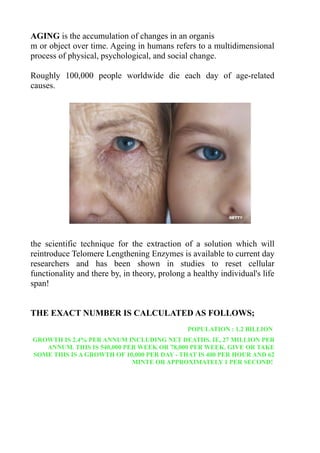
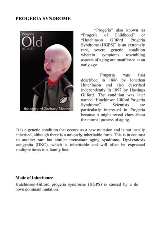
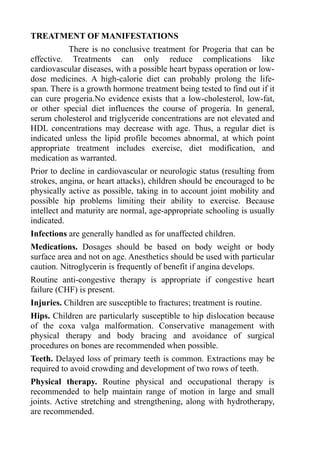
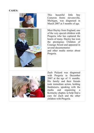
1. Bhargavi Saragadam
MSC II – Semester
Seminar on Progeria Syndrome
Department of Human Genetics
ANDHRA UNIVERSITY
2. AGING is the accumulation of changes in an organis
m or object over time. Ageing in humans refers to a multidimensional
process of physical, psychological, and social change.
Roughly 100,000 people worldwide die each day of age-related
causes.
the scientific technique for the extraction of a solution which will
reintroduce Telomere Lengthening Enzymes is available to current day
researchers and has been shown in studies to reset cellular
functionality and there by, in theory, prolong a healthy individual's life
span!
THE EXACT NUMBER IS CALCULATED AS FOLLOWS;
POPULATION : 1.2 BILLION
GROWTH IS 2.4% PER ANNUM INCLUDING NET DEATHS. IE, 27 MILLION PER
ANNUM. THIS IS 540,000 PER WEEK OR 78,000 PER WEEK. GIVE OR TAKE
SOME THIS IS A GROWTH OF 10,000 PER DAY - THAT IS 400 PER HOUR AND 62
MINTE OR APPROXIMATELY 1 PER SECOND!
3. TELOMERE THEORY
Telomeres (structures at the ends of chromosomes) have
experimentally been shown to shorten with each successive cell
division.
Shortened telomeres activate a mechanism that prevents further cell
multiplication. This may be an important mechanism of ageing in
tissues like bone marrow and the arterial lining where active cell
division is necessary.
In model organisms and laboratory settings, researchers have been
able to demonstrate that selected alterations in specific genes can
extend lifespan. At present, the biological basis of aging is unknown.
4. PROGERIA SYNDROME
“Progeria” also known as
“Progeria of Childhood” or
“Hutchinson Gilford Progeria
Syndrome (HGPS)” is an extremely
rare, severe genetic condition
wherein symptoms resembling
aspects of aging are manifested at an
early age.
Progeria was first
described in 1886 by Jonathan
Hutchinson and also described
independently in 1897 by Hastings
Gilford. The condition was later
named “Hutchinson Gilford Progeria
Syndrome”. Scientists are
particularly interested in Progeria
because it might reveal clues about
the normal process of aging.
It is a genetic condition that occurs as a new mutation and is not usually
inherited, although there is a uniquely inheritable form. This is in contrast
to another rare but similar premature aging syndrome, Dyskeratosis
congentia (DKC), which is inheritable and will often be expressed
multiple times in a family line.
Mode of Inheritance
Hutchinson-Gilford progeria syndrome (HGPS) is caused by a de
novo dominant mutation.
5. INCIDENCE
The disorder has a very low
incidence and occurs in one per
eight million live births and male
predominance with M:F ratio of
1.5:1 and a strong racial
susceptibility for Caucasians who
represent 97% of patients.
DISEASE CHARACTERISTICS
• Clinical features develop in childhood and resemble some features of
accelerated aging.
• Children with HGPS appear normal at birth. Profound failure to
thrive occurs during the first year.
• Characteristic faces, partial alopecia, loss of subcutaneous fat,
stiffness of joints, bone changes and abnormal tightness of the skin.
• Death usually occurs as a result of complications of cardiac or
cerebrovascular disease generally between 6 and 20 years of age,
with an average life span of approximately 13 years.
6. CLINICAL DIAGNOSIS
The diagnosis of Hutchinson-Gilford progeria syndrome (HGPS,
progeria) is based on recognition of common clinical features and the
presence of the LMNA p.Gly608Gly mutation.
The clinical manifestations include the following abnormalities,
which are almost always present after age three years:
• Growth
• Short stature and
stunted growth
• Weight distinctly
low for height
• Head
disproportionately
large for face
• Body fat
• Diminished
subcutaneous fat
• Prominent scalp
veins
8. • CVS. Severe, progressive
atherosclerosis with
widely variable age of
clinical manifestation
resulting in myocardial
infarction and stroke
• Other
• Prominent eyes
• Lagophthalmos
• Wide-based,
shuffling gait
• Failure to complete
secondary sexual
development
9. The following features are frequently present:
• Body fat. Prominent superficial veins
• Skin
• Thin, taut, dry, wrinkled skin that is brown-spotted in
various areas
• "Sclerodermatous" skin over lower abdomen and proximal
thighs, in which irregular bumps reflect underlying
lipodystrophy
• Loss of eyebrows and sometimes eyelashes
• Dystrophic nails
• Skeletal system. Persistently patent anterior fontanel
• Other
• Pinched nose, beaked nasal tip
• Faint nasolabial cyanosis
• Thin lips
• Protruding ears; lack of ear lobes
• Thin, high-pitched voice
Individuals having most of these features are considered to have the
classic Hutchinson-Gilford progeria syndrome. Individuals with either
more or less severe features are considered to have atypical progeria.
10. TESTING:
The clinical diagnosis of HGPS is based on recognition of
common clinical features and detection of the p.Gly608Gly mutation
in exon 11 of the LMNA gene, which is present in all individuals with
HGPS. Molecular genetic testing for this mutation is clinically
available.
Urinary hyaluronic acid. Although urinary hyaluronic acid has
been reported to be increased in most children with HGPS [Brown et
al 1990], the measurement is now regarded as unreliable [Gordon et al
2003] and is not recommended for diagnosis.
Molecular Genetic Testing
GeneReviews designates a molecular genetic test as clinically
available only if the test is listed in the GeneTests Laboratory
Directory by either a US CLIA-licensed laboratory or a non-US
clinical laboratory. GeneTests does not verify laboratory-submitted
information or warrant any aspect of a laboratory's licensure or
performance. Clinicians must communicate directly with the
laboratories to verify information.
11. Gene.
The p.Gly608Gly mutation in exon 11 of the LMNA gene is present
in all individuals with HGPS.
Clinical methods
• Confirmatory diagnostic testing
• Prenatal diagnosis
Clinical testing
• Targeted mutation analysis and sequence analysis can be used to
identify p.Gly608Gly, the recurrent de novo LMNA mutation in
exon 11 that defines HGPS.
12. Table 1. Molecular Genetic Testing Used in Hutchinson-Gilford
Progeria Syndrome
Mutation Test
Mutations
Test Method Detection Availab
Detected
Rate1 ility
Sequence
analysis
LMNA Clinical
Targeted p.Gly608Gly point 100% Testing
mutation mutation
analysis
13. 1. Hutchinson-Gilford progeria syndrome (HGPS, progeria) is defined
by the presence of the LMNA p.Gly608Gly mutation.
Gen Prot H
Chromos
e ein G
omal Locus Specific
Sym Na M
Locus
bol me D
Human Intermediate
Filament Database LMNA
(lamin C1)
Human Intermediate
Filament Database LMNA
(lamin A) L
Lam
LM Human Intermediate M
1q21.2 in-
NA Filament Database LMNA N
A/C
(lamin C2) A
The LMNA mutations
database
IPN Mutations, LMNA
Leiden Muscular
Dystrophy pages (LMNA)
Data are compiled from the following standard references: gene
symbol from HGNC; chromosomal locus, locus name, critical region,
complementation group from OMIM; protein name from UniProt.
14. PRENATAL TESTING
Prenatal diagnosis for HGPS is possible
by analysis of DNA extracted from fetal
cells obtained by amniocentesis usually
performed at approximately 15 to 18
weeks' gestation or chorionic villus
sampling (CVS) at approximately ten to
12 weeks' gestation. The disease-causing
allele of an affected family member must
be identified before prenatal testing can
be performed.
Note: (1) Because HGPS has thus far not
been reported to recur in families,
prenatal testing would only be performed
because of the (unlikely) possibility of
germline mosaicism in one of the parents.
(2) Gestational age is expressed as
menstrual weeks calculated either from
the first day of the last normal menstrual
period or by ultrasound measurements.
Preimplantation genetic diagnosis (PGD)
may be available for families in which the
disease-causing mutation has been
identified in an affected family member.
For laboratories offering PGD.
Note: Because HGPS has thus far not
been reported to recur in families, PGD
would only be performed because of the
(unlikely) possibility of germline
mosaicism in one of the parents.
15. MANAGEMENT
A regular diet is recommended; however, if the lipid profile
becomes abnormal, treatment includes exercise, diet modification, and
medication as warranted.
Age-appropriate schooling is usually recommended. Appropriate
medication dosage is based on body weight or body surface area
rather than age. Anesthetics should be used with caution.
Nitroglycerin is frequently of benefit if angina develops. Routine anti-
congestive therapy is appropriate if congestive heart failure (CHF) is
present.
Hip dislocation is best managed conservatively with physical therapy
and body bracing; surgery involving bones should be avoided if
possible.
Dental extractions may be required to avoid dental crowding. Routine
physical and occupational therapy, active stretching and strengthening
exercises, and hydrotherapy are recommended. Surveillance includes:
annual or semi-annual electrocardiogram (ECG), echocardiogram, and
carotid duplex scans; annual lipid profiles and dental examination and
x-ray; physical and occupational therapy multiple times per week; and
hip x-ray every few years to evaluate for avascular necrosis and
progressing coxa valga. Children with HGPS should avoid being in
the midst of large crowds with much taller/larger peers because of the
risk of injury.
16. GENETIC COUNSELING
Almost all individuals with HGPS have the disorder as the result
of a de novo dominant mutation. Because HGPS is caused by a de
novo mutation, the risk to the sibs of a proband is small. One instance
of apparent somatic and germline mosaicism has been reported. Thus,
the recurrence risk may be on the order of one in 500, as in other de
novo dominant mutations. Prenatal testing is available; however,
because of the low risk of recurrence, prenatal testing would only be
performed because of the (limited) possibility of germline mosaicism
in one of the parents.
Genetic counseling is the process of providing individuals and
families with information on the nature, inheritance, and implications
of genetic disorders to help them make informed medical and personal
decisions. The following section deals with genetic risk assessment
and the use of family history and genetic testing to clarify genetic
status for family members. This section is not meant to address all
personal, cultural, or ethical issues that individuals may face or to
substitute for consultation with a genetics professional. To find a
genetics or prenatal diagnosis clinic, see the GeneTests Clinic
Directory.
17. RISK TO FAMILY MEMBERS
Parents of a proband
• All probands with HGPS have the disorder as the result of a de
novo mutation.
• Parents of probands are not affected.
Sibs of a proband
• Because HGPS is caused by a de novo mutation, the risk to the
sibs of a proband is small.
• One instance of apparent somatic and germline mosaicism has
been reported [Wuyts et al 2005]. Therefore, the recurrence risk
may be on the order of one in 500, as in other de novo dominant
mutations.
• With the exception of two sets of identical twins with HGPS, the
authors are unaware of any convincing cases of a family with
more than one sib with classic HGPS.
Offspring of a proband. Individuals with HGPS do not reproduce.
Other family members of a proband. Because HGPS occurs as the
result of a de novo mutation, other family members of a proband are
not at increased risk.
18. GENETICALLY RELATED (ALLELIC) DISORDERS
More than ten other diseases and conditions with mutations or
variations in the LMNA gene have been identified. See OMIM
150330.
Progeroid laminopathy. The term "progeroid laminopathy" can be
used to describe phenotypes that resemble HGPS in which an LMNA
mutation other than p.Gly608Gly has been identified.
Approximately 10% of individuals with
clinically diagnosed HGPS have
uniparental isodisomy (UPD) of
chromosome 1 (including the LMNA
gene), a mosaic rearrangement of
chromosome 1, and a deletion involving
the LMNA gene locus [Eriksson et al
2003]. These individuals are
hypothesized to have somatic changes
that occur in vivo or in vitro, deleting the
mutated LMNA and providing a growth
advantage to that clone of cells.
Penetrance: Penetrance is complete.
Differential Diagnosis
The following are other syndromes that include some features of
premature aging:
• Neonatal progeroid syndrome (Weidemann-Rautenstrauch
syndrome)
• Acrogeria
• allermann-Streif syndrome
• Gerodermia osteodysplastica
• Berardinelli-Seip lipodystrophy ( generalized lipodystrophy)
• Petty-Laxova-Weidemann progeroid syndrome
• Ehlers-Danlos syndrome, progeroid form
• Werner syndrome.
19. EVALUATIONS FOLLOWING INITIAL DIAGNOSIS
To establish the extent of disease in an individuals diagnosed with
Hutchinson-Gilford progeria syndrome (HGPS), the following
evaluations are recommended:
• Establishement of vascular status using baseline
electrocardiogram (ECG), echocardiogram, and carotid duplex
scans for stenosis and for intimal thickness
• Skeletal x-ray to evaluate for characteristic findings:
acroosteolysis, clavicular resorption, and coxa valga
• Dual-energy x-ray absorptiometry (DEXA) to assess bone
mineral density
• Standard goniometry to assess global joint mobility
• Nutritional assessment to optimize caloric intake
20. TREATMENT OF MANIFESTATIONS
There is no conclusive treatment for Progeria that can be
effective. Treatments can only reduce complications like
cardiovascular diseases, with a possible heart bypass operation or low-
dose medicines. A high-calorie diet can probably prolong the life-
span. There is a growth hormone treatment being tested to find out if it
can cure progeria.No evidence exists that a low-cholesterol, low-fat,
or other special diet influences the course of progeria. In general,
serum cholesterol and triglyceride concentrations are not elevated and
HDL concentrations may decrease with age. Thus, a regular diet is
indicated unless the lipid profile becomes abnormal, at which point
appropriate treatment includes exercise, diet modification, and
medication as warranted.
Prior to decline in cardiovascular or neurologic status (resulting from
strokes, angina, or heart attacks), children should be encouraged to be
physically active as possible, taking in to account joint mobility and
possible hip problems limiting their ability to exercise. Because
intellect and maturity are normal, age-appropriate schooling is usually
indicated.
Infections are generally handled as for unaffected children.
Medications. Dosages should be based on body weight or body
surface area and not on age. Anesthetics should be used with particular
caution. Nitroglycerin is frequently of benefit if angina develops.
Routine anti-congestive therapy is appropriate if congestive heart
failure (CHF) is present.
Injuries. Children are susceptible to fractures; treatment is routine.
Hips. Children are particularly susceptible to hip dislocation because
of the coxa valga malformation. Conservative management with
physical therapy and body bracing and avoidance of surgical
procedures on bones are recommended when possible.
Teeth. Delayed loss of primary teeth is common. Extractions may be
required to avoid crowding and development of two rows of teeth.
Physical therapy. Routine physical and occupational therapy is
recommended to help maintain range of motion in large and small
joints. Active stretching and strengthening, along with hydrotherapy,
are recommended.
21. Surveillance
The following are appropriate:
• ECG, echocardiogram, and carotid duplex scans annually or
semi-annually to monitor for cardiovascular disease (Children
may experience severe carotid artery atherosclerotic blockage
prior to any significant ECG changes.)
• Yearly lipid profiles
• Yearly dental examination and x-ray
• Physical and occupational therapy multiple times per week
• Hip x-rays every few years to evaluate for avascular necrosis and
progressing coxa valga
Agents/Circumstances to Avoid
Children should avoid being in the midst of large crowds with much
taller and larger peers because of the increased risk of injury.
22. CASES:
This beautiful little boy
Cameron froms stevensville,
Michigan, was diagnosed in
March 2007 at 5 months of age.
Meet Hayley from England, one
of the very special children with
Progeria who has captured the
hearts of many. Hayley has won
the prestigious Children of
Courage Award and appeared in
several documentaries
and other media stories about
Progeria.
Zach Pickard was diagnosed
with Progeria in December
2007 at the age of 11 months.
His family and their friends
took immediate action, holding
fundraisers, speaking with the
media and organizing a
Kentucky chapter, to help find a
cure for Zach and the other
children with Progeria.
23. The series of portraits depicts
the 24 year old Leon Botha, one
of the world’s longest surviving
Progeria sufferers.
He is an Artist.
While the average life expectancy of a child with Progeria is less than
14 years, it is believed that the oldest case ever recorded 26 years of
age. Not true. The oldest case ever recorded was a Japanese man who
lived with the disease for 45 years.
In this particular case, the child did not show signs of growth
retardation until around 12 years of age. It was noted, however, that
his head was larger than normal at the age of one and he did
experience hair loss in childhood, but enjoyed a rather normal life for
12 years. By age 20, this particular subject had total Alopecia and
aging began to accelerate. The subject died at the age of 45 from
myocardial infarction.
24. PROGERIA CASES IN INDIA
Progeria is a debilitating, rare illness and genetic disorder with just 45
odd cases in the world. The disease which infects one in four lakh
people, is present in India too. Bisul khan and Razia Khatooon,’s
family in Chhapra, Bihar; has seven children, of which five are
Progeria patients.
Out Of the five, three daughters,
Guriya, Rehana and Rubina are
dead; having passed away at the
ages of 17,24 and 13 respectively.
Two sons Ikramul (23) and Ali
Hosain (22) are still alive, but
their medical ages are 70 and 66.
Two children Sanjita (21 year
old) and Gulab Shah(7 year old)
are normal.
The sad thing is that the villagers
ostracized the Progeria-stricken
family from Chhapra in 2003
because the children were
considered bad omen.
Doctors Apurba Ghosh, director of the Institute of Children Health,
Kolkata and Dr Chandan Chatterjee from Switzerland, were the first
to diagnose this disorder. Rubena’s Pneumonia treatment convinced
the medical team that the kids are affected with Progeria. Bisul Khan’s
family in India, is the ONLY FAMILY in the world that has more than
one case of progeria (five to be prescise). Dr. Ghosh says, “There are
only 40 known cases world-wide, all isolated and seemingly
random.On average, a progeria child will die by 17. They develop
striking physical symptoms - premature baldness, heart disease,
thinning bones and arthritis.”The family of Progeria was a curious
case to the medical team of ICH and Chandan Chattopadhyay, who
found out that the family had a defective gene, known as Amino AC.T
25. There is a trust called by S B Devi Charity home which takes care of
the Progeria family’s needs, financial problems and medical
requirements. It is to the credit of good medication and regular
psychological sessions that Ali and Ikramul have been able to survive
for so long. Though death is imminent, the children enjoy their lives
and have fun. They can eat only in small measure(like 3-4 year olds
do). Paa is a Progeria based movie.
Ikramul and Ali suffer from astro-arthritis and cannot bend their legs
or sit properly. Their bodies are very weak and their liver and heart are
under-developed. The longest lifespan for progeria in the world is 23.
Unfortunately, both the kids are nearing that age. They are born in an
extremely poor father, who earns Rs 2,500 a month as a security
guard, in a private company. They are dependent on their mother,
Rajia, for eating, bathing and virtually their every need.
Razia's family is the only family in the world, which had five cases of
Progeria.
She has already lost her two sons and a daughter to this disease and is
hoping for a miracle to save her two sons.
26. Nature is a mutable cloud which is
always and never the same
Thank You