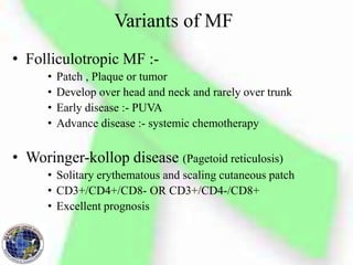
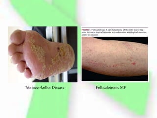
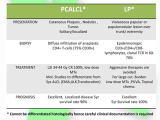
Primary Cutaneous
Lymphoma
Dr. Shashank Bansal
MD Radiotherapy PG
BBCI Guwahati
Definition
• Accumulation of malignant Lymphoid cells in
the skin without evidence of extracutaneous
disease at the time of presentation.
• Important to distinguish it from Nodal
Lymphoma with skin infiltration
Classification
Incidence
• In western countries annual incidence 1/100000 .
• Cutaneous T cell lymphoma (CTCL) constitutes 75-80% of all
cases (1)
– Mycosis fungoides is most common CTCL
• Cutaneous B cell Lymphoma (CBCL) constitutes 20-25% of
all cases. (1)
• Incidence in PBCR , KUD :- NHL 1.66 % of total cancer
(2012-2014)
• BBCI in year 2015 of all cases presented 2.30% was
diagnosed with NHL.
(1) Willemze R, Jaffe ES, Burg G et al. WHO–EORTC classification for cutaneous lymphomas. Blood 2005;
105: 3768–3785.
Mycosis Fungoides
• Most common type of CTCL
• Most common in Males (M:F::1.6-2:1)
• Median age of presentation 55 to 60 yrs
• Precise Etiology remains Elusive.
Mycosis Fungoides first described in 1806 by Jean-Louis-Marc Alibert
in his Atlas of Dermatoses “ description des maladies de la peau”
• Cutaneous disease
• Leukemic CTCL and Sezary Syndrome
• Extracutaneous disease
• Cutaneous disease :- Pruritis in involved area
• Evolution of skin lesion
– Premycotic Phase
• Red scaled macular or patch like lesion in sun shielded areas
• Unstable and Regress
• Biopsy not diagnostic b/o paucity of Lymphocytes
– Patch Phase
• Any size lesion without induration or significant elevation
above the surrounding uninvolved skin
• Poikiloderma may be present
– Plaque Phase ( dense infiltration by malignant cells )
• Any size lesion that is elevated or indurated
• Crusting or poikiloderma may be present
– Tumor Phase
• Plaque may evolve into cutaneous tumor
• Any solid or nodular mass > 1 cm in diameter with
evidence of deep infiltration in the skin and /or vertical
growth
• MF may also progress or present with
erythroderma
• Leukemic CTCL and Sezary Syndrome (SS)
– In rare CTCL cases circulating malignant T-cells are
identified in peripheral blood.
– Most commonly occurs in association with erythroderma
but may occur in patient with minimal cutaneous disease
– CD4+/CD7- or CD4+/CD26- immunophenotype
– Disease burden is explained by B staging.
– Distinction between Erythrodermic MF with leukemic
involvement and SS is a controversy
– SS defined as combination of erythroderma with B2
disease.
Cerebriform nuclei of Sezary cell
• Extracutaneous disease
– Involvement of regional lymph node or
extracutaneous organ system
– Most commonly involved organ are Lungs , Oral
cavity , Pharynx ,CNS.
REVISED ISCL/EORTC MYCOSIS FUNGOIDES STAGING
NCI-VA Classification Dutch classification
Number of atypical lymphocytes with
cerebriform nuclei (cerebriform cells) in
the paracortical areas
Use nuclear diameter of the cerebriform
cells to distinguish dermatopathic
lymphadenopathy
LN1 :-occasional and isolated atypical
lymphocytes not arranged in clusters
Grade 1 :- dermatopathic
lymphadenopathy(DL) Alone
LN2:- many atypical lymphocytes occurring
singly or in three to six cell clusters
Grade 2:- DL with isolated cerebriform
mononuclear cells with nuclei greater than
7.5 micron.
LN3 :- atypical lymphocytes arranged in
aggregates with nodal architecture essentially
preserved.
Grade 3 :- many large cerebriform
mononuclear cells and partial distortion of
the normal LN architecture
LN4 :- partial or complete replacement of
LN architecture by atypical lymphocytes or
frankly neoplastic cells
Grade 4 :- complete effacement of LN by
frankly neoplastic cells
Molecular pathology of
mycosis fungoides .
Activated skin-homing T cells
extravasate through the dermal
capillaries due to interactions of
cutaneous lymphocyte antigen (CLA)
and chemokine receptor 4 (CCR4) with
their respective ligands on the dermal
capillaries, E-selectin, and chemokine
ligand 17 (CCL17). T cells then migrate
to the epidermis and interact with
antigen-presenting dendritic cells
Diagnosis
• History and physical examination
» Duration, Evolution, % Area involved
» Presence or Absence of cutaneous tumor
» Evaluation of Lymphatic system
» Apropriate images of cutaneous lesion as guide for therapy
response .
• Skin Biopsy
» From minimum two distinct sites
» Excisional biopsy from LN (priority should be given to
largest LN with SUV max.)
• Laboratory and imaging studies
» CBC
» LFT
» LDH
» Peripheral blood examination (Extracutaneous disease)
» Immunophenotype (CD 2+,3+,4+,5+,45RO+,8-,30-)
» PCR for T-cell receptor gene reassortment
» Bone marrow biopsy (Extracutaneous disease)
» CT scan Chest, Abdo. ,Pelvis (Except patch/plaque/limited
disease)
» PET-CT > CT Scan.
Diagnosis contd…
DIAGNOSTIC CRITERIA FOR EARLY STAGE MYCOSIS
FUNGOIDES
• The majority of patients present with early
stage disease (30% IA, 25% IB, 11% IIA, 16%
IIB, 3% IIIA, 8% IIIB, 6% IVA, and 1% IVB)
• Only 7% of patients presented with peripheral
blood involvement
Treatment
Local Superficial Irradiation
• Used in early Stage 1a MF
• Single abutting Radiation fields are used
• Treatment fields should be designed to encompass the
entirety of the lesion (determined by visual inspection,
palpation, and/or appropriate imaging)
• 1- to 2-cm margin is given
• lead or Cerrobend cut out are used to conform field borders
• EORTC recommends that the 80% isodose line is set at the
deep border of the dermis approx 4.5mm
• S/E :- mild dermatitis, local alopecia, and pigmentation
changes.
• Radical dose 30-36Gy and 8Gy in 2 # for Palliation.
TSEBT (Total Skin Electron Beam Therapy )
• Technique :-
• Accomplished with 6- to 9-MeV electrons
• Patient standing behind a polycarbonate screen ~3.8 meters from
the linear accelerator head
• Treatment is provided in “cycles,” with one cycle composed of
treatment of the patient in six different positions over 2 days (three
positions each day)
• A dual-field technique is used to deliver treatment to a superior and
inferior field by angling the gantry 16 to 17.5 degrees above and
below horizontal.
• Six position treatment deliver maximum dose to a depth of 1mm,
80% dose to 6-7mm.
• A 3.2-mm polycarbonate screen (Lexan) is placed in front of the
patient which serves to attenuate and scatter the incident electrons,
resulting in an electron energy of 3.9 MeV at the skin surface
• Dose and Fractionation
• TSEBT provided with a relatively protracted course of 2
cycles/ wk for 9wks which provide a total dose of 36Gy
to the skin surface
• Complete response is dependent on radiation dose
DOSE (Gy) CR (%)
10 18
10-20 55
20-25 66
25-30 75
30-36 94
• Lower dose yields impressive overall response
,overall survival, PFS, Relapse free survival
rate
• Relapse rates are even higher in patient in
which CR is obtained and the absolute benefit
of CR relative to the increased side effect at
higher dose of TSEBT is unclear.
Supplemental treatment
• Required for soles, perineum and scalp
• Accomplished by use of 120 kV superficial photon with HVL
4.2 mm of Al. , or low energy (6 MeV) Electron with 1cm
bolus to treat soles and perineum (1Gy/#) and scalp is treated
with an angled electron reflector above the patient
Side Effects
• m/c flaring up of pruritis and erythema
• Acute s/e :-
» Xerosis
» Dry desquamation
» Extremity edema, Blister, Bullae Formation ,alopecia , nail
changes .
» Hypohydrosis
» Dryness of Nose
» Gyanaecomastia (rare)
• Chronic s/e :-
» Cataract
» Chronic xerosis
» Persistent alopecia
» Dystrophic nails
» Secondry skin cancer
» melanomas
Prevention is done
by additional
shielding of Eye
,Lips, Testes and
Fingernails.
Eye shield to protect Cornea
and Lens
Nail shields to protect Nail bed
1) Testicular shield to reduce dose to testicles lined by 1.5 cm wax
2) Hand Gloves are used after certain fractions to reduce dose to
finger.
Radiation Dermatitis Late effects:skin atrophy and
telangiectasia
• Clinical Efficacy :-
• T1 lesion :-
– CR > 90 % , 15 Relapse free survival (RFS) 40%
– Recurrence is limited to <5 % of skin amenable to salvage therapy
• T2 lesion :-
– CR 76-90 % , Relapse free survival 50% @ 5yr, 10% @ 10yr
– Adjuvant PUVA therapy improves RFS to 85 % @5 yr
– Adjuvant Nitrogen Mustard :- RFS 40 % @ 10 yr
• T3 lesion :-
– Less effective but better results than other therapies
– CR 44-54 %
– Adjuvant Nitrogen Mustard may increase durability of response
• T4 lesion :-
– 70-100 % response rates (T4N0)
– 5 yr PFS 25-69%
– In Extracutaneous and peripheral circulation involvement PFS
decreases further .
Palliative Radiotherapy
• Given in symptomatic patients with visceral
and nodal disease .
• Dose :- 12-30 Gy
• Similar dose can also be used in patients with
recurrence in extremities
Systemic therapies (options)
• Skin directed therapy should be tried in early
diseases but in advance cases systemic
therapies can be considered
• Oral Bexarotene 54% response rates in refractory CTCL
• Denileukin diftitox ( fusion protein IL2 and diphtheria
toxin) 30% response rate in rCTCL.
• Histone deacetylase inhibitor (vorinostat and
romidepsin)
• Alemtuzumab (Monoclonal antibody CD52)
• Single agent Chemotherapy (liposomal doxirubicin ,
methotrexate )
• Autologus or Allogenic Stem Cell Transplant
– May be considered in patients refractory to other therapies
– Additional studies required
• Transformed disease (Large cell transformation)
– Occurs in 8-39% patients
– identified by >25 % large cells in samples
– High level of beta2 micro globulin and LDH > risk of LCT
– Treatment options
• Systemic chemotherapy
• Local radiation for localized disease
.
Variants of MF
• Folliculotropic MF :-
• Patch , Plaque or tumor
• Develop over head and neck and rarely over trunk
• Early disease :- PUVA
• Advance disease :- systemic chemotherapy
• Woringer-kollop disease (Pagetoid reticulosis)
• Solitary erythematous and scaling cutaneous patch
• CD3+/CD4+/CD8- OR CD3+/CD4-/CD8+
• Excellent prognosis
Woringer-kollop Disease Folliculotropic MF
• Granulomatous slack skin
• Cutaneous infiltration of malignant T-cell
• Associated with increased incidence of secondry
lymphomas (HL)
• Hypopigmented MF
• Presents at earlier age
• Development of hypopigmented patches in trunk
/extremity
Granulomatous slack skin Hypopigmented MF
Non MF CTCL
• CD30+ Lymphoproliferative disorder
– Median age at diagnosis 61 yr
– 5yr disease specific survival 92%
– Localisation of disease in head and neck region is
poor prognostic factor
– Types :-
• Primary Cutaneous Anaplastic Large Cell Lymphoma
• Lymphomatoid papulosis
PCALCL* LP*
PRESENTATION Cutaneous Plaques , Nodules ,
Tumor.
Solitary/localized
Violaceous papular or
papulonodular lesion over
trunk/ extremity
BIOPSY Diffuse infiltration of anaplastic
CD4+ T-cells (75% CD30+)
Epidermotropic
CD3+/CD4+/CD8-
lymphocytes, clonal TCR in 60-
70%
TREATMENT LSI 34-44 Gy CR 100%, low dose
MTx
Mol. Studies to differentiate from
Sys ALCL (EMA,ALK,Translocation)
Aggressive therapies are
avoided
For large cut. Burden
Low dose MTx ,PUVA, Topical
chemo.
PROGNOSIS Excellent, Localized disease 5yr
survival rate 90%
Excellent
5yr Survival rate 100%
* Cannot be differentiated histologically hence careful clinical documentation is required
Primary Cutaneous
Anaplastic Large
Cell Lymphoma
Lymphomatoid papulosis
Non MF CTCL
EXTRANODAL
NK-TCL,Nasal type
SUBCUTANEOUS
PANNICULITIS like -
TCL
PRESENTATION Originate in nasal cavity may
present with cutaneous lesion
Subcutaneous nodule/plaques
commonly over legs/trunk
BIOPSY Dense Lymphoid infiltrate
exhibiting epidermotropic
component CD3-/CD2+/CD56+
Lymphoid infiltrate composed of
CD8+ cytotoxic T-cell
TREATMENT Extended Field RT (50Gy)
CR 94.5% ,
Adjuvant L-Asparaginase prevent
racurrence
Combination of RT and
Corticosteroids
NOTE Directly associated with EBV Heamophagocytic syndrome
predicts a worse prognosis
Cutaneous B- Cell Lymphoma
PCFCL PCMZBCL
PRESENTATION Clustered Plaques and
Tumor on scalp and
forehead of trunk
Isolated or multifocal
violaceous plaques /
nodules on trunk
/extremities
BIOPSY Lymphoid infiltrate with
varying proportion of
centrocytes, centroblasts ,
reactive T-cells .
Composed of
lymphoplasmacytoid cells ,
Plasma cells, centroblast like
and immunoblast like cells
IMMUNOPHENOTYPE CD20+
CD79a+
CD20+
CD79a+
CD5- / CD10-
PCFCL :- PRIMARY CUTANEOUS FOLLICLE CENTRE LYMPHOMA
PCMZBCL:- PRIMAY CUTANEOUS MARZINAL ZONE B-CELL LYMPHOMA
TREATMENT PCFCL PCMZBCL
LOCAL SUPERFICIAL
IRRADIATION
Dose 20-54 Gy
Margin 0.5-5cm
CR 99%, RR 30%
Dose 30-45 Gy
Field Margin 1-5cm
CR 99% ,RR 46 %
SURGERY (EXCISION) CR 98%, RR 40% CR 99%, RR 43 %
INF ALFA CR 100 % , RR 29 % CR 100 %, RR 25 %
RITUXIMAB Overall response 100%
CR 80%
Overall response 60 %
CHEMOTHERAPY MULTIAGENT (CHOP)
CR 85%, RR 48%
MULTIAGENT (CHOP)
CR 85%, RR 57%
TREATMENT OPTION BASED ON ISCL/EORTC LITERATURE REVIEW, NO RANDOMISE
DATA TO ESTABLISH TREATMENT PROTOCOL
Follow-up
• The frequency of follow-up visits depends on the type
of PCL and the stage of disease. It may vary from
every 6 or 12 months in patients with indolent types
of PCL and stable disease or patients in complete
remission .
• 4–6 weeks in patients with active or progressive
disease. Follow-up visits should focus on history and
physical examination, and additional testing
(histology, blood examination, imaging, etc.) should
only be carried out if required.
Conclusion
• A correct histopathologic diagnosis is
important in determination of appropriate
management
• Due to rarity of disease numerous question
regarding patho-physiology and optimal
treatment remain unanswered and international
collaborations are required for better
understanding.
Primary cutaneous lymphoma

Primary cutaneous lymphoma

  • 1.
    Primary Cutaneous Lymphoma Dr. ShashankBansal MD Radiotherapy PG BBCI Guwahati
  • 3.
    Definition • Accumulation ofmalignant Lymphoid cells in the skin without evidence of extracutaneous disease at the time of presentation. • Important to distinguish it from Nodal Lymphoma with skin infiltration
  • 4.
  • 5.
    Incidence • In westerncountries annual incidence 1/100000 . • Cutaneous T cell lymphoma (CTCL) constitutes 75-80% of all cases (1) – Mycosis fungoides is most common CTCL • Cutaneous B cell Lymphoma (CBCL) constitutes 20-25% of all cases. (1) • Incidence in PBCR , KUD :- NHL 1.66 % of total cancer (2012-2014) • BBCI in year 2015 of all cases presented 2.30% was diagnosed with NHL. (1) Willemze R, Jaffe ES, Burg G et al. WHO–EORTC classification for cutaneous lymphomas. Blood 2005; 105: 3768–3785.
  • 6.
    Mycosis Fungoides • Mostcommon type of CTCL • Most common in Males (M:F::1.6-2:1) • Median age of presentation 55 to 60 yrs • Precise Etiology remains Elusive.
  • 7.
    Mycosis Fungoides firstdescribed in 1806 by Jean-Louis-Marc Alibert in his Atlas of Dermatoses “ description des maladies de la peau”
  • 8.
    • Cutaneous disease •Leukemic CTCL and Sezary Syndrome • Extracutaneous disease
  • 9.
    • Cutaneous disease:- Pruritis in involved area • Evolution of skin lesion – Premycotic Phase • Red scaled macular or patch like lesion in sun shielded areas • Unstable and Regress • Biopsy not diagnostic b/o paucity of Lymphocytes – Patch Phase • Any size lesion without induration or significant elevation above the surrounding uninvolved skin • Poikiloderma may be present – Plaque Phase ( dense infiltration by malignant cells ) • Any size lesion that is elevated or indurated • Crusting or poikiloderma may be present
  • 10.
    – Tumor Phase •Plaque may evolve into cutaneous tumor • Any solid or nodular mass > 1 cm in diameter with evidence of deep infiltration in the skin and /or vertical growth • MF may also progress or present with erythroderma
  • 12.
    • Leukemic CTCLand Sezary Syndrome (SS) – In rare CTCL cases circulating malignant T-cells are identified in peripheral blood. – Most commonly occurs in association with erythroderma but may occur in patient with minimal cutaneous disease – CD4+/CD7- or CD4+/CD26- immunophenotype – Disease burden is explained by B staging. – Distinction between Erythrodermic MF with leukemic involvement and SS is a controversy – SS defined as combination of erythroderma with B2 disease.
  • 13.
  • 14.
    • Extracutaneous disease –Involvement of regional lymph node or extracutaneous organ system – Most commonly involved organ are Lungs , Oral cavity , Pharynx ,CNS.
  • 15.
    REVISED ISCL/EORTC MYCOSISFUNGOIDES STAGING
  • 16.
    NCI-VA Classification Dutchclassification Number of atypical lymphocytes with cerebriform nuclei (cerebriform cells) in the paracortical areas Use nuclear diameter of the cerebriform cells to distinguish dermatopathic lymphadenopathy LN1 :-occasional and isolated atypical lymphocytes not arranged in clusters Grade 1 :- dermatopathic lymphadenopathy(DL) Alone LN2:- many atypical lymphocytes occurring singly or in three to six cell clusters Grade 2:- DL with isolated cerebriform mononuclear cells with nuclei greater than 7.5 micron. LN3 :- atypical lymphocytes arranged in aggregates with nodal architecture essentially preserved. Grade 3 :- many large cerebriform mononuclear cells and partial distortion of the normal LN architecture LN4 :- partial or complete replacement of LN architecture by atypical lymphocytes or frankly neoplastic cells Grade 4 :- complete effacement of LN by frankly neoplastic cells
  • 17.
    Molecular pathology of mycosisfungoides . Activated skin-homing T cells extravasate through the dermal capillaries due to interactions of cutaneous lymphocyte antigen (CLA) and chemokine receptor 4 (CCR4) with their respective ligands on the dermal capillaries, E-selectin, and chemokine ligand 17 (CCL17). T cells then migrate to the epidermis and interact with antigen-presenting dendritic cells
  • 18.
    Diagnosis • History andphysical examination » Duration, Evolution, % Area involved » Presence or Absence of cutaneous tumor » Evaluation of Lymphatic system » Apropriate images of cutaneous lesion as guide for therapy response . • Skin Biopsy » From minimum two distinct sites » Excisional biopsy from LN (priority should be given to largest LN with SUV max.)
  • 19.
    • Laboratory andimaging studies » CBC » LFT » LDH » Peripheral blood examination (Extracutaneous disease) » Immunophenotype (CD 2+,3+,4+,5+,45RO+,8-,30-) » PCR for T-cell receptor gene reassortment » Bone marrow biopsy (Extracutaneous disease) » CT scan Chest, Abdo. ,Pelvis (Except patch/plaque/limited disease) » PET-CT > CT Scan. Diagnosis contd…
  • 20.
    DIAGNOSTIC CRITERIA FOREARLY STAGE MYCOSIS FUNGOIDES
  • 21.
    • The majorityof patients present with early stage disease (30% IA, 25% IB, 11% IIA, 16% IIB, 3% IIIA, 8% IIIB, 6% IVA, and 1% IVB) • Only 7% of patients presented with peripheral blood involvement
  • 22.
  • 23.
    Local Superficial Irradiation •Used in early Stage 1a MF • Single abutting Radiation fields are used • Treatment fields should be designed to encompass the entirety of the lesion (determined by visual inspection, palpation, and/or appropriate imaging) • 1- to 2-cm margin is given • lead or Cerrobend cut out are used to conform field borders • EORTC recommends that the 80% isodose line is set at the deep border of the dermis approx 4.5mm • S/E :- mild dermatitis, local alopecia, and pigmentation changes. • Radical dose 30-36Gy and 8Gy in 2 # for Palliation.
  • 24.
    TSEBT (Total SkinElectron Beam Therapy ) • Technique :- • Accomplished with 6- to 9-MeV electrons • Patient standing behind a polycarbonate screen ~3.8 meters from the linear accelerator head • Treatment is provided in “cycles,” with one cycle composed of treatment of the patient in six different positions over 2 days (three positions each day) • A dual-field technique is used to deliver treatment to a superior and inferior field by angling the gantry 16 to 17.5 degrees above and below horizontal. • Six position treatment deliver maximum dose to a depth of 1mm, 80% dose to 6-7mm. • A 3.2-mm polycarbonate screen (Lexan) is placed in front of the patient which serves to attenuate and scatter the incident electrons, resulting in an electron energy of 3.9 MeV at the skin surface
  • 28.
    • Dose andFractionation • TSEBT provided with a relatively protracted course of 2 cycles/ wk for 9wks which provide a total dose of 36Gy to the skin surface • Complete response is dependent on radiation dose DOSE (Gy) CR (%) 10 18 10-20 55 20-25 66 25-30 75 30-36 94
  • 29.
    • Lower doseyields impressive overall response ,overall survival, PFS, Relapse free survival rate • Relapse rates are even higher in patient in which CR is obtained and the absolute benefit of CR relative to the increased side effect at higher dose of TSEBT is unclear.
  • 30.
    Supplemental treatment • Requiredfor soles, perineum and scalp • Accomplished by use of 120 kV superficial photon with HVL 4.2 mm of Al. , or low energy (6 MeV) Electron with 1cm bolus to treat soles and perineum (1Gy/#) and scalp is treated with an angled electron reflector above the patient
  • 31.
    Side Effects • m/cflaring up of pruritis and erythema • Acute s/e :- » Xerosis » Dry desquamation » Extremity edema, Blister, Bullae Formation ,alopecia , nail changes . » Hypohydrosis » Dryness of Nose » Gyanaecomastia (rare) • Chronic s/e :- » Cataract » Chronic xerosis » Persistent alopecia » Dystrophic nails » Secondry skin cancer » melanomas Prevention is done by additional shielding of Eye ,Lips, Testes and Fingernails.
  • 32.
    Eye shield toprotect Cornea and Lens Nail shields to protect Nail bed
  • 33.
    1) Testicular shieldto reduce dose to testicles lined by 1.5 cm wax 2) Hand Gloves are used after certain fractions to reduce dose to finger.
  • 34.
    Radiation Dermatitis Lateeffects:skin atrophy and telangiectasia
  • 35.
    • Clinical Efficacy:- • T1 lesion :- – CR > 90 % , 15 Relapse free survival (RFS) 40% – Recurrence is limited to <5 % of skin amenable to salvage therapy • T2 lesion :- – CR 76-90 % , Relapse free survival 50% @ 5yr, 10% @ 10yr – Adjuvant PUVA therapy improves RFS to 85 % @5 yr – Adjuvant Nitrogen Mustard :- RFS 40 % @ 10 yr • T3 lesion :- – Less effective but better results than other therapies – CR 44-54 % – Adjuvant Nitrogen Mustard may increase durability of response • T4 lesion :- – 70-100 % response rates (T4N0) – 5 yr PFS 25-69% – In Extracutaneous and peripheral circulation involvement PFS decreases further .
  • 36.
    Palliative Radiotherapy • Givenin symptomatic patients with visceral and nodal disease . • Dose :- 12-30 Gy • Similar dose can also be used in patients with recurrence in extremities
  • 37.
    Systemic therapies (options) •Skin directed therapy should be tried in early diseases but in advance cases systemic therapies can be considered • Oral Bexarotene 54% response rates in refractory CTCL • Denileukin diftitox ( fusion protein IL2 and diphtheria toxin) 30% response rate in rCTCL. • Histone deacetylase inhibitor (vorinostat and romidepsin) • Alemtuzumab (Monoclonal antibody CD52) • Single agent Chemotherapy (liposomal doxirubicin , methotrexate )
  • 38.
    • Autologus orAllogenic Stem Cell Transplant – May be considered in patients refractory to other therapies – Additional studies required • Transformed disease (Large cell transformation) – Occurs in 8-39% patients – identified by >25 % large cells in samples – High level of beta2 micro globulin and LDH > risk of LCT – Treatment options • Systemic chemotherapy • Local radiation for localized disease .
  • 39.
    Variants of MF •Folliculotropic MF :- • Patch , Plaque or tumor • Develop over head and neck and rarely over trunk • Early disease :- PUVA • Advance disease :- systemic chemotherapy • Woringer-kollop disease (Pagetoid reticulosis) • Solitary erythematous and scaling cutaneous patch • CD3+/CD4+/CD8- OR CD3+/CD4-/CD8+ • Excellent prognosis
  • 40.
  • 41.
    • Granulomatous slackskin • Cutaneous infiltration of malignant T-cell • Associated with increased incidence of secondry lymphomas (HL) • Hypopigmented MF • Presents at earlier age • Development of hypopigmented patches in trunk /extremity
  • 42.
    Granulomatous slack skinHypopigmented MF
  • 44.
    Non MF CTCL •CD30+ Lymphoproliferative disorder – Median age at diagnosis 61 yr – 5yr disease specific survival 92% – Localisation of disease in head and neck region is poor prognostic factor – Types :- • Primary Cutaneous Anaplastic Large Cell Lymphoma • Lymphomatoid papulosis
  • 45.
    PCALCL* LP* PRESENTATION CutaneousPlaques , Nodules , Tumor. Solitary/localized Violaceous papular or papulonodular lesion over trunk/ extremity BIOPSY Diffuse infiltration of anaplastic CD4+ T-cells (75% CD30+) Epidermotropic CD3+/CD4+/CD8- lymphocytes, clonal TCR in 60- 70% TREATMENT LSI 34-44 Gy CR 100%, low dose MTx Mol. Studies to differentiate from Sys ALCL (EMA,ALK,Translocation) Aggressive therapies are avoided For large cut. Burden Low dose MTx ,PUVA, Topical chemo. PROGNOSIS Excellent, Localized disease 5yr survival rate 90% Excellent 5yr Survival rate 100% * Cannot be differentiated histologically hence careful clinical documentation is required
  • 46.
    Primary Cutaneous Anaplastic Large CellLymphoma Lymphomatoid papulosis
  • 47.
    Non MF CTCL EXTRANODAL NK-TCL,Nasaltype SUBCUTANEOUS PANNICULITIS like - TCL PRESENTATION Originate in nasal cavity may present with cutaneous lesion Subcutaneous nodule/plaques commonly over legs/trunk BIOPSY Dense Lymphoid infiltrate exhibiting epidermotropic component CD3-/CD2+/CD56+ Lymphoid infiltrate composed of CD8+ cytotoxic T-cell TREATMENT Extended Field RT (50Gy) CR 94.5% , Adjuvant L-Asparaginase prevent racurrence Combination of RT and Corticosteroids NOTE Directly associated with EBV Heamophagocytic syndrome predicts a worse prognosis
  • 48.
    Cutaneous B- CellLymphoma PCFCL PCMZBCL PRESENTATION Clustered Plaques and Tumor on scalp and forehead of trunk Isolated or multifocal violaceous plaques / nodules on trunk /extremities BIOPSY Lymphoid infiltrate with varying proportion of centrocytes, centroblasts , reactive T-cells . Composed of lymphoplasmacytoid cells , Plasma cells, centroblast like and immunoblast like cells IMMUNOPHENOTYPE CD20+ CD79a+ CD20+ CD79a+ CD5- / CD10- PCFCL :- PRIMARY CUTANEOUS FOLLICLE CENTRE LYMPHOMA PCMZBCL:- PRIMAY CUTANEOUS MARZINAL ZONE B-CELL LYMPHOMA
  • 49.
    TREATMENT PCFCL PCMZBCL LOCALSUPERFICIAL IRRADIATION Dose 20-54 Gy Margin 0.5-5cm CR 99%, RR 30% Dose 30-45 Gy Field Margin 1-5cm CR 99% ,RR 46 % SURGERY (EXCISION) CR 98%, RR 40% CR 99%, RR 43 % INF ALFA CR 100 % , RR 29 % CR 100 %, RR 25 % RITUXIMAB Overall response 100% CR 80% Overall response 60 % CHEMOTHERAPY MULTIAGENT (CHOP) CR 85%, RR 48% MULTIAGENT (CHOP) CR 85%, RR 57% TREATMENT OPTION BASED ON ISCL/EORTC LITERATURE REVIEW, NO RANDOMISE DATA TO ESTABLISH TREATMENT PROTOCOL
  • 50.
    Follow-up • The frequencyof follow-up visits depends on the type of PCL and the stage of disease. It may vary from every 6 or 12 months in patients with indolent types of PCL and stable disease or patients in complete remission . • 4–6 weeks in patients with active or progressive disease. Follow-up visits should focus on history and physical examination, and additional testing (histology, blood examination, imaging, etc.) should only be carried out if required.
  • 51.
    Conclusion • A correcthistopathologic diagnosis is important in determination of appropriate management • Due to rarity of disease numerous question regarding patho-physiology and optimal treatment remain unanswered and international collaborations are required for better understanding.