Presentation1, x ray film reading of the chest.
•Download as PPTX, PDF•
32 likes•2,407 views

Health &medicine

Recommended
More Related Content
What's hot
What's hot (20)
Similar to Presentation1, x ray film reading of the chest.
Similar to Presentation1, x ray film reading of the chest. (20)
More from Abdellah Nazeer
More from Abdellah Nazeer (20)
Recently uploaded
Recently uploaded (20)
Presentation1, x ray film reading of the chest.
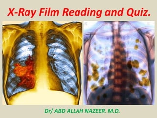
- 1. X-Ray Film Reading and Quiz. Dr/ ABD ALLAH NAZEER. M.D.
- 3. These are PA and lateral films of RML pneumonia (arrows). Note the indistinct borders, air bronchograms, and silhouetting of the right heart border.
- 5. PA and Lateral films of RUL pneumonia.
- 7. Left upper pulmonary opacity representing pneumonia with pleural effusion.
- 9. Upper lobe round pneumonia with resolution on follow-up study.
- 11. Old TB infection with consolidation and cystic changes.
- 15. T.B of Chest radiogram in HIV-seronegative patients.
- 17. A right sided tension pneumothorax with right sided lucency and leftward mediastinal shift.
- 19. Large cavitating lesion in right hemithorax with soft tissue density in lower zone laterally.
- 24. A) Frontal chest radiograph and (B) axial CT image of an adult with an aspergilloma (open black arrow) forming in an old tuberculosis cavity. Note the characteristic air crescent sign (white arrow).
- 26. Note bilateral flattening of the diaphragms and significant hyperinflation as demonstrated by visualization of 11 posterior ribs.
- 28. Popcorn Calcification due 2 Hamartoma
- 30. Anterior mediastinal mass(T-cell Lymphoma).
- 32. Note in these images that the hilum can be seen through the mass. It is not a hilar mass. The lateral shows nothing abnormal posteriorly. The CT sections demonstrate the mass in the anterior mediastinum (arrows) at the aortopulmonary window which was a thymoma.
- 36. A, lateral, and aortogram of a saccular aortic aneurysm, another cause of a middle mediastinal mass.
- 38. left sided pulmonary arterial aneurysm (arrow).
- 40. Mass above the aortic arch can be seen to be posterior to the aorta on the lateral. It does not silhouette out the superior margin of the aorta. A chest CT was performed to further evaluate the mass. This CT scan shows that the mass is posterior to the aorta, smoothly marginated, low density, and associated with the esophagus. It is an esophageal duplication cyst.
- 42. The mass projects above the clavicles, therefore it is not an anterior structure. MRI shows the mass is extra pleural and associated with the spinal nerves. It is a schwannoma, a benign tumor of the nerve sheath.
- 44. Lateral film of an intraparenchymal mass. Note acute margins like "A" in the diagram on the right. Both "B" and "C" have obtuse margins. "B" demonstrates a pleural mass while "C" is an extrapleural chest wall mass.
- 46. PA and lateral of hiatal hernia. Can you see the air-filled "mass" posterior to the heart?
- 48. Silicosis Egg shell calcification of lymph nodes Other findings include: Diaphragmatic pleural calcification Multiple cavities with fluid levels.
- 50. Alveolar microlithiasis. Subpleural and peribronchovascular microspheres of calcium with lower lobe predominance.
- 52. Alveolar proteinosis. Bilateral pulmonary opacities. CT shows the typical crazy paving pattern.
- 54. Right upper zone well defined large cyst likely hydatid cyst.
- 58. Chest X-ray revealed increased cardiothoracic ratio with linear sharp calcification in left heart border, related to hydatid cyst.
- 60. Chest X-ray showing multiple cannon ball opacities in both lung fields.
- 62. Multiple lung cannon ball metastasis in both lung fields.
- 66. Pulmonary edema.
- 68. Perforated Duodenal Ulcer with air under diaphragm.
- 70. Right hilar mass lesion with upper lobe collapse.
- 72. Left Ventricular Aneurysm. There is an unusual contour of the left heart border (white arrow) and an abnormal protuberance in the region of the left ventricle (red arrow). Enhanced CT scan at level of heart shows very large left ventricular aneurysm partially filled with clot
- 74. The patient is rotated. The radiograph is over penetrated. The radiograph was performed during expiration. The radiograph is under penetrated.
- 76. Identify the object labeled "3" in the above image.
- 78. Right Lower Zone Consolidation with upper zone consolidates as well.
- 80. HYDATID CYST.
- 82. Peripheral primitive neuro ectodermal tumors (PNETs).
- 84. Lung abscess (Koch's on histopathology).
- 86. Osteopetrosis.
- 88. Rasmussen Aneurysm in a chronic healed tuberculous cavity.
- 90. Hiatus hernia.
- 92. Pneumothorax.
- 96. Pulmonary edema (Bat Wing appearance). Chronic eosinophilic granuloma (Anti-bat wing appearance).
- 97. Thank You.