
Recommended
More Related Content
Similar to Bronchiectasis.pptx
Similar to Bronchiectasis.pptx (20)
More from ChintanBanugariya1
Recently uploaded
Recently uploaded (20)
Bronchiectasis.pptx
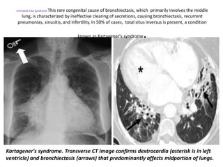
- 1. Kartagener's syndrome. Transverse CT image confirms dextrocardia (asterisk is in left ventricle) and bronchiectasis (arrows) that predominantly affects midportion of lungs. Immobile Cilia Syndrome This rare congenital cause of bronchiectasis, which primarily involves the middle lung, is characterized by ineffective clearing of secretions, causing bronchiectasis, recurrent pneumonias, sinusitis, and infertility. In 50% of cases, total situs inversus is present, a condition known as Kartagener's syndrome.
- 2. Mounier-Kuhn's syndrome. Enlarged mainstem bronchi (black arrows) and distal bronchiectasis (white arrows).
- 3. Mounier-Kuhn syndrome, also known as tracheobronchomegaly, is a rare congenital abnormality of the trachea and main bronchi characterized by cystic dilatation of the tracheobronchial tree and recurrent respiratory infections.
- 4. Mounier-Kuhn Syndrome. Two axial CT images of the thorax demonstrate marked dilatation of the trachea (T) and right (R) and left (L) main bronchi in this patient with Mounier-Kuhn syndrome. Notice the bronchiectasis (red arrows and red circle) in which the bronchi are larger than their accompanying blood vessel and there is tram=tracking of thickened bronchial walls seen in profile.
- 5. Elderly male with COPD and upper lobe bronchiectasis and scarring. New hemoptysis. Questionable soft tissue nodule within a left upper lobe bullous (arrow). Axial CT scan on lung windows. Mycetoma within the left upper lobe bullous (arrow).
- 6. Coronal reformat demonstrating bilateral upper lobe bullae, scarring and bronchiectasis with a fungus ball on the left (arrow).
- 7. MPR in MIP demonstrates hypervascular area of bronchiectasis with multiple cysts containing air mucus at postero basal segment of left lower lobe.
- 8. MPR (oblique section) in MIP demonstrates area of bronchiectasis at posterior basal segment.
- 9. MPR (axial section) in MIP demonstrates areas of bronchiectasis at posterior basal segments at both sides.
- 10. HRCT features of NSIP include extensive ground-glass areas in the lung (black arrows) and traction bronchiectasis. This bronchiectasis frequently shows a parallel course through the lung, well depicted by sagittal reconstruction in D (black dotted arrows). There is no honeycombing in the lung. Cystic bronchiectasis is generally well documented by MPR images
- 11. Cystic bronchiectasis in middle lobe. Chest x ray and MDCT (axial, coronal and sagittal).
- 12. T1-weighted magnetic resonance imaging showing appearance a) before and b) after contrast medium in a 43-year-old cystic fibrosis patient. The post-contrast images demonstrate extensive bronchial wall enhancement and permit differentiation of a thickened wall from intrabronchial secretions, with intrabronchial fluid having an air–fluid level (arrow).
- 13. Diagnostic approach to bronchiectasis.