SlideShare a Scribd company logo
1 of 282
Download to read offline
CT Chest Fundamentals
December 2020
Dr. Emad Efat
MB BCh, MSc
Faculty of Medicine, Menoufia University
CT Chest - Tutorials
1.Computed tomography (CT) Chest – Types
2.CT Chest – Anatomy
3.CT Chest - Abnormalities
CT Chest - Types
1.Standard or conventional CT:
 Slice thickness: 3-10 mm
 Scans a large volume, very quickly
 Covers the full lung
 +/- contrast
Indications
 Chest x- ray (CXR) abnormality
 Lung cancer staging
 F/U metastases
 Pleural and mediastinal abnormalities
 Empyema
CT Chest - Types
2. High-resolution computed tomography (HRCT)
 Narrow x-ray beam collimation: 1-1.3 mm vs. conventional 3-10 mm
 Cross sections are further apart: 10 mm
 High definition images of lung parenchyma: vessels, airspaces,
airway and interstitium
 No contrast
HRCTSTANDARD CT
CT Chest – Types - HRCT
Indications
 Diffusely abnormal CXR
 Normal CXR with abnormal PFT’s
 Baseline for patients with diffuse lung disease
 Solitary pulmonary nodules
 Reversible (active) vs. non-reversible (fibrotic) lung
disease
 Hemoptysis
 Lung biopsy guide
 Follow up known lung disease
CT Chest - Types
3. Low Dose CT:
 Uses low doses of radiation -- as much as 30 to 50 percent less
than regular CT
 Detail is decreased
 Uses:
Screening
ongoing trials
Follow up
infections
metastases
post lung
transplant
Axial images show pulmonary nodule in
standard dose (right) and ultra-low-dose scan
protocols in a 64-year-old patient with
prostate cancer.
CT Chest - Types
4. CT angiography:
Contrast injected into peripheral vein
Indications:
 Pulmonary embolism
 Aortic aneurysms
 Aortic dissection
 Arteriovenous malformation
 Evaluate superior vena
cava syndrome
Risks
Iodinated contrast: –
Allergic/ nephrotoxic
Chest CT angiography shows acute
pulmonary embolism (arrow).
CT Chest - Types
5. CT with contrast: Indications:
Evaluation of the mediastinum
(lymph nodes, infection)
Infection of the chest wall
Evaluation of suspected cancer
Pleural thickening, pleural
nodules, empyema and
evaluation of metastatic or
primary malignancy of the pleura
Pulmonary Lobar Collapse
Helpful to evaluate lung abscess,
although it should not be
performed routinely
Differentiate between enlarged
Axial intravenous contrast-
enhanced CT in a patient with
passive atelectasis of the right
lower lobe due to a large pleural
effusion. Note the dense
homogeneous enhancement of
the collapsed right lower lobe.
lymph nodes and the vascular structures
CT Chest - Types
6. Paired inspiratory and expiratory chest CT scans:
The quantitative analysis is done by relative difference in density
(Hounsfield unit) in inspiratory and expiratory scans
Indication : obstructive airway diseases including:
 obliterative bronchiolitis
 hypersensitivity pneumonitis (HP)
 cryptogenic organising
pneumonia (COP)
(formerly BOOP)
 bronchial asthma
 sarcoidosis
 emphysema
 bronchiectasis
CT Chest Anatomy – views
Three views:
1. Axial view
2. Coronal view
3. Sagittal view
CT Chest Anatomy – window settings
Three Windows:
1. Mediastinal or Soft Tissue windows
2. Lung windows
3. Bone windows
CT Chest Anatomy – Hounsfield unit
Hounsfield units (HU), a parameter generated from standard CT, are
related to the density of the structure of interest.
CT Chest Anatomy – mediastinum
 The mediastinum is defined as the tissue compartment located
between the two lungs, posterior to the sternum, anterior to the
vertebral column, and extending from the thoracic inlet to the
diaphragm.
 As an aid to understanding regional anatomy, the mediastinum can
be divided into four compartments, respectively, from superior to
inferior:
(a) the supraaortic or superior mediastinum;
(b) the region of the aortic arch and aortopulmonary window
(APW);
(c) the pulmonary arteries, subcarinal space, and azygoesophageal
recess; and
(d) the heart and paracardiac mediastinum.
CT Chest Anatomy- mediastinum- compartments
(a) The supraaortic mediastinum;
Right Left
CT Chest Anatomy- mediastinum- compartments
(a) The supraaortic mediastinum;
Right Left
CT Chest Anatomy- mediastinum- compartments
(a) The supraaortic mediastinum;
Right Left
CT Chest Anatomy- mediastinum- compartments
(a) The supraaortic mediastinum;
Right Left
CT Chest Anatomy- mediastinum- compartments
(b) The region of the aortic arch;
Right Left
CT Chest Anatomy- mediastinum- compartments
(b) The region of the azygos arch and aortopulmonary window (APW);
AA, ascending aorta DA, descending aorta
Right Left
CT Chest Anatomy- mediastinum- compartments
(c) The main pulmonary artery, subcarinal space, and azygoesophageal
recess;
LPA, left pulmonary artery PA, pulmonary artery RB & LB, right & left main bronchi
Right
Left
CT Chest Anatomy- mediastinum- compartments
(c) The right pulmonary artery (RPA) and azygoesophageal recess;
Right Left
AER, azygo-
esophageal
recess
CT Chest Anatomy- mediastinum- compartments
(d) The heart and paracardiac mediastinum;
AR, aortic root LA, left atrium LAA, left atrial appendage
Right
Left
CT Chest Anatomy- mediastinum- compartments
(d) The heart and paracardiac mediastinum;
Right Left
CT Chest Anatomy- mediastinum- compartments
(d) The heart;
LVO, left ventricular
outflow
Right Left
CT Chest Anatomy- mediastinum- compartments
(d) The heart;
Right
Left
Right Left
CT Chest Anatomy – The fissures
 Each lung has an oblique fissure and the right lung has a horizontal
fissure.
 There are numerous accessory fissures
 In conventional CT, the fissures are visualized as lucent bands
devoid of vascularity, whereas they appear as sharp lines in high-
resolution CT
Schematic drawings of the lungs illustrating the normal fissures (right
major, minor and left major) in coronal, sagittal and axial projections.
CT Chest Anatomy – The fissures
The right lung:
 The oblique fissures (the major fissures or greater fissures):
begins roughly at the spinous process of the T4 level of the thoracic
spine and ends at the anterior costophrenic angle
 The horizontal fissure (the minor fissure): running horizontally
from the edge of right lung towards the right hilum, at
approximately the level of the anterior 4th rib
The oblique
fissures (white
arrow) and The
horizontal fissure
(green arrow) .
CT Chest Anatomy – The fissures
 The oblique fissures (the major fissures or greater fissures):
begins between the spinous processes of vertebrae T3 and T4 and
ends about 5 cm posterior to the anterior costophrenic angle
The left lung
(a) axial view for left lung. (b) sagittal view for left lung; (c) coronal view
for left lung
CT Chest Anatomy – lung segments
(a) At level of trachea (lung apex );
CT Chest Anatomy – lung segments
(a) At level of trachea (upper lobe );
CT Chest Anatomy – lung segments
(a) At level of main stem bronchi ( at hilum ); anatomy on the right
like the left;
CT Chest Anatomy – lung segments
(a) At level below hilum and main stem bronchi;
CT Chest Anatomy- Bronchial tree- Bronchial nomenclatures
CT Chest Anatomy- Bronchial tree
CT Chest Anatomy - thoracic lymph node
Supraclavicular nodes: 1.Low cervical, supraclavicular and sternal
notch nodes
Superior Mediastinal Nodes 2-4
2R & 2L Upper Paratracheal
3A Pre-vascular & 3P Retrotracheal
4R & 4L Lower Paratracheal
Aortic Nodes 5-6
5. Subaortic & 6. Para-aortic
Inferior Mediastinal Nodes 7-9
7. Subcarinal
8. Paraesophageal
9. Pulmonary Ligament
N1 nodes: 10. Hilar & 11. interlobar
12. Lobar & 13. segmental &
14. subsegmental
CT Chest Anatomy - thoracic lymph node
3A.Pre-vascular
3P.Pre-vertebral
 Mediastinal lymph nodes are not seen in the scan if they are
normal, Nodes which are seen are pathologically enlarged
CT Chest Anatomy - thoracic lymph node
CT Chest Abnormalities - Trachea
The trachea should be central or slightly to the right.
 Causes of tracheal deviation:
 Ipsilateral (To pull): Collapse and Fibrosis
 Contralateral (To push): Apical mass , Pleural effusion and
Pneumothorax
Wegener granulomatosis: HRCT findings:
 The trachea and main bronchi may be diffusely or focally
circumferentially thickened;
 Subglottic stenosis, bronchial stenosis
 Peripheral bronchial narrowing, lobar and segmental atelectasis, or
bronchiectasis may also be present.
 Coexistent pulmonary nodules and masses, which are sometimes
cavitary
 Consolidation and ground-glass opacities
CT Chest Abnormalities – Trachea- Wegener granulomatosis
Laryngotracheal computed tomography of Wegener granulomatosis ; (A)
Subglottic stenosis (red arrow). (B) 3D reconstruction of subglottic stenosis
(white arrow). (C)–(E) Axial images showing a granulomatous lesion (*)
(yellow arrows) partially obstructing the tracheal lumen.
CT Chest Abnormalities - Trachea
Tracheomalacia:
Collapse of the trachea more than 70% during the expiration, may be
congenital or acquired
 In inspiratory CT, a dilated trachea (> 3 cm), especially with posterior
bowing of the membranous portion (thus becoming circular) may
indicate over compliance of the trachea and suggest the diagnosis.
 During expiration, the posterior membranous trachea bows
anteriorly, producing an upside down U-shaped air column on
transverse CT termed the “frown” sign
“frown” sign
CT Chest Abnormalities - Trachea
Relapsing polychondritis (RPC):
 An autoimmune disease affects
cartilaginous structures (e.g. the
nose, ear, and
laryngotracheobronchial tree.
 On CT, RPC is characterized by:
 Increased attenuation and
thickening of the anterior and
lateral walls of the large airways
 Concomitant destruction of the
cartilaginous tracheobronchial
rings with sparing of the posterior
wall.
 Tracheomalacia and large airways
stenosis may also be present
A deformity of the ear that followed
recurrent acute attacks; and saddle
nose, which usually follows repeated,
painful nasal chondritis
CT Chest Abnormalities - Trachea
Mounier- Kuhn syndrome:
a congenital defect or atrophy of the
elastic and smooth muscle tissues of the
trachea and main bronchi, resulting in
dilatation
 Diagnostic criteria: if
Trachea exceed diameter > 3.0 cm
Right mainstem diameter > 2.4 cm
Left mainstem diameter > 2.3 cm
Secretions are poorly mobilized, leading
to the chronic accumulation of secretions:
Recurrent infections,
Bronchiectasis,
Rarely pulmonary fibrosis
Two axial CT images of the thorax
demonstrate marked dilatation of
the trachea (T)
and right (R) and left (L) main
bronchi. Notice the bronchiectasis
(red arrows and red circle) and there
is tram=tracking of thickened
bronchial walls.
CT Chest Abnormalities - Trachea
Saber-sheath trachea:
 Diffuse coronal narrowing of
the intrathoracic portion of the
trachea with the concomitant
widening of the sagittal
diameter.
 Occur almost exclusively in men
with chronic obstructive
pulmonary disease (COPD) Saber-sheath trachea
 Other tracheal Disease which causes Tracheal Narrowing and Wall
Thickened: Amyloidosis, Sarcoidosis, Inflammatory bowel disease
and Tracheobronchopathia osteochondroplastica.
CT Chest Abnormalities - The lung hilum
CT is helpful in the diagnosis of endobronchial lesions, hilar and
parahilar masses, and hilar vascular lesions.
Hilar enlargement: May be unilateral or bilateral, symmetrical or
asymmetrical
CT Chest Abnormalities - The lung hilum
1.Lymph Node enlargement:
 Contrast-enhanced
computed tomography
(CECT) is helpful to
differentiate lymph nodes
from vascular structures as
lymph nodes usually do not
take contrast.
 Mild enhancement may be
seen in tuberculosis, fungal
infection, lymphoma,
metastatic lung cancer, and
sarcoidosis.
Thoracic CT images from the patient
with Tubercular Myocarditis at the
time of diagnosis, radiologically
bulky right hilar lymphadenopathy
(yellow arrow) was evident.
The lung hilum- Lymph Node enlargement
 Calcification within lymph nodes
usually suggest granulomatous
disease like TB, histoplasmosis,
sarcoidosis, silicosis,
nontuberculous mycobacterial
infection
 Enlargment of discrete lymph
nodes, most commonly seen in
sarcoidosis. Tumors and
inflammatory processes may
produce a large mass of
confluent lymph nodes.
Enlarged hilar lymph nodes in
sarcoidosis (red arrows)
CT Chest Abnormalities - The lung hilum
 CT pulmonary
angiography (CTPA)
is the best method
for demonstrating
emboli.
 HRCT is useful for
demonstrating lung
disease, which may
account for
secondary
pulmonary
hypertension.
Pulmonary Arterial Hypertension. Frontal radiograph
of the chest shows an enlarged main pulmonary
artery (red arrow) and a markedly enlarged right
pulmonary artery (white arrow) with rapid
attenuation in the size of the vessels in the lung
periphery. On the contrast-enhanced CT, the main
pulmonary artery (P) is much larger than the aorta (A).
Normally, they should roughly be about the same size.
2.Arterial enlargement:
CT Chest Abnormalities - The lung hilum
CT thorax, lung window showing
spiculated right hilar mass ( red
arrow)
3. Malignancy:
 CT enables staging of the
disease
 Squamous cell carcinoma
(SCC ) and small cell
carcinoma are typically central
 Central SCC often causes lung
collapse and/or obstructive
pneumonitis.
 Cavitation is a frequent
finding in SCC.
 In Small cell carcinoma, Direct infiltration of adjacent structures is
more common. Necrosis and haemorrhage are both common.
CT Chest Abnormalities - lung fields
Lung abnormalities:
Abnormal whiteness
(increased density):
 Consolidation
 Interstitial
 Atelectasis
 Nodule or mass
Abnormal blackness
(decreased density):
 Cyst
 Emphysema
 Cavity
CT Chest Abnormalities - lung fields
Four patterns of
increased density:
 Consolidation
 Lobar
 Diffuse
 Multifocal ill-
defined
 Atelectasis
 Nodule or mass
 Solitary Pulmonary
Nodule
 Multiple Masses
 Interstitial
 Reticular
 Fine Nodular
lung field abnormalities - Consolidation
Consolidation:
 Increased attenuation of the lung parenchyma causing obscuration
of pulmonary vessels. Reflect diseases that are primarily alveolar,
interstitial, or mixed.
 Without significant loss of volume. Air bronchograms can be found
 Causes:
 transudate, e.g. pulmonary oedema secondary to heart failure
 pus, e.g. bacterial pneumonia
 blood, e.g. pulmonary haemorrhage
 cells, e.g. bronchoalveolar carcinoma
 protein, e.g. alveolar proteinosis
 fat, e.g. lipoid pneumonia
 gastric contents, e.g. aspiration pneumonia
 water, e.g. drowning
lung field abnormalities - Consolidation
Air bronchogram:
Refers to the phenomenon of air-filled bronchi (dark) being
made visible by the opacification of surrounding alveoli
(grey/white).
Pneumococcal
pneumonia
produces
consolidation in
the right upper
lobe with
multiple air
bronchograms
(black
branching
structures)
lung field abnormalities - Consolidation
 Bat's wing or butterfly pulmonary opacities:
A bilateral perihilar distribution of consolidation.
 Reverse bat wing pulmonary opacities:
Peripheral or subpleural consolidation, sparing the perihilar region
Bat's wing opacitiesReverse bat wing opacities
lung field abnormalities - Consolidation
Reverse bat wing opacitiesBat's wing opacities
 Chronic eosinophilic pneumonia
 Cryptogenic organising
pneumonia (COP)
 Bronchoalveolar carcinoma
 Pulmonary contusion: in the
setting of trauma
 Pulmonary haemorrhage with or
without infarction: in the setting
of Pulmonary embolism.
 Pulmonary vasculitis
 Aspiration
 Pulmonary oedema (especially
cardiogenic)
 Pneumonia
 aspiration pneumonia
 pneumocystis pneumonia (PCP)
 viral pneumonia
 lipoid pneumonia
 Inhalation injury
 noxious gas
 liquid
 pulmonary alveolar proteinosis
 Pulmonary haemorrhage
(e.g. Goodpasture syndrome)
 Lymphoma/leukaemia
 Bronchoalveolar carcinoma
lung field abnormalities - Consolidation
Lymphoma: Imaging Findings:
 Hilar or mediastinal lymphadenopathy:
 A soft tissue attenuating mass, with smooth or lobulated margins
which conforms to surrounding structures. Cystic/low density
areas are common.
 Calcification is usually seen following therapy
 Pleural effusions (Mostly small, unilateral, and exudative)
 Destructive rib or vertebral body lesion
 Pericardial effusion
A large soft tissue mass in the
anterior mediastinum, which
arises in the thymus (yellow
arrow). There is associated
paratracheal adenopathy (red
arrow).
CT Chest Abnormalities - Consolidation - Lymphoma
 Parenchymal lung involvement:
 Multiple nodules
 A mass or mass-like consolidation
greater than 1 cm with or without
cavitations or bronchograms
 Masses of pleural origin
 Nodules <1 cm
 Peribronchial or perivascular
thickening
 Alveolar or interstitial infiltrates
 Segmental or lobar atelectasis
Large left upper lobe mass, with patchy
opacity peripherally. Truncation of the
left upper lobe bronchus. Extends to the
mediastinum with enlarged mediastinal
nodes
lung field abnormalities - Consolidation
CT scan obtained with pulmonary
window setting in the right middle
lobe shows a focal area of
consolidation with what may be tiny
nodules (yellow arrow).
Tuberculosis (TB): Primary pulmonary tuberculosis:
Imaging Findings:
 Patchy or lobar consolidation
 Ipsilateral hilar and
mediastinal (paratracheal)
lymphadenopathy, usually
right sided.
 Caseating granuloma
(tuberculoma) which usually
calcifies (known as a Ghon
lesion)
 Pleural effusions
 Calcification of nodes
 Atelectasis
 Cavitation (uncommon)
CT Chest Abnormalities - Consolidation - Tuberculosis
Classical CT pattern showing a large
thick-wall cavity, tree-in bud and
bronchial wall thickening.
Post-primary pulmonary Tuberculosis: Imaging Findings:
 Patchy consolidation
 “tree-in-bud” sign:
Poorly defined linear and
nodular opacities
 Ground-glass opacities
 Bronchial wall thickening
 Interlobular septal
thickening
 Cavitation, Aspergillomas,
fibrosis and Bronchiectasis
 Pleural effusion
 Hilar nodal enlargement
 Lobar consolidation,
tuberculoma, calcifications
and miliary TB
Consolidation- Tuberculosis
Tree-in-bud sign or pattern: usually represents endobronchial spread
of infection.
Consolidation- Tuberculosis- Tree-in-bud sign
Causes:
 Infective bronchiolitis
(pulmonary tuberculosis, e.g.
mycobacterium tuberculosis
infection, atypical mycobacterial
infections, e.g. mycobacterium
avium (MAIC), viral pneumonia,
fungal pneumonia, e.g.
aspergillus, allergic
bronchopulmonary aspergillosis
(ABPA), pneumocystis
pneumonia)
 Congenital (cystic fibrosis,
immotile cilia syndrome, e.g.
Kartagener syndrome, yellow
nail syndrome )
 Connective tissue disorders
(rheumatoid arthritis (RA),
Sjögren syndrome)
 Bronchial (obliterative
(constrictive) bronchiolitis,
diffuse panbronchiolitis,
follicular bronchiolitis)
 Neoplastic ( bronchioloalveolar
cell carcinoma, distant
metastatic disease (e.g. breast,
liver, ovary, prostate, kidney),
primary pulmonary lymphoma,
chronic lymphocytic leukemia )
CT Chest Abnormalities - Consolidation - Tuberculosis
Tuberculoma and Miliary Tuberculosis: Imaging Findings:
 Tuberculoma and miliary
tuberculosis are rare
 Miliary deposits are seen both
in primary and post-primary
tuberculosis. It appear as 1-3
mm diameter nodules, which
are uniform in size and
uniformly distributed
 Tuberculomas are usually
found as single nodules
and they may include a
cavity or a calcification
with sharp margins. They
are usually found in the
upper lobes
Miliary Tuberculosis
Tuberculoma. Axial lung (A) and mediastinal
window (C) CT images show a soft tissue density
nodule in the right upper lobe of the lung.
lung field abnormalities - Consolidation
Aspergillomas:
 Mass-like fungus balls of Aspergillus fumigatus, occur in patients
with normal immunity but with pre-existing cavities:
 pulmonary tuberculosis
 pulmonary sarcoidosis
 bronchiectasis
 bronchogenic cyst
 pulmonary sequestration
 Pneumocystis pneumonia (PCP)
associated pneumatocoeles
 Imaging Findings: Air crescent sign:
Rounded or ovoid soft tissue attenuating
masses located in a surrounding cavity
and outlined by a crescent of air.
Differential diagnosis (DD); hydatid cyst,
bronchogenic carcinoma and PCP.
Aspergilloma - A solid
mass within a cavity and
the “air crescent sign“
can be shown in this CT
image.
CT Chest Abnormalities - Consolidation - Tuberculosis
lung field abnormalities - Consolidation
Aspiration Pneumonitis and Pneumonia: Imaging Findings:
 An infiltrate, frequently in
the posterior segment of an
upper lobe and the superior
or posterior basal segments
of a lower lobe (The right
lower lobe is the most
frequent location).
 Aspiration-related lung
abscess
 Interstitial or nodular
infiltrates, pleural effusion,
and other changes may be
slowly progressive.
 Aspirated material can be
demonstrated on CT scans
Ct shows centrilobular nodules with
surrounding ground-glass opacities and
subpleural non-segmental
consolidations (yellow arrow) at the
dorsal portions of the right lung. Note
that the lumens of segmental bronchi
are filled with aspirated materials (red
arrow).
lung field abnormalities - Consolidation
Consolidation due to Lung infarction: Imaging Findings:
 Wedge-shaped (less often rounded) pleural-based consolidation
(Hampton hump)
 With or without cavitation
 Convex borders with a halo
sign due to adjacent
haemorrhage.
 Usually no air-bronchogram
 Plate-like (subsegmental,
discoid) atelectasis
 Pleural effusion
 Elevated hemidiaphragm
 "Melting Ice Cube Sign":
rapid peripheral resolution
of a Hampton's hump
 Feeding vessel sign
(Hampton hump); CT with lung
windowing shows a focal subpleural
area of consolidation in the left
lower lobe (arrows). There are also
small bilateral pleural effusions
Consolidation- Lung infarction
Feeding vessel sign:
 The feeding vessel sign consists of a distinct vessel leading directly
to a nodule or a mass. This sign has been considered highly
suggestive of septic embolism
 Also occurs in:
 pulmonary metastasis
 lung cancer
 Pulmonary arteriovenous
malformations (PAVM's)
 pulmonary infarction
 pulmonary vasculitis
 angioinvasive pulmonary
aspergillosis
 granuloma Feeding vessel sign. A patient with
bronchial carcinoma. Pulmonary artery
(yellow arrow) leading directly to the mass
is seen.
Consolidation- Lung infarction
The halo sign (HS): typically seen on HRCT, ground glass opacity
surrounding a pulmonary nodule or mass and represents
haemorrhage, It is typically seen in angioinvasive aspergillosis.
Causes:
 Neoplasms (e.g. adenocarcinoma,
squamous cell carcinoma, Kaposi
sarcoma, lymphoma and lung
metastases)
 Inflammatory (e.g. Fungal,
mycobacterial, and viral infections)
 Others: Wegener granulomatosis,
eosinophilic lung disease,
pulmonary endometriosis,
pulmonary embolism, organizing
pneumonia, HP, iatrogenic injury,
pulmonary pseudoaneurysm
Metastatic tumor; The CT halo
sign depicts peri-lesion ground-
glass attenuation (red arrow).
Consolidation- Lung infarction
Reversed halo sign: Central ground-glass opacity surrounded by
denser consolidation of crescentic or ring shape, at least 2 mm thick
relatively specific for cryptogenic organizing pneumonia ; causes:
 wegener’s and lymphomatoid granulomatosis
 sarcoidosis
 tuberculosis
 pneumocystis pneumonia
 pulmonary mucormycosis
 paracoccidiodomycosis
 invasive aspergillosis
 neoplastic (metastasis)
 pulmonary infarction
 lipoid pneumonia
 schistosomiasis
Fairy Ring Sign; Like Reversed halo sign but with normal central lung
parenchyma, seen sarcoidosis
Reversed halo sign: central ground-glass
density surrounded by peripheral
consolidation (yellow arrow).
lung field abnormalities - Consolidation
Klebsiella pneumonia (aka Friedländer’s pneumonia):
Imaging Findings:
 usually involves one of the
upper lobes
 ground glass opacities
 consolidation
 intralobular reticular opacities
 interlobular septal thickening
 centrilobular nodules
 cavitation
 necrotising pneumonia
 Pleural effusion and/or
empyema
 Bulging fissure sign
Transverse thin-section CT of right lower
lobe showing consolidation (arrowhead)
and intralobular reticular opacity
(arrows) with peripheral distribution.
Pleural effusion was also present
Consolidation- Klebsiella pneumonia
Bulging fissure sign: refers to lobar consolidation where the affected
portion of the lung is expanded causing displacement of the adjacent
fissure ; causes:
 pneumococcal pneumonia
 klebsiella pneumonia
 pseudomonas pulmonary infection
 staphylococcus aureus
 legionella pneumophila
 mycobacterium tuberculosis
 lung adenocarcinoma
 lung abscess
 pulmonary hemorrhage
Klebsiella pneumonia: coronal CT
image shows necrotising
consolidation (black arrow) in the
right upper lobe with the bulging
fissure sign (white arrow)
lung field abnormalities - Consolidation
Necrotising pneumonia (NP): refers to a pneumonia characterised by
the development of the necrosis within infected lung tissue.
 Causative pathogens include ( Staphylococcus aureus, Klebsiella
pneumonia, Enterobacter, Nocardai, Actinomyces, Pseudomonas,
Pneumococcus, Haemophilus influenza)
 CT with may be better as it allows appreciation of low attenuation
and non enhancement within the necrosed portions (representing
liquifaction) of the affected area of infection (consolidation).
Necrotizing
pneumonia.
heterogeneous
enhancing
consolidation with
smooth air
bronchograms and
cavities in the right
upper lobe
Consolidation - Cardiogenic pulmonary edema
Consolidation due to Congestive Heart Failure (CHF) :
Stage I CHF – Redistribution:
 Redistribution of blood flow from the lower to the upper. This is
know as cephalization because the pulmonary veins of the
superior zone dilate due to increased pressure.
 Increased artery-to-bronchus ratio at hilar level (normally they are
equal)
 Cardiomegally
Significant enlargement of
segmental / subsegmental
pulmonary arteries (NB
pulmonary artery-to-
bronchus ratio >1) and
Mosaic attenuation pattern
Consolidation - Cardiogenic pulmonary edema
Stage II CHF - Interstitial edema Characterized by:
1. Thickened interlobular septa: Septal lines, also known as Kerley
lines, become prominent. They usually occur when pulmonary
capillary wedge pressure reaches 20-25 mmHg.
Smooth septal
thickening (red arrow)
in the lower
right lobe, increased
vascular diameter
(yellow arrow) and
bilateral pleural
effusion (black arrow)
in a patient with
congestive heart
failure. Please observe
the aneurysm on the
descending aorta
Consolidation - Cardiogenic pulmonary edema
2. Bronchial wall thickening will also be seen, and is the basis of
peribronchial cuffing seen on chest x-rays.
Interstitial pulmonary edema.
(A) Axial chest CT demonstrates
smooth thickening of
interlobular septae (white
arrow). This is the CT equivalent
of Kerley B lines seen on chest
x-rays. (B) Magnified view from
the RLL in the same patient
demonstrates bronchial wall
thickening (green arrow), which
is the CT equivalent of
peribronchial cuffing seen on
radiographs. Also, note that the
adjacent pulmonary artery
branch (red arrow) is slightly
larger than the bronchus.
Consolidation - Cardiogenic pulmonary edema
3. Fluid in the major or minor fissure produces thickening of the
fissure and 'Phantom' or 'vanishing' tumours
4. Peribronchovascular interstitial thickening
5. ground-glass opacities
6. mosaic pattern of attenuation
7. pleural effusion
8. lymph nodes may be enlarged
congestive heart failure presenting
ground-glass opacities and smooth
interlobular septa thickening, a mosaic
pattern of attenuation and bilateral
pleural effusion
Thickening of the right major
fissure from subpleural edema
Consolidation - Cardiogenic pulmonary edema
Pulmonary pseudotumor or Phantom or vanishing tumours:
it is caused by a loculated pleural effusion trapped in the pleural
fissures
Axial projection (A) and Sagittal projection (B) of chest CT. Greater fluid
collection within the major and minor fissures with the second collection
pictured into the accessory superior fissure of the right lower lobe.
Consolidation - Cardiogenic pulmonary edema
Stage III CHF - Alveolar edema Characterized by:
 Peribronchial nodules
 Alveolar edema with perihilar consolidations and air
bronchograms (Bat's wing or butterfly pulmonary opacities)
 Pleural effusion
 An enlarged cardiac silhouette
High-resolution computed
tomography scans, showing
slices acquired at the aortic arch
level, using a pulmonary
window, in a patient with acute
myocardial infarction and
Cardiogenic pulmonary edema
presenting consolidations,
ground-glass opacities, smooth
interlobular septal thickening
and bilateral pleural effusion.
lung field abnormalities - Consolidation
Adult Respiratory Distress Syndrome ( ARDS ), three phases:
Fibrotic phaseFibroproliferative phaseExudative phase
 persistent ground-
glass densities
 coarse reticulation
 air cysts (arrowheads)
 traction
bronchiectasis
 alveolar opacities
 reticular opacities
 Bronchiectasis (arrows)
 honeycombing
 signs of pulmonary
hypertension
 consolidations
 ground-glass opacities
 thickening of interlobular
septa (crazy paving)
 posterior compressive
atelectasis
lung field abnormalities - Consolidation
Bronchopneumonia: bilateral and predominantly in the lung bases
late stagesEarly stages
 Extends peripherally along the bronchus to involve
the entire pulmonary lobule
 Multifocal heterogeneous confluent consolidation
= patchwork quilt; eventually coalesce.
 Exudates fill airways = no air bronchograms
 25-75% form abscess
 Empyema and parapneumonic effusion common
 Multiple foci of opacity
 Begins centrally in and
around lobular bronchi
 Peribronchial thickening
and poorly defined
bronchovascular nodules
 May result in a tree-in-bud
appearance
lung field abnormalities - Consolidation
Atypical pneumonia (AP) characterised by:
 Focal ground-glass opacity in a lobular distribution (Diffuse and
bilateral)
 Diffuse ground glass nodules in a centrilobular pattern
 Bronchial wall thickening
 pleural effusion may present
30-year-old woman with
Mycoplasma pneumoniae
pneumonia. CT shows
bronchial wall thickening
(arrows). Lobular areas of
consolidation and ground-
glass attenuation are also
seen.
lung field abnormalities - Consolidation
(AP) Legionella pneumonia:
 Multifocal and bilateral patchy infiltrate (ground glass and/or
consolidation), Middle and lower zone predominance
 Pleural effusions can be common
 Associated hilar adenopathy
may be present
 Cavitation and a masslike
appearance
Legionella pneumonia. (A) Typical non-
segmental lesion in the right middle
lobe and the right lower lobe. (B) sharp-
bordered fissures (arrow), which is a
characteristic feature of non-segmental
distribution. (C) Segmental lesions in
the right lower lobe. (D) consolidation
and ground-glass opacity
lung field abnormalities - Consolidation
(AP) Mycoplasma pneumonia characterised by:
 Areas of ground-glass attenuation and air-space consolidation
Mycoplasma pneumoniae pneumonia.
HRCT shows bilateral lobular areas of
consolidation, patchy ground-glass
opacities, interlobular septal thickening
 lobular distribution
 Centrilobular nodules
 Thickening of the
peribronchovascular
interstitium
 Interlobular septal
thickening
 Mosaic perfusion
 lower lobe
predominance
lung field abnormalities - Consolidation
(AP) Chlamydia pneumonia characterised by:
 Areas of ground-glass attenuation and air-space consolidation
 lobular distribution
 Findings are often limited to a single lobe.
Chlamydia pneumonia. an
acinar pattern on a
background of ground-glass
attenuation. Right pleural
effusion is also present.
 lower lobe involvement occurring more frequently
 bronchovascular bundle thickening.
 Centrilobular nodules
 Up to 25% of patients may have a small to moderate-sized pleural
effusion
 lymphadenopathy: uncommon
lung field abnormalities - Consolidation
Respiratory syncytial
pneumonia. bilateral ill-
defined centrilobular nodules
(arrows) and bronchial wall
thickening (arrowhead).
(AP) Viral pneumonia characterised by:
 Patchy, unilateral, or bilateral consolidations and ground-glass
opacity or poorly defined centrilobular nodules.
 Peribronchial thickening
 Thickened interlobular septa
 Areas of atelectasis or air trapping
 Pleural effusion, hilar
lymphadenopathy and
pneumothorax are
uncommon findings.
lung field abnormalities - Consolidation
(AP) Covid-19 pneumonia: The primary findings of
COVID-19 on chest radiograph and CT are those of
atypical pneumonia or organizing pneumonia.
 Normal in up to 63% in the early stages
 Ground glass (68.5%), coarse horizontal linear
opacities, and consolidation.
 Crazy paving
 These are more likely to be multifocal, bilateral,
peripheral and in the lower zones
 Vascular dilatation and bronchovascular
thickening
 Traction Bronchiectasis
 Atypical CT findings
(mediastinal lymphadenopathy,
pleural effusion, multiple tiny
pulmonary nodules, tree-in-bud,
pneumothorax, cavitation)
(COVID-19).
A, ground-glass
opacity (GGO),
B, crazy-paving
pattern, and,
C, consolidation
lung field abnormalities - Consolidation
Pulmonary fungal disease: pulmonary aspergillosis characterised by:
 Aspergillomas (mycetomas)
 ABPA (next).
 Chronic necrotizing or Semi-invasive aspergillosis:
 upper lobe predominant opacities
 pleural thickening
 Cavitation with or without a central mycetoma
 Often there are multiple cavities, often thick-walled
Axial CT image
demonstrates a
cavitary lesion
containing fungal
ball in a patient with
HIV and semi-
invasive aspergillosis
lung field abnormalities - Consolidation
Pulmonary fungal disease: pulmonary aspergillosis characterised by:
 Airway-invasive aspergillosis:
 tracheobronchitis: tracheal or bronchial wall thickening
 Bronchiolitis: centrilobular and tree-in-bud opacities
 Bronchopneumonia: perihilar consolidations
Bronchial wall thickening,
tree-in-bud nodules, and
peribronchial
consolidation in sagittal
CT image of a patient with
airway-invasive
aspergillosis
leading to bronchitis,
bronchiolitis, and
bronchopneumonia
lung field abnormalities - Consolidation
Pulmonary fungal disease: pulmonary aspergillosis characterised by:
 Angioinvasive aspergillosis:
 Solitary or multiple pulmonary nodules/masses
 halo sign and/or Reversed halo sign
angioinvasive aspergillosis.
ground-glass density
surrounding a nodule (halo
sign)
lung field abnormalities - Consolidation
E-Cigarette or Vaping Product-Associated Lung Injury:
1. Lipoid pneumonia: basilar predominant ground glass opacities,
nodular, crazy paving or tree-in-bud opacities,
2. Diffuse alveolar damage: dependent ground glass opacities and
consolidation (acute/exudative phase), followed by reticulation
and traction bronchiectasis (organizing/proliferative phase)
Lipoid pneumoniaDiffuse alveolar damage
lung field abnormalities - Consolidation
E-Cigarette or Vaping Product-Associated Lung Injury:
Acute eosinophilic pneumonia
3. Acute eosinophilic pneumonia: ground glass opacities and
consolidation, often with pleural effusions and septal thickening
lung field abnormalities - Consolidation
E-Cigarette or Vaping Product-Associated Lung Injury:
4. Organizing pneumonia: peripheral or perilobular ground glass
opacities and/or consolidation, sometimes with the atoll/reverse
halo sign
Organizing
pneumonia
lung field abnormalities - Consolidation
E-Cigarette or Vaping Product-Associated Lung Injury:
5. Diffuse alveolar hemorrhage:
centrilobular ground glass opacities,
nodules, and/or consolidation with
subpleural sparing, followed by septal
thickening (reflecting lymphatic
engorgement with blood products)
6. Respratory bronchiolitis interstitial lung
disease:
upper lobe predominant centrilobular
ground glass nodules (next)
7. Hypersensitivity pneumonitis (next)
8. Giant cell interstitial pneumonia:
fibrosis seen as peripheral reticulation,
ground glass opacity, and traction
bronchiectasis
lung field abnormalities - Consolidation
Wegener's granulomatosis characterized by:
 Nodules or mass lesions, which may cavitate
 Fleeting focal infiltrates (lung consolidation )
 Ground-glass opacities
Wegener's
granulomatosis.
Multiple pulmonary
nodules. The ground-
glass halo is
compatible with
perinodular capillaritis
and alveolar
hemorrhage ( yellow
arrow). There was also
a large cavitating mass
as well ( red arrow)
lung field abnormalities - Consolidation
Eosinophilic pneumonia (EP):
Chronic EPAcute EP
 Non-segmental air-space consolidation with
peripheral predominance (reversed batwing)
 Middle or upper zone predilection
 Lymph nodes may be enlarged
 During resolution, wavy lines paralleling
chest wall
 Less common: ground-glass opacities,
pulmonary nodules, reticulation, lobar
atelectasis and pleural effusion
 Bilateral ground-glass areas
 Interlobular septal thickening
 Pleural effusions
 Air-space consolidation
 Thickening of bronchovascular
bundles
 Ill-defined centrilobular nodules
lung field abnormalities - Consolidation
Septic emboli: CT findings:
 Multiple peripheral parenchymal
nodules, these may have a
dependent, lower zone
predication
 Cavitation or air bronchogram in
more than 89%, cavities are thin-
walled and may have no fluid level
 Wedge-shaped subpleural
consolidation with apex of lesion
directed toward pulmonary hilum
 Feeding vessel sign = pulmonary
artery leading to nodule (67%)
 Hilar and mediastinal adenopathy
can occur
 Pleural effusion is rare
CT scan reveals bilateral
peripheral nodules and wedge-
shaped consolidation with
various size. Some of the lesions
show connection with
pulmonary vessels (so-called
feeding vessel sign, arrows),
and this suggests septic emboli.
Also note right pleural effusion.
lung field abnormalities - Consolidation
 Two types intralobar and extralobar
 Pulmonary tissue without normal
connection to bronchial tree
 Anomalous arterial blood supply,
from aorta
 CT angiography is used because it
shows the vascularization of the
sequestered tissue in great detail
 A solid mass that may be
homogeneous or heterogeneous,
sometimes with cystic changes.
 An infected sequestration may be
associated with a parapneumonic
effusion, and may contain one or
more air-fluid levels.
Pulmonary sequestration:
Axial CT angiographic image
showing anaomalous vessels
(red arrow) arising from the
descending aorta and
supplying the enhancing left
lower lobe sequestered lung
segment (green arrow)
lung field abnormalities - Interstitial disease
The secondary pulmonary lobule:
 The smallest functional unit of the lung.
 Each lobule is demarcated by interlobular septae, which contain
lymphatics and pulmonary veins.
 The lobule is supplied centrally by a terminal bronchiole and
accompanying centrilobular pulmonary artery, which are together
known as the
bronchovascular
bundle.
 A second set of
lymphatics also runs
with the
bronchovascular
bundle.
Interstitial disease- secondary pulmonary lobule
 Centrilobular area is the central part of the secondary lobule. It is
usually the site of diseases, that enter the lung through the airways
( i.e. hypersensitivity pneumonitis, respiratory bronchiolitis,
centrilobular emphysema ).
 Perilymphatic area is the peripheral part of the secondary lobule.
It is usually the site of diseases, that are located in the lymphatics
of the interlobular
septa ( i.e. sarcoid,
lymphangitic
cinomatosis, pulmonary
edema). These diseases
are usually also located
in the central network
of lymphatics that
surround the broncho-
vascular bundle.
Interstitial disease- secondary pulmonary lobule
Centrilobular
area in blue
(left) and
perilymphatic
area in yellow
(right)
Normal interlobular septa
(solid black arrows) and
centrilobular arteries
(open white arrows) are
clearly visible. Interlobular
septa are normally 0.1 mm
thick and can be seen in
the lung periphery,
particularly along the
anterior and mediastinal
pleural surfaces
lung field abnormalities - Interstitial disease
What is the dominant HR pattern?
1- LINEAR ABNORMALITIES
2-NODULES
3 - GROUND GLASS OPACITY
4 - CONSOLIDATION
1-AREAS OF DECREASED ATTENUATION WITH WALLS
(CYSTS ; HONEYCOMB ; BRONCHIECTASIS)
2-AREAS OF DECREASED ATTENUATION WITHOUT WALLS
(EMPHYSEMA,MOSAIC ATTENUATION)
A- High attenuation (CT scan findings manifesting as
increased opacity)
B- Low attenuation (CT scan findings manifesting as
decreased opacity)
lung field abnormalities - Interstitial disease
 Fine "ground-glass" (1-2 mm):
e.g. interstitial pulmonary
oedema
 Medium "honeycombing"
(3-10 mm): commonly seen in
pulmonary fibrosis
 Coarse (> 10 mm):
cystic Spaces caused by
parenchymal destruction, e.g.
usual interstitial pneumonia
(UIP), pulmonary sarcoidosis,
Pulmonary Langerhans cell
histiocytosis (PLCH)
Reticular Pattern: results from the summation or superimposition of
irregular linear opacities.
lung field abnormalities - Interstitial disease
Causes of Reticular Pattern:
 Pulmonary edema (heart
failure, fluid overload,
nephropathy)
 Infection ( viral, mycoplasma,
Pneumocystis, malaria )
 Post-infectious scarring
(tuberculosis, histoplasmosis,
coccidioidomycosis)
 Mitral valve disease
 Collagen vascular disorders
 Granulomatous disease
( pulmonary sarcoidosis,
eosinophilic granuloma )
 Drug reactions (e.g.
amiodarone )
 Pulmonary neoplasms
( lymphangitis carcinomatosis,
pulmonary lymphoma )
 Inhalational lung disease
(asbestosis, silicosis, coal
workers pneumoconiosis,
hypersensitivity pneumonitis,
chronic aspiration pneumonia)
 Idiopathic (usual interstitial
pneumonia,
lymphangioleiomyomatosis,
tuberous sclerosis,
neurofibromatosis,
amyloidosis )
Interstitial disease- Reticular pattern
linear and reticular opacities:
Represents thickening of interstitial fibers of lung by
- fluid
or
- fibrous tissue
or
- infiltration by cells
Interstitial disease- Reticular pattern
Linear Pattern:
1. Thickened
interlobular septa
2. Peribronchovascular
interstitial thickening
3. Intralobular Lines
4. Thickened Fissures
5. Subpleural lines
6. Parenchymal bands
Interstitial disease- Reticular pattern- Linear Pattern
Interlobular Septal thickening:
IrregularNodularSmooth
 UIP
 Sarcoidosis
 Asbestosis
 HP
 lymphangitic
carcinomatosis
 Sarcoidosis
 lymphangitic
carcinomatosis
 lymphoproliferative
disorders (LIP,
lymphoma, leukaemia)
 Silicosis, coal worker's
pneumoconiosis (CWP)
 Kaposi sarcoma
 Pulmonary oedema,
haemorrhage
 Lymphoma, leukaemia
 lymphangitic carcinomatosis
 lymphocytic interstitial
pneumonia (LIP), non
specific interstitial
pneumonia (NSIP)
Interstitial disease- Reticular pattern
 Sarcoidosis
 Pulmonary interstitial
oedema
 Certain types of
pneumonias – pneumonitis:
 mycoplasma pneumonia
 acute eosinophilic
pneumonia
 lymphoid interstitial
pneumonia (LIP)
 covid-19 pneumonia
 Microscopic polyangiitis
 Lymphangitis
carcinomatosis
Lymphangitic Carcinomatosis. A thin-
section CT shows both smooth and nodular
thickening of the bronchovascular
structures (arrows) that represents
lymphatic tumor surrounding the axial
interstitium.
Peribronchovascular interstitial thickening : Causes:
Interstitial disease- Reticular pattern
Honeycomb cysts:
 an irreversible finding in
interstitial lung disease
 small (3 to 10 mm) cystic spaces
with thick (1 to 3 mm) walls
 usually posterior subpleural and
basal in distribution
 frequently seen in UIP and
chronic HP and occasionally in
sarcoidosis.
 additional signs:
thickened interlobular and
intralobular lines
parenchymal bands
areas of ground glass opacity
Idiopathic Pulmonary Fibrosis (IPF). The
HRCT scan shows basal and peripheral
reticular opacities with honeycombing
and traction bronchiectasis.
irregularity of lung interfaces (between broncho-vascular bundles or
fissures or pleural surfaces and lung)
Interstitial disease- Reticular pattern- Honeycomb cysts
 Macrocystic e.g UIP
 Mixed macrocystic
and Microcystic e.g
UIP
 Combined
emphysema and
honeycombing e.g
desquamative
interstitial
pneumonia (DIP) and
Pulmonary
Langerhans cell
histiocytosis (PLCH)
Types:
 Microcystic e.g fibrotic nonspecific interstitial pneumonia (NSIP)
lung field abnormalities - Interstitial disease
Nodular pattern:
 Homogenous and contain no air bronchograms
 Nodular opacities may be:
 Miliary nodules: <2 mm
 Pulmonary micronodule: 2-7 mm
 Pulmonary nodule: 7-30 mm
 Pulmonary mass: >30mm
Morphology:
 Solid calcified pulmonary nodules
 Ground glass pulmonary nodules
 Partly solid or non-solid: may represent:
malignancy: primary or metastases
atypical adenomatous hyperplasia
focal interstitial fibrosis
aspergillosis
focal pulmonary haemorrhages
solid
nodule
ground
glass
nodule
partly
solid
nodule
Interstitial disease- Nodular pattern
Nodular distribution:
 Random distribution:
Nodules involve the pleural
surfaces and fissures.
 Centrilobular distribution:
Unlike perilymphatic and random
nodules, centrilobular nodules
spare the pleural surfaces. The
most peripheral nodules are
centered 5-10mm from fissures
or the pleural surface.
 Perilymphatic distribution:
nodules are seen in relation to
pleural surfaces, interlobular
septa and the
peribronchovascular interstitium.
Interstitial disease- Nodular pattern
Nodular distribution:
PerilymphaticCentrilobularRandom
Sarcoidosis, silicosis,
coal-worker's
pneumoconiosis,
lymphangitic spread
of carcinoma, LIP,
amyloidosis
Infectious bronchiolitis, diffuse
panbronchiolitis, respiratory
bronchiolitis, HP, LIP,
pulmonary edema, vasculitis,
plexogenic lesions of
pulmonary hypertension,
metastatic neoplasms
Hematogenous
metastases, Miliary
tuberculosis, Miliary
fungal infections, PLCH
(early nodular stage),
Sarcoidosis (when very
extensive)
Interstitial disease- Nodular pattern
Causes of Miliary opacities :
 Infection
 tuberculosis
 fungal (often febrile)
 healed varicella pneumonia
 viral pneumonitis
 nocardosis
 salmonella
 Miliary metastases
 thyroid carcinoma
 renal cell carcinoma
 breast carcinoma
 malignant melanoma
 pancreatic neoplasms
 osteosarcoma
 trophoblastic disease
 Sarcoidosis
 Pneumoconioses
 silicosis
 coal workers pneumoconiosis
 Pulmonary haemosiderosis
 Hypersensitivity pneumonitis
 Langerhans cell histiocytosis
( PLCH )
 pulmonary alveolar
proteinosis
Interstitial disease- Nodular pattern
Causes of Calcified pulmonary nodules:
 Healed infection
 Calcified granulomata, e.g.
 Thoracic histoplasmosis
 Recovered miliary TB
 Healed varicella pneumonia
 Pneumoconioseses
 Silicosis
 Coal worker's pneuomconiosis
 Pulmonary hamartomas
 Metastatic pulmonary calcification
 Chronic renal failure
 Multiple myeloma
 Secondary hyperparathyroidism
 Massive osteolytic metastases
 IV calcium therapy
 Pulmonary haemosiderosis
 Idiopathic pulmonary
haemosiderosis
 Mitral stenosis
 Goodpasture syndrome
 Pulmonary alveolar
microlithiasis
 Sarcoidosis
 Calcified pulmonary
metastases
 Pulmonary amyloidosis
 Pulmonary hyalinizing
granuloma
 Calcifying fibrous
pseudotumour of lung
Interstitial disease- Nodular pattern
 A reticulonodular pattern
results from a combination
of reticular and nodular
opacities.
 A differential diagnosis
should be developed based
on the predominant
pattern.
 If there is no predominant
pattern, causes of both
nodular and reticular
patterns should be
considered.
 Causes: the same disorders as reticular patterns
Reticulonodular pattern:
Sarcoidosis. a “reticulonodular pattern”
characterised by the presence of
thickening of the interlobular septae and
bronchovascular bundles, perilymphatic
and perifissural micronodules and
architectural distortion
Interstitial disease- High attenuation
Ground-glass opacification/opacity (GGO): a hazy area of increased
attenuation in the lung with preserved bronchial and vascular
markings.
Aetiology:
 Normal expiration
 Partial filling of air spaces
 Partial collapse of alveoli
 Interstitial thickening
 Inflammation
 Oedema
 Fibrosis
 Neoplasm
Symmetric perihilar ground-glass
opacity, representing pulmonary
hemorrhage in a patient with
Wegener’s granulomatosis.
Interstitial disease- High attenuation
Ground-glass opacification/opacity (GGO) and consolidation: causes:
ChronicAcute
 Hypersensitivity pneumonitis
 Smoking related interstitial lung disease
(respiratory bronchiolitis-associated
interstitial lung disease (RB-ILD), DIP)
 Idiopathic interstitial pneumonias (Non-
specific interstitial pneumonia (NSIP),
rarely usual interstitial pneumonia)
 Bronchioloalveolar carcinoma
 Cryptogenic Organizing Pneumonia
(COP)
 Lymphoid interstitial pneumonia (LIP)
 Eosinophilic pneumonia (chronic)
 Exogenous lipoid pneumonia
 Alveolar proteinosis
 Sarcoidosis
 Edema
 Diffuse alveolar damage
(DAD)/acute respiratory distress
syndrome (ARDS)/acute
interstitial pneumonia (AIP)
 Infections (bacterial, viral,
Pneumocystis jiroveci,
Mycoplasma pneumoniae)
 Hemorrhage
 Hypersensitivity pneumonitis
 Eosinophilic pneumonia (acute)
 Radiation pneumonitis (acute)
Interstitial disease- High attenuation
Ground-glass opacity (GGO) and consolidation: distribution:
Diffuse/symmetricPatchyFocal
Edema
DAD/ARDS/AIP
Infections (viral,
atypical)
Interstitial pneumonias
Hemorrhage
Bronchoalveolar cell
carcinoma
Alveolar proteinosis
Infection
Sarcoid
Hypersensitivity
pneumonitis
Organizing pneumonia
Bronchoalveolar cell
carcinoma
Hemorrhage
Eosinophilic pneumonia
Infection
Aspiration
Hemorrhage
Bronchoalveolar
cell carcinoma
Infarct
Interstitial disease- High attenuation
Causes:
 Pulmonary alveolar proteinosis (PAP)
 Edema (heart failure, ARDS, AIP)
 Infection (PCP, viral, Mycoplasma, bacterial)
 Pulmonary hemorrhage
 Cryptogenic organizing pneumonia (COP)
 Neoplasm
(bronchoalveolar
carcinoma (BAC))
 Sarcoidosis
 NSIP
Crazy paving: a combination of ground-glass opacity with superimposed
interlobular septal thickening and intralobular reticular thickening
Interstitial disease- Low attenuation
Low attenuation
pattern
Interstitial disease- Low attenuation
Air containing spaces:
1. Blebs appear as small air spaces (<1-2 cm) within the layers of the
visceral pleura or subpleural, located most frequently at the lung
apices. They have thin walls (less than 1 mm thick).
2. Bulla: thin wall (<1 mm), usually larger than blebs (>2 cm)
3. Pneumatocele are rounded thin wall air space that represent
distended airspaces distal to a check-valve obstruction of a
bronchus or bronchiole, caused by acute pneumonia, trauma, or
aspiration of hydrocarbon fluid and is usually transient
4. Cyst
5. Cavity
Interstitial disease- Low attenuation
A lung cyst:
An air filled structure and occurs without associated pulmonary
emphysema with perceptible wall typically 1 mm in thickness but can
be up to 4 mm. The diameter of a lung cyst is usually < 1 cm.
Aetiology:
 Sjogren syndrome
 light chain deposition disease
 Amyloidosis
Others:
 Birt-Hogg-Dubé syndrome
 Pulmonary trauma
 Congenital cystic lung disease
(congenital pulmonary airway
malformation, pulmonary
sequestration, bronchogenic cyst)
 Tracheobronchial papillomatosis
 Hydatid Cyst
Interstitial disease:
 Pulmonary Langerhans cell
histiocytosis (PLCH)
 lymphangioleiomyomatosis with
or without tuberous sclerosis
 Interstitial pneumonia (DIP, LIP)
 Pneumatocele
 Sarcoidosis
 Neurofibromatosis
 Cystic bronchiectasis
 PCP
 Honeycombing in UIP
Interstitial disease- Low attenuation- Cystic lung
 Early stage:
Small irregular or stellate
nodules in centrilobular location
 Late stage (more common):
Bizarre shaped Cysts
Upper and mid lobe
predominance.
Recurrent pneumothorax.
 Other common findings:
GGO: Upper and mid lobe with
sparing of the costophrenic
recesses
Mosaic attenuation
Emphysema
Desquamative interstitial
pneumonia (DIP)-like change
Pulmonary Langerhans cell histiocytosis (PLCH):
Interstitial disease- Low attenuation- Cystic lung
 features tend to be diffuse with mid to lower lobe predominance
 thickening of bronchovascular bundles
 interstitial thickening along lymph channels
 small but variable sized pulmonary nodules (can be centrilobular or
subpleural, and often ill-defined)
 ground-glass change
 scattered thin walled cysts:
usually deep within
the lung parenchyma
size range from 1-30 mm
typically abuts vessels
(i.e. is perivascular or subpleural)
differentiate LIP from malignant lymphoma
 mediastinal lymphadenopathy
 honeycombing
Lymphocytic interstitial pneumonitis (LIP): HRCT features:
LIP.There is a background
of ground-glass
opacification and a few
thin-walled cystic
airspaces
Interstitial disease- Low attenuation- Cystic lung
 General/radiograph
chylothorax: chylous pleural effusion
evidence of hyperinflation
diffuse bilateral reticulonodular
densities
recurrent pneumothoraces
 HRCT
thin walled cysts of variable sizes
surrounded by normal lung
parenchyma, seen throughout the lung
interlobular septal thickening
may show a dilated thoracic duct
haemorrhages may be seen as areas of
increased attenuation
Lymphangioleiomyomatosis (LAM): is a rare multi-system disorder
that can occur either sporadically or in association with the tuberous
sclerosis complex (TSC), It affects women of child-bearing age
CT images demonstrate
innumerable small regular lung
cysts diffusely distributed
throughout the lungs.
Interstitial disease- Low attenuation
Pulmonary emphysema: morphologic subtypes;
ParaseptalPanlobularCentrilobular
 Adjacent to the pleura
and interlobar fissures
 It can lead to the
formation of subpleural
bullae and spontaneous
pneumothorax
 In alpha-1-antitrypsin
deficiency
 Affects the whole
secondary lobule
 Lower lobe
predominance
 Most common type
 Affects the centrilobular
portion of the lobule
 Upper lobe
predominance
 Up to 1 cm in diameter
Interstitial disease- Low attenuation- emphysema
Pulmonary emphysema: In all three subtypes, the emphysematous
spaces are not bounded by any visible wall
High-resolution CT (HRCT)
shows subpleural bullae
consistent with paraseptal
emphysema. Red mark
shows the size of a normal
acinus
Panlobular emphysema.
large bullae in both inferior
lobes due to uniform
enlargement and
destruction of the alveoli
walls causing distortion of
the pulmonary architecture
Centrilobular
emphysema. low
attenuation areas
without walls located
centrally in the acini.
Red element shows the
size of a normal acinus
Interstitial disease- Low attenuation- emphysema
 An expansion of the alveolar spaces with a diameter over 1 cm and
a wall thickness less than 1 mm.
 Giant bullae in 1 or both upper lobes occupying at least one-third of
the hemithorax
 More in the paraseptal location.
Bilateral
bullous
emphysema
Vanishing Lung Syndrome (Giant Bullous Emphysema):
Interstitial disease- Low attenuation- emphysema
HRCT would typically show:
 Centrilobular and/or
paraseptal emphysema:
often upper zone
predominant
 Pulmonary fibrosis of the
lower lobes: can be of UIP
or NSIP pattern
 Complications:
 pulmonary hypertension
 lung cancer
A
HRCT scan at
the level of
the aortic
arch.
paraseptal
emphysema
B
HRCT scan at
the level of
the dome of
the right
hemi-
diaphragm.
UIP pattern
Combined pulmonary fibrosis and emphysema (CPFE): characterised
by the coexistence of usual interstitial pneumonia (UIP) or nonspecific
interstitial pneumonia (NSIP) with emphysema in smokers.
Interstitial disease- Low attenuation- emphysema
Axial (A) and coronal (B) CT show
hyperinflated left upper lobe (arrows)
with attenuated lung markings and
herniation across the midline
 Rates of occurrence :
 Left upper lobe - 41%
 Right middle lobe - 34%
 Right upper lobe - 21%
 CT can provide details about the
involved lobe and its vascularity,
as well as information about the
remaining lung.
 A hyperlucent, hyperexpanded
lobe with a paucity of vessels
 Midline substernal lobar herniation
and compression of the remaining lung.
 Usually, the mediastinum is significantly
shifted away from the side of the abnormal lobe.
 Compressive atelectasis of neighbouring lobes
Congenital Lobar Emphysema: progressive overinflation of one or
more lobes of a neonate lung.
Interstitial disease- Low attenuation- emphysema
Pulmonary Interstitial emphysema (PIE ): Much more common in
neonates, rare in adults . PIE occurs almost in association with
mechanical ventilation.
 CT features :
 lines and dots intermingled with large gaseous inclusions is typical,
representing peribronchovascular bundles compressed by the air-
filled interstitium
 Shows cystic radiolucencies in affected segment
A: Multiple cystic,
predominantly round
images in association with
linear (arrow heads) and
punctate (arrows) images
– lines and dots pattern.
B: Cystic mass with
regular, well defined
borders (pseudocyst).
lung field abnormalities - Interstitial disease
Smoking-related interstitial lung disease (SR-ILD):
 Respiratory bronchiolitis (RB): Poorly defined centrilobular ground-
glass nodules in the upper zones.
 Respiratory bronchiolitis ILD (RB-ILD)
 Desquamative interstitial pneumonitis (DIP)
 Pulmonary Langerhans cell histiocytosis (PLCH)
 Idiopathic pulmonary fibrosis (IPF)
 Combined pulmonary fibrosis and emphysema (CPFE)
 Acute eosinophilic pneumonia (AEP)
 Rheumatoid-arthritis-associated interstitial lung disease (RA-ILD)
 Diffuse alveolar hemorrhage (DAH) in Goodpasture syndrome
 Pulmonary alveolar proteinosis
Respiratory bronchiolitis in the upper lungs,
with bilateral centrilobular ground-glass
nodules (red arrow), patchy ground-glass
opacities (white solid arrow) and bronchial
wall thickening (white dashed arrow).
Interstitial disease- SR-ILD
Respiratory bronchiolitis ILD (RB-ILD):
 CT features :
 Poorly defined centrilobular ground-glass nodules
 Patchy ground-glass opacities
 Bronchial wall thickening
 Reticulation occasionally (no traction bronchiectasis or
honeycombing)
 Centrilobular emphysema
 Air-trapping
 Upper lobe
(left) shows diffuse faint
centrilobular ground-glass
nodules (red arrows). High
resolution CT 1 year after
smoking cessation (right)
shows complete resolution
of the centrilobular nodules.
Interstitial disease- SR-ILD
Desquamative interstitial pneumonitis (DIP):
 CT features :
 Lower lobe (60%), Diffuse (20%) and Patchy (20%)
 Ground-glass opacity (widespread, bilateral and symmetrical)
 bronchial wall thickening and centrilobular emphysema
 Peripheral cystic spaces (dilated bronchioles and alveolar ducts)
 Reticular opacities
 Traction bronchiectasis
 Honeycombing is
uncommon
Desquamative interstitial
pneumonia: diffuse ground-
glass opacities predominantly
in the mid-lower lung. No
traction bronchiectasis or
architectural distortion is seen.
Interstitial disease- Low attenuation
Mosaic attenuation: is used to describe density differences between
affected and non-affected lung areas. There are patchy areas of black
and white lung.
Obliterative bronchiolitis in a patient with cystic fibrosis. HRCT at the level of the
carina at (a) inspiration and (b) expiration reveals at expiration a “mosaic
attenuation pattern” secondary to air-trapping (b) which is not revealed on
inspiration (a)
Interstitial disease- Low attenuation - Mosaic attenuation
Causes:
Parenchymal disease
Occlusive vascular
disease
Obstructive small
airways disease
 high attenuation
regions are abnormal
and represent
ground-glass opacity
 e.g. hypersensitivity
pneumonitis,
pulmonary edema,
Sarcoidosis, ARDS,
Pneumocystis
jiroveci, NSIP,
Bronchoalveolar
carcinoma
 low attenuation
regions are
abnormal and
reflect relative
oligaemia,
 e.g. chronic
pulmonary
embolism,
pulmonary
hypertension
 low attenuation
regions are abnormal
which become more
evident in expiratory
CT scans,
 e.g. Bronchiolitis
obliterans, asthma,
bronchiectasis, cystic
fibrosis,
hypersensitivity
pneumonitis
lung field abnormalities - Interstitial disease
Hypersensitivity pneumonitis (HP) - (acute):
 Homogeneous ground-glass
and alveolar opacities :
usually bilateral and
symmetric but sometimes
patchy
concentrated in the middle
part and base of the lungs
or in a bronchovascular
distribution
 Airspace Consolidation
 Small (< 5 mm diameter)
ill-defined centrilobular
nodules
There is homogeneous bilateral and
symmetric alveolar opacities and numerous
centrilobular ground-glass alveolar nodules.
No evidence of fibrosis.
lung field abnormalities - Interstitial disease
Hypersensitivity pneumonitis (HP) - (Subacute): The CT demonstrates:
 Diffuse soft centrilobular ground-glass nodules (3-5 mm)
 Patchy ground-glass opacities predominantly involving the middle
and lower lung zones
 Lobular areas of mosaic attenuation
 Air trapping may be seen on expiratory scans
 Headcheese sign
Subacute HP: Inspiratory
axial CT image showing
ground-glass opacities and
lobular areas of mosaic lung
attenuation.
Interstitial disease - Hypersensitivity pneumonitis
 There is a combination of:
lung consolidation
ground glass opacities
normal lung
hyperinflated/air trapped
lung (mosaic attenuation)
 Relatively specific for HP,
can occasionally be seen in
other conditions including
RB-ILD, DIP, LIP, follicular
bronchiolitis, sarcoidosis,
and atypical infections
Headcheese sign in patient with subacute
hypersensitivity pneumonitis showing
combination of three lung attenuations - areas
of mosaic lung attenuation (blue arrow),
ground-glass opacities (red arrow) and normal
lung attenuation (green arrow).
Head cheese sign: a mixed infiltrative and obstructive process.
lung field abnormalities - Interstitial disease
Hypersensitivity pneumonitis (HP) - (chronic): HRCT demonstrates:
 Findings of acute or
subacute HP
 Reticulation and traction
bronchiectasis,
bronchiolectasis, and
honeycombing due to
fibrosis
 N.B. There is often a middle
or upper zone
predominance of CT
findings with sparing of the
lung bases, unlike NSIP or
UIP which show a lower
zone predominance.
Chronic HP. bilateral reticulation, traction
bronchiectasis (red arrow), and traction
bronchiolectasis (green arrows). Also evident
are subpleural cysts consistent with mild
honeycombing (yellow arrows). Area of
ground-glass opacity with superimposed
reticulation is present in right middle lobe.
lung field abnormalities - Interstitial disease
Sarcoidosis; classified by
chest x-ray into 5 stages :
 stage 0: normal chest
radiograph
 stage I: hilar or
mediastinal nodal
enlargement only
 stage II: nodal
enlargement and
parenchymal disease
 stage III: parenchymal
disease only
 stage IV: end-stage lung
(pulmonary fibrosis)
Interstitial disease- Sarcoidosis
HRCT demonstrates:
1. Nodal changes :
 Bilateral hilar and mediastinal
lymphadenopathy, usually
symmetrical: Garland triad, also
known as the 1-2-3 sign is
bilateral hilar and right
paratracheal lymphadenopathy.
 Dystrophic calcification of
involved lymph nodes:
Calcification can be amorphous,
punctate, popcorn like, or
eggshell.
CT with mediastinal windowing shows
bilateral hilar (arrows) and subcarinal
(asterisk) lymphadenopathy.
Sarcoidosis. CT shows precarinal
lymphadenopathy with eggshell
calcification (arrow).
Interstitial disease- Sarcoidosis
2. Parenchymal changes:
Sarcoidosis and TB are often
termed the “great mimicker”
as their radiologic
manifestations can simulate
numerous diseases
A. Typical HRCT findings:
i. Irregular nodular
thickening <10 mm, in a
perilymphatic distribution
with upper and middle
zone predominance.
ii. Sarcoid cluster, galaxy
signs, Fairy ring
iii.Mosaic attenuation and
air-trapping
Sarcoidosis: hilar lymphadenopathy
and small nodules along
bronchovascular bundles (yellow
arrow) and along fissures (red
arrows)
Interstitial disease- Sarcoidosis - Parenchymal changes
iv. "galaxy sign":
a large nodule (represents
innumerable coalescent
granulomas), usually with
irregular boundaries, encircled
by a rim of numerous tiny
satellite nodules. Also seen in
tuberculosis and lung
carcinoma
v. “sarcoid cluster sign”:
rounded or long clusters of
many small nodules that are
close to each other but, in
contrast to those of the “sarcoid
galaxy”, not confluent.
galaxy sign
sarcoid cluster
Interstitial disease- Sarcoidosis - Parenchymal changes
B. Atypical HRCT findings:
 Large nodules, 1-3 cm in
diameter, and masses >3 cm
may cavitate and very
seldom calcify
 Pseudoalveolar sarcoidosis :
Ground-glass opacity and
lung consolidation
C. Less common findings:
 Paving pattern
 Calcified micronodules
 Halo sign and reversed halo
sign
 Miliary opacities: rare
Atypical pattern of sarcoidosis.
Axial HRCT: large spiculated
nodules in Right upper lobe (red
arrow).
Interstitial disease- Sarcoidosis - Parenchymal changes
D. Pulmonary fibrosis (stage IV):
linear bands of fibrosis
traction bronchiectasis
Honeycombing
pulmonary cysts
E. Complications:
Mycetomas: in apical
bullous disease
Pulmonary hypertension Irregular
dense bands
(red arrow)
Traction
bronchiectasis
(green arrow)
Honeycomb cysts,
Mycetomas (blue arrow)
and hilar and mediastinal
calcified adenopathy.
lung field abnormalities - Interstitial disease
Usual interstitial pneumonia (UIP): HRCT criteria for a UIP pattern
ATS/ERS/JRS/ALAT International IPF guidelines
Inconsistent with UIP pattern
(Any one of the following seven
features present)
Possible UIP pattern
(All three features
present)
UIP pattern
(All four features
present)
 Upper or mid-lung predominance
 Peribronchovascular predominance
 Extensive ground glass abnormality
(i.e. more than reticular
abnormality)
 Diffuse mosaic attenuation / air-
trapping (bilateral in ≥3 lobes)
 Profuse micronodules (bilateral,
predominantly upper lobes)
 Discrete cysts (multiple, bilateral,
away from honeycombing)
 Consolidation in bronchopulmonary
segment(s) or lobe(s)
 Subpleural, basal
predominance
 Reticular
abnormality
 Absence of
features listed as
"inconsistent with
UIP pattern" (see
third column)
 Subpleural, basal
predominance
 Reticular
abnormality
 Honeycombing
+/- traction
bronchiectasis
 Absence of
features listed as
"inconsistent with
UIP pattern" (see
third column)
lung field abnormalities - Interstitial disease
Usual interstitial pneumonia (UIP):
(A and B) UIP pattern, with
extensive honeycombing:
axial and coronal HRCT
images show basal
predominant, peripheral
predominant reticular
abnormality with multiple
layers of honeycombing
(arrows).
(C and D ) Possible UP
pattern: axial and coronal
images show peripheral
predominant, basal
predominant reticular
abnormality with a moderate
amount of ground glass
abnormality, but without
honeycombing.
lung field abnormalities - Interstitial disease
Non-specific interstitial pneumonia (NSIP): HRCT findings
 Ground-glass opacities:
dominant feature
mostly bilateral
basal or diffuse distribution
mostly subpleural
immediate subpleural sparing
- a relatively specific sign
 Bilateral irregular reticulation
 lung volume loss: particularly
lower lobes
 In advanced disease:
traction bronchiectasis
consolidation
microcystic honeycombing:
relatively less common
NSIP: peribronchovascular and
basilar predominant distribution of
ground-glass opacity with associated
traction bronchiectasis (blue arrows).
The areas of immediate subpleural
sparing (red arrows) are specific to
NSIP.
lung field abnormalities - Interstitial disease
Bronchiectasis: HRCT findings:
1. Bronchial dilatation and increased bronchoarterial ratio producing
the so-called signet-ring sign: diameter of a bronchus greater than
1.5 times that of the adjacent pulmonary artery branch
Bronchiectasis. Signet-
Ring Sign, The bronchi
(red arrows) are larger
than their corresponding
arteries (green arrows).
Interstitial disease- Bronchiectasis
2. Tram-track sign: the thickened non-tapering (parallel) walls of
cylindrical bronchiectasis
3. Distortions of normal bronchial shape, such as varicoid (string of
pearls) or cystic morphology
Normal bronchus (arrow) (A), cylindric bronchiectasis with lack of bronchial tapering
(arrow) (B), varicose bronchiectasis with string-of-pearls appearance (arrow) (C), and
cystic bronchiectasis (arrow) (D)
Interstitial disease- Bronchiectasis
5. Cystic bronchiectasis: severe form with
cyst-like bronchi that extend to the pleural
surface, which end in large clusters of
grape-like cysts, (cluster of grapes sign).
Air-fluid levels are commonly present
6. Mucus impaction (finger-in-glove sign)
7. Air-trapping and mosaic perfusion
8. Tree-in-bud sign
Bilateral severe bronchiectasis, resembling grapes.
Black arrow points to
bronchus visible in
peripheral 1 cm of lung
4. Visualisation of bronchi within 1 cm of the costal pleura.
Interstitial disease- Bronchiectasis
Finger in glove sign: Indicates mucoid impaction within an obstructed
bronchus or dilated bronchi with secretions, Bronchiectasis is a
common cause.
 Characterized by branching tubular
or fingerlike opacities
 Originate from the hilum and are
directed peripherally
 Aetiology:
Non-bstructive: allergic
bronchopulmonary aspergillosis
(ABPA), asthma, cystic fibrosis
Obstructive: neoplasms
(bronchial hamartomas, lipomas,
bronchogenic carcinoma,
carcinoid), congenital
CT shows dilated and impacted
central bronchi in the left lower
lobe (arrow).
(bronchial atresia, intralobar sequestration, bronchogenic cysts)
Interstitial disease- Bronchiectasis
Allergic bronchopulmonary aspergillosis (ABPA):
 Centrilobular nodules: dilated and opacified bronchioles
 Fleeting pulmonary alveolar opacities: common
 Bronchiectasis
 central, upper lobe saccular
bronchiectasis
 bronchial wall thickening: common
 mucoid impaction results in a
bronchocoele, the finger in glove sign
 this may give a Y, V or toothpaste-like
configuration
 high attenuation mucus
+/- (calcification) in impacted mucus
 chronic disease: pulmonary fibrosis,
predominantly in the upper lobe
 cavitation: 10%
ABPA. central bronchiectasis. It
is identified by the 'signet ring'
(short, thick arrow) and 'string
of pearls' (long, thin arrow)
appearances. Mucoid
impaction and dilated bronchi
are also present
Interstitial disease- Bronchiectasis
Traction bronchiectasis:
 An aetiological sub type of bronchiectasis
 There is irreversible dilatation of bronchi and bronchioles due to
traction of surrounding parenchymal fibrosis
 Distribution: There may be a predilection for the upper lobes where
there is less supporting cartilage.
Usual interstitial
pneumonia.
Bibasilar and
subpleural
reticulation and
traction
bronchiectasis
are
seen in areas of
fibrosis (arrows)
Interstitial disease- Bronchiectasis - Location
lung field abnormalities - Interstitial disease
Pneumocystis pneumonia (PCP): HRCT findings
 Ground-glass pattern:
a principal finding
predominantly involving
perihilar or mid zones
 Reticular opacities or septal
thickening
 Crazy paving
 Cysts (or pneumatoceles):
typically involving upper lobes
have bizarre shapes and thick
walls
increased risk of pneumothorax
 Uncommon: lymphadenopathy,
pleural effusion, consolidation
and nodules (granulomas)
Pneumocystis jiroveci
pneumonia (PJP): CT shows a
combination of ground glass
opacities and pneumatoceles
lung field abnormalities - Interstitial disease
Lymphangitic carcinomatosis: HRCT findings:
 Irregular, nodular, and/or
smooth interlobular septal
thickening
 Thickening of the
peribronchovascular
interstitium and fissures
 Mediastinal and/or hilar
lymphadenopathy
 Pleural effusions (pleural
carcinomatosis), especially
laminar effusion
 Nodular opacities
 A helpful sign is that the
overall lung and lobular
architecture is preserved
Lymphangitic carcinomatosis:
unilateral interstitial edema (blue circle) with
a pleural effusion (red arrow), thickening and
irregularity of the bronchovascular bundles
(yellow arrow) and thickening of the
interlobular septa (green arrow).
lung field abnormalities - Interstitial disease
Silicoproteinosis. Numerous
bilateral airspace nodules, some
of them confluent (green arrows)
with areas of consolidation.
Calcified mediastinal and hilar
lymph nodes (red arrows) are
also evident.
Silicosis:
1. Acute silicosis (silicoproteinosis):
 Bilateral nodular/ground-glass
opacities with a centrilobular
distribution.
 Multifocal patchy ground glass
opacities
 Consolidation
 Crazy-paving appearance:
 DD-Alveolar proteinosis
 Punctate calcifications
superimposed in areas of
consolidation
 Calcified lymph nodes
Interstitial disease - Silicosis
HRCT shows numerous small nodules
and pseudoplaque formation
 Multiple small nodules:
2-5 mm in diameter
Well-defined and uniform in
shape and attenuation with
perilymphatic distribution
Predominantly located in the
upper lobe and posterior
portion of the lung
Subpleural nodules, if they are
confluent may resemble pleural
plaques
Nodules may Calcify
 Lymph node enlargement:
Eggshell calcification is common,
DD: Sarcoidosis
2. Classic or chronic simple silicosis (common type):
Eggshell calcification
Interstitial disease - Silicosis
3. Classic or chronic complicated silicosis (progressive massive
fibrosis (PMF), or conglomerate silicosis): HRCT findings:
Focal soft-tissue masses:
diameter >1 cm
irregular margins
may calcify + cavitate
(ischemic necrosis/TB)
commonly involving
apical and posterior
segments of the upper
lobes
surrounded by areas of
emphysematous change
with progressive fibrosis,
these large opacities
migrate towards hila
PMF. Coronal CT
scan obtained with
mediastinal
window shows
bilateral
conglomerate
masses with
calcifications
(arrows).
PMF. a cavitary
conglomerate
mass in the left
upper lobe.
Note the
paracicatricial
emphysema
Interstitial disease - Silicosis
4. Complicated silicosis by tuberculous (Silicotuberculosis):
 Asymmetric nodules or consolidation, cavitation
cavitation in a silicotic conglomerate may be due to tuberculosis,
anaerobic infection or ischemia
CT a) axial and b) coronal. Micronodular pattern with conglomerate formation and
an extensive cavitation is shown in a patient with silicotuberculosis (arrows).
lung field abnormalities - Interstitial disease
Distribution of Interstitial disease within the lung:
CentralPeripherallower zoneUpper zone
 Sarcoidosis
 Cardiogenic
pulmonary
edema
 Asbestosis
 Rheumatologic
diseases
 Eosinophilic
pneumonia
 COP
 UIP
Asbestosis
 Rheumatologic
diseases
 DIP
 COP
 UIP
 NSIP
 Aspiration
 Pulmonary edema
 lipoid pneumonia
 lymphangitic
carcinomatosis
 Alveolar
hemorrhage
 Panlobar
emphysema
 Pneumoconiosis (silica
or coal)
 Paraseptal and
centrilobular
emphysema
 RB-ILD
 PLCH
 Chronic HP
 Berylliosis
 Cystic fibrosis
 ABPA
 Eosinophilic pneumonia
 Sarcoidosis
 Tuberculosis
 Ankylosing spondylitis
 Neurofibromatosis
Diffuse
 Hypersensitivity pneumonitis
(HP)
 LAM
 Diffuse pneumonia
 Sarcoidosis
 lymphangitic carcinomatosis
lung field abnormalities - Atelectasis
CT scans show direct and indirect signs of lobar collapse:
 Direct signs include:
 Displacement of fissures
 Opacification of the collapsed lobe.
 Indirect signs include the following:
 Displacement of the hilum
 Mediastinal shift toward the side of collapse
 Loss of volume in the ipsilateral hemithorax
 Elevation of the ipsilateral diaphragm
 Crowding of the ribs
 Compensatory hyperlucency of the remaining lobes
 Atelectasis can be sub-categorized by morphology as follows:
 linear (plate, band, discoid, subsegmental) atelectasis
 Subsegmental and segmentalatelectasis
 lobar or whole lung atelectasis
 Round atelectasis
lung field abnormalities - Atelectasis
Complete atelectasis: Characterized by:
 Opacification of the entire hemithorax
 An ipsilateral shift of the mediastinum.
CT: Demonstrates the mediastinal shift to the left and the collapsed
left lung (A) surrounded by pleural effusion. A central hilar mass (M)
with complete obliteration of the left bronchus and main left
pulmonary artery can be noted ( arrows)
lung field abnormalities - Atelectasis
Increased density in the upper medial
aspect of the right hemithorax
 Appears as a right paratracheal opacity
 Elevation of the minor fissure, appears
concave laterally.
 Elevation of the right hilum
 Hyperinflation of the right middle and
lower lobe
 Right diaphragmatic tenting (next)
 The Golden S-sign (or reverse S-sign of
Golden) (next)
 Non-specific signs :
 Elevation of the hemidiaphragm
 Crowding of the right sided ribs
 Shift of the mediastinum and trachea to the right
Lobar Atelectasis: Right upper lobe collapse:
Post contrast axial CT scan at
the level of the tracheal
bifurcation shows a mass
obliterating the right upper
lobe bronchus and Right
upper lobe atelectasis.
Lobar Atelectasis - Right upper lobe collapse
Post-contrast sagittal
reformat images in lung
window settings show
collapse of the right upper
lobe with pulling up of the
otherwise horizontal minor
fissure which now appears
concave superiorly
Right upper lobe collapse: Coronal CT
reveals the Golden S-sign; a reverse S
shaped curve of the horizontal fissure. The
supero-lateral concave segment of the S is
formed by the elevated horizontal fissure
(red arrow). The infero-medial convex
segment is formed by the central tumour or
lymph node enlargement.
lung field abnormalities - Atelectasis
Right middle lobe collapse:
 Axial and coronal images: a triangular opacity along the right heart
border, with the apex pointing laterally, is a characteristic finding.
This appearance resembles a tilted ice-cream cone.
 Sagittal image: obliquely oriented triangular opacity with apex
pointed toward hilum
 Non-specific signs may be subtle or absent due to its small size
 Right middle lobe syndrome are the combination of: right middle
lobe collapse and bronchiectasis
coronal SagittalAxial
Right
lower
lobe
collapse:
increase
density
at right
lower
lobe
lung field abnormalities - Atelectasis
Right lower lobe collapse:
 It collapses downwards, posteriorly and medially towards the
posterior mediastinum and spine.
 The right hilum is depressed
 Non-specific signs :
Elevation of the hemidiaphragm
Shift of the mediastinum to right
Crowding of the right sided ribs
Coronal CT image revealed a
large right hilar mass (yellow
arrow) resulting in right lower
lobe atelectasis (red arrow)
Lobar Atelectasis - Right lower lobe collapse
 On the Sagittal view there is posterior displacement of the oblique
fissure.
 There is opacity seen at the level of the ‘mediastinal wedge’, which
is the region of the posterior costophrenic sulcus
Sagittal reformatted CT image
demonstrating that the right
lower lobe collapses posteriorly
(black arrows). This causes the
so called ‘mediastinal wedge’.
The horizontal fissure can be
seen on the image separating
the right upper and middle
lobes (white arrow). RLL, right
lower lobe; RML, right middle
lobe; RUL, right upper lobe.
lung field abnormalities - Atelectasis
Left upper lobe collapse:
 The left upper lobe predominantly
lies in the anterior and superior part
of the left hemithorax. It collapses
medially and anteriorly
 A wedge-shaped triangular opacity,
apex at the hilum and base at the
chest wall
 Endobronchial obstruction
 Elevation of the left hilum
 Hyperinflation of the left lower lobe
 Non-specific signs :
Elevation of the hemidiaphragm
Axial CT slice demonstrating
left upper lobe collapse. The
collapsed lung is draped over
the anterior aspect of the
hemithorax (yellow arrow).
Note the left pleural effusion
in this patient (red arrow)Crowding of the left sided ribs
Shift of the mediastinum to left
‘Peaked' or 'tented‘ hemidiaphragm: Juxtaphrenic peak (next)
Lobar Atelectasis - Left upper lobe collapse
Juxtaphrenic peak (Kattan's sign):
 It is a small sharply defined shadow which projects cranially from
the medial two thirds of the diaphragmatic surface usually close to
the crest of the diaphragm
 commonly seen in upper lobe collapse but may also be seen in
middle lobe collapse
Coronal CT image revealed left
upper lobe collapse. Juxtaphrenic
peak sign (red arrow)
left upper lobe collapse: Coronal CT image
revealed triangular opacity with upward
displacement of the major fissure (red
arrow). Juxtaphrenic peak sign (blue arrow)
Lobar Atelectasis - Left upper lobe collapse
Lingular collapse:
 The lingular segment of the left upper lobe is analogous to the
middle lobe
 It does not have its own bronchus, and is therefore commonly
collapsed together with the left upper lobe
 The lingula collapses inferiorly and medially
Right middle lobe and
lingula distribution of
nontuberculous
mycobacterial infection.
Axial (A) and coronal (B)
CT images show
advanced cylindrical
bronchiectasis and
atelectasis of the right
middle lobe and lingula
in a “Lady Windermere
syndrome” distribution.
lung field abnormalities - Atelectasis
Left lower lobe collapse:
 Triangular opacity in the posteromedial aspect of left lung
 left hilum will be depressed
 Non-specific signs indicating left sided atelectasis :
Elevation of the hemidiaphragm
Crowding of the left sided ribs
Shift of the mediastinum to left
Coronal CT reformatted image
demonstrating left lower lobe
collapse.
Complete collapse of the left lower lobe
due to a mucous plug
lung field abnormalities - Atelectasis
Round atelectasis; also known as folded lung or Blesovsky syndrome:
 Usually 2.5–8 cm peripheral round,
oval or fusiform lesion of soft tissue
density, with air bronchogram
 Subpleural location, acute angle
between the mass and the pleura,
thickening of adjacent pleura
 Typically found in lower lung lobes,
particularly in posterior regions
 The volume of the affected lobe is
reduced
 Pleural effusion
 Comet tail sign (next)
Sagittal projections. Round
atelectasis with “comet tail”
sign. Displacement of the
oblique fissure and reduced
volume of the lower lobes.
 Causes: asbestosis is the most common cause,pneumoconiosis,
exudative pleuritis, tuberculosis, hemothorax, cardiac surgery, in
chronically dialyzed patients
Atelectasis - Round atelectasis
Comet tail sign:
 Consists of a curvilinear opacity that extends from a subpleural
"mass" toward the ipsilateral hilum.
 The bronchovascular bundles appear to be pulled into the mass and
resemble a comet tail.
 Adjacent pleural thickening
 On administration of IV contrast, homogenous enhancement is
seen. This, however may also be seen in carcinomas and hence
cannot be used as a differentiating feature.
(a) shows a round atelectasis (black
asterisk) and stretched vessel and
bronchus (white arrows): these
combinations-reproducing a "comet
tail". On axial CT image (b,
mediastinal window), a dorsal pleural
plaque with calcification is also
appreciable (white arrowhead)
lung field abnormalities - Atelectasis
Segmental atelectasis:
 Collapse of one or several segments of a lung lobe.
 It is a morphological subtype of lung atelectasis.
 Its radiographic appearance can range from being a thin linear to a
wedge shaped opacity.
CT: the segmental
atelectasis is located
within the anterior
segment of the left upper
lobe. Bronchial wall
thickening with partial
narrowing of the left
upper bronchus and
adjacent bullae indicates
the compressive nature of
the atelectasis.
lung field abnormalities - Atelectasis
Plate-like/subsegmental/discoid atelectasis:
 Seen in smokers, elderly, after abdominal surgery, patients in the
ICU and in pulmonary embolism .
 linear shadows of increased density at the lung bases, usually
horizontal, measure 1-3 mm in thickness and are only a few
centimetres long.
Atelectasis;
Plate-like
consolidations
lung field abnormalities - Atelectasis
Cicatrisation atelectasis:
 Atelectasis can be the result of fibrosis of lung tissue.
 This is seen after radiotherapy and in chronic infection, especially
TB.
Contrast
enhanced chest
tomography
(CECT) scan of
chest showing loss
of lung volume on
left side with
lower lobe
cicatricial collapse
and consolidation
with air-
bronchogram
lung field abnormalities - Nodules and Masses
A solitary pulmonary nodule:
 Defined as a discrete, well-marginated, rounded opacity less than
or equal to 3 cm in diameter that is completely surrounded by lung
parenchyma, does not touch the hilum or mediastinum, and is not
associated with adenopathy, atelectasis, or pleural effusion.
 Lesions larger than 3 cm are considered masses and are treated as
malignancies until proven otherwise.
A 1.5-cm coin lesion
in the left upper lobe
in a patient with
prior colonic
carcinoma.
Transthoracic needle
biopsy findings
confirmed this to be
a metastatic deposit
Differential diagnosis:
 Congenital
 Arteriovenous malformation
 Lung cyst and Intrapulmonary
Bronchogenic Cyst
 Bronchial atresia with mucoid
impaction
 Miscellaneous
 Hydatid cyst
 Pulmonary infarct
 Intrapulmonary lymph node
 Mucoid impaction
 Pulmonary haematoma
 Pulmonary amyloidosis
 Fungal infection
 Atelectasis
 Wegener granulomatosis
 Neoplastic
 Malignant
• Bronchogenic carcinoma
• Solitary metastasis
• Lymphoma
• Carcinoid tumours
• Sarcoma
 Benign
• Pulmonary hamartoma
• Pulmonary chondroma
 Inflammatory
 Granuloma (e.g. TB)
 lung abscess
 Rheumatoid nodule
 Plasma cell granuloma
 Round pneumonia
Nodules and Masses- solitary pulmonary nodule
lung field abnormalities - Nodules and Masses
Solitary pulmonary nodule: benign versus malignant:
Suggests Malignant noduleSuggests Benign noduleFeature
Calcification
 > 10 mm < 5 mmSize
 Irregular, lobulated or
spiculated
 well-defined, SmoothMargin
 thick, irregular walls thin, smooth wallCavitation
Solitary pulmonary nodule: benign versus malignant
Suggests Malignant noduleSuggests Benign noduleFeature
 Nonsolid, ground-glass Dense, SolidDensity
 doubles in 1-18 months
(average 4-8 months)
 less than one month or more
than months, or remains the
same size for 2 years
Growth
 enhance more than 20 HU absence of significant lung
nodule enhancement (≤15
HU)
Contrast
enhancement
 low or no fluorine 18 (18F)–labeled fluorodeoxyglucose (FDG)
uptake at PET/CT suggest benignity
Positron
emission
tomography
(PET)/CT
lung field abnormalities - Nodules and Masses
A. Uncomplicated cysts:
well-circumscribed
fluid attenuation
lesions
homogenous content
hypodense content
and smooth,
hyperdense walls
enhancement after
contrast injection
Hydatid Cyst: CT features: Hydatid
Cyst:
Large cyst
with thick
wall is
seen at
right
upper lobe
Hydatid
Cysts:
multiple
hydatid cysts
in both lungs
as well-
demarcated
cystic masses
Nodules and Masses - Hydatid Cyst
B. Complicated cysts:
1. Meniscus sign or air
crescent sign: crescents
of air between the
pericyst and the endocyst
due to Bronchial erosion
2. “Inverse crescent sign”:
air crescents along the
posterior aspect of lesion
3. Cumbo sign or onion peel
sign: air lining between
the endocyst and pericyst
has the appearance of an
onion peel
Air crescent sign
Cumbo sign:
Double air arc
Inverse crescent
sign
Nodules and Masses - Hydatid Cyst- Complicated cysts
4. “Whirl” or the “serpent
sign”: collapsed
membranes After
expectoration of cyst fluid
5. “Air bubble sign”: Small
intracystic air foci can be
seen at the periphery of
cyst
6. “Water lily sign”: With
complete collapse, the
crumpled endocyst
appears as a wavy
membrane floating on
fluid
Water lily or Camelot sign
Air bubble sign
Whirl sign: air and
fluid with multiple
curvilinear
hyperattenuating
membranes in
dependant part
Nodules and Masses - Hydatid Cyst- Complicated cysts
7. “Empty cyst sign”: cyst after
complete expectoration of
fluid and membranes
8. “Ring enhancement sign”:
Increases in the cyst wall
thickness with enhancement
due to infection
9. Consolidation adjacent to
the cyst (ruptured cyst)
10.Other less common
thoracic hydatid
manifestations include:
invasion of the
mediastinum, pericardium,
chest wall, cardiovascular
system, or inferior vena cava
Ring enhancement
sign
Empty cyst sign
An infected
cavitary lesion
with adjacent
parenchymal
consolidation
lung field abnormalities - Nodules and Masses
Two Lesions, the larger of the two
having a large central cavity and air-
fluid level
Other causes :
 Hyperdense pulmonary mass:
(a pulmonary mass with internal
calcification)
 Cavitating pulmonary mass
(Pulmonary cavity) : (gas-filled
areas of the lung in the center of
the mass. They are typically
thick walled and their walls
must be greater than 2-5 mm.
They may be filled with air as
well as fluid and may also
demonstrate air-fluid levels).
A Pulmonary mass:
An area of pulmonary opacification that measures more than 3 cm.
The commonest cause for a pulmonary mass is lung cancer.
lung field abnormalities - Nodules and Masses
Pulmonary hamartoma. A well
defined lobulated soft tissue mass
occupying the anterior basal
segment of left lower lobe and
shows popcorn calcification and no
significant enhancement. No fat
could be identified within the lesion.
Hyperdense pulmonary mass: They include:
 Granuloma: most common
 Pulmonary hamartoma
 Bronchogenic cyst
 Bronchogenic carcinoma
 Pulmonary metastases
 Carcinoid tumours
 Dystrophic calcification:
 Papillary thyroid carcinoma
 Giant cell tumour of bone
 Synovial sarcoma
 Treated pulmonary metastases
 Bone forming / cartilage
mineralisation:
 Osteosarcoma
 Chondrosarcoma
lung field abnormalities - Cavities
Pulmonary cavities :
Are gas-filled areas of
the lung in the center
of a nodule, mass or
area of consolidation.
They are typically thick
walled and their walls
must be greater than
2-5 mm.
They may be filled with
air as well as fluid and
may also demonstrate
air-fluid levels.
pulmonary tuberculosis. Cavitating
lesion with air-fluid level, in the left
upper lobe with widespread patchy,
linear and nodular opacities (tree in
bud appearance) along with
consolidation.
lung field abnormalities - Cavities
Pulmonary cavities: A helpful mnemonic is CAVITY:
 I: infection (bacterial/fungal)
 Pulmonary abscess
 Cavitating pneumonia
 Pulmonary tuberculosis
 Septic pulmonary emboli
 T: trauma - pneumatocoeles
 Y: youth (not true "cavity")
 Congenital cystic
adenomatoid malformation
(CCAM)
 Pulmonary sequestration
 Bronchogenic cyst
 C: cancer
 Bronchogenic carcinoma:
(especially squamous cell
carcinoma)
 Cavitatory metastasis(es):
 Squamous cell carcinoma
 Adenocarcinoma, e.g.
gastrointestinal tract, breast
 Sarcoma
 A: autoimmune; granulomas:
 Wegener's granulomatosis
 Rheumatoid nodules.
 V: vascular (both bland and
septic pulmonary embolus)
lung field abnormalities - Cavities
 Classified by CT as:
 Type I (large (2-10 cm) variable cysts with at least one dominant
cyst) and II (smaller (< 2 cm) uniform cysts) CCAM demonstrate a
multicystic (air-filled) lesion.
 Type III ( microcysts ) CCAM: Least common types, can appear as a
consolidation.
 The cysts may be completely or partially fluid filled, in which case
the lesion may appear solid or with air fluid levels.
 Large lesions may cause mass effect with resultant, mediastinal
shift, and depression and even inversion of the diaphragm.
Congenital pulmonary airway malformations (CPAM) or (CCAM):
Type I
CPAM:
air-filled,
thin-walled
spaces of
varying size.
Type II
CPAM:
several small
cysts (arrow)
in the right
lower lobe.
Pleural disease - Pneumothorax
Pneumothorax; HRCT benefits:
1. CT is the most reliable imaging study for the diagnosis, but it is not
recommended for routine use
2. Confirm the diagnosis of pneumothorax in mechanically ventilated
patients
3. Detect underlying emphysema, parenchymal and pleural diseases
Moderate left-sided pneumothorax. (A) Axial and ( B ) coronal CT demonstrating
subpleural blebs ( blue arrow ). Red arrows indicate pneumothorax
Pleural disease – Pneumothorax - HRCT benefits
4. Determine the exact size of the pneumothorax
5. Detect occult (a pneumothorax that is seen only on CT and not a
conventional chest x-ray) pneumothoraces, blebs, bullae, cysts and
pneumomediastinum
a)
Anteroposterior
supine
radiograph
shows no
abnormality.
b)
CT scan shows
the existence of
an occult
pneumothorax
on the right
side.
Tension pneumothorax (green
arrow) and subcutaneous
emphysema (red arrow). CT
shows increased volume of the
right hemithorax, reduction of
ipsilateral pulmonary volume and
shifts the mediastinum to the left
Pleural disease – Pneumothorax - HRCT benefits
6. Distinguish between a large bulla and a pneumothorax
Chest radiograph shows unilateral
hyperlucency affecting the entire right lung
(white arrows). (b) CT demonstrates a large
bulla (star) on the right side causing
considerable compression of the mediastinum
CT shows large asymmetric upper
lobe bullae. Chest tube is located
peripherally in right pleural space.
Note presence of air in pleural
space surrounding anterior bulla
on right (arrow) and parallel
configuration of bulla wall with
chest wall. This is double-wall
sign of pneumothorax.
7. "Double wall" sign described in
cases with ruptured bulla causing
pneumothorax (air outlining both
sides of the bulla wall parallel to the
chest wall).
Ct chest fundamentals

More Related Content

What's hot

lung hrct patterns
lung hrct patterns lung hrct patterns
lung hrct patterns Satish Naga
 
Ascites and Pleural Effusion
 Ascites and Pleural Effusion Ascites and Pleural Effusion
Ascites and Pleural EffusionMedia Genie
 
Radiology day 3 mediastinal anatomy
Radiology day 3   mediastinal anatomyRadiology day 3   mediastinal anatomy
Radiology day 3 mediastinal anatomyVibhay Pareek
 
Presentation1.pptx radiological imaging of mediastinal masses .
Presentation1.pptx radiological imaging of mediastinal masses .Presentation1.pptx radiological imaging of mediastinal masses .
Presentation1.pptx radiological imaging of mediastinal masses .Abdellah Nazeer
 
Hrct chest technique and interpretation
Hrct chest  technique and interpretationHrct chest  technique and interpretation
Hrct chest technique and interpretationthambimbbs
 
Interpretation of chest CT
Interpretation of chest CTInterpretation of chest CT
Interpretation of chest CTSakiru Isa
 
The mediastinum BY Dr Nikhil Bansal
The mediastinum BY Dr Nikhil BansalThe mediastinum BY Dr Nikhil Bansal
The mediastinum BY Dr Nikhil BansalNikhil Bansal
 
Signs in Chest Xray
Signs in Chest Xray Signs in Chest Xray
Signs in Chest Xray Archana Koshy
 
Interstitial lung diseases- HRCT
Interstitial lung diseases- HRCTInterstitial lung diseases- HRCT
Interstitial lung diseases- HRCTNavdeep Shah
 
Presentation1, ultrasound examination of the chest.
Presentation1, ultrasound examination of the chest.Presentation1, ultrasound examination of the chest.
Presentation1, ultrasound examination of the chest.Abdellah Nazeer
 
Interstitial lung diseases radiology
Interstitial lung diseases radiologyInterstitial lung diseases radiology
Interstitial lung diseases radiologyShrikant Nagare
 
Role of hrct in interstitial lung diseases pk upload
Role of hrct in interstitial lung diseases pk uploadRole of hrct in interstitial lung diseases pk upload
Role of hrct in interstitial lung diseases pk uploadDr pradeep Kumar
 
Diagnostic Imaging of Chest Trauma
Diagnostic Imaging of Chest TraumaDiagnostic Imaging of Chest Trauma
Diagnostic Imaging of Chest TraumaMohamed M.A. Zaitoun
 
Diagnostic Imaging of Diffuse Lung Lesions
Diagnostic Imaging of Diffuse Lung LesionsDiagnostic Imaging of Diffuse Lung Lesions
Diagnostic Imaging of Diffuse Lung LesionsMohamed M.A. Zaitoun
 
HRCT High attenuation pattern
HRCT High attenuation pattern HRCT High attenuation pattern
HRCT High attenuation pattern Sakher Alkhaderi
 
Radiological imaging of pleural diseases
Radiological imaging of pleural diseases Radiological imaging of pleural diseases
Radiological imaging of pleural diseases Pankaj Kaira
 
Presentation1.pptx, radiological imaging of diffuse lung disease.
Presentation1.pptx, radiological imaging of diffuse lung disease.Presentation1.pptx, radiological imaging of diffuse lung disease.
Presentation1.pptx, radiological imaging of diffuse lung disease.Abdellah Nazeer
 

What's hot (20)

lung hrct patterns
lung hrct patterns lung hrct patterns
lung hrct patterns
 
Ascites and Pleural Effusion
 Ascites and Pleural Effusion Ascites and Pleural Effusion
Ascites and Pleural Effusion
 
Radiology day 3 mediastinal anatomy
Radiology day 3   mediastinal anatomyRadiology day 3   mediastinal anatomy
Radiology day 3 mediastinal anatomy
 
Presentation1.pptx radiological imaging of mediastinal masses .
Presentation1.pptx radiological imaging of mediastinal masses .Presentation1.pptx radiological imaging of mediastinal masses .
Presentation1.pptx radiological imaging of mediastinal masses .
 
Basics of CT chest
Basics of CT chestBasics of CT chest
Basics of CT chest
 
CT CHEST ANATOMY
CT CHEST ANATOMYCT CHEST ANATOMY
CT CHEST ANATOMY
 
Hrct chest technique and interpretation
Hrct chest  technique and interpretationHrct chest  technique and interpretation
Hrct chest technique and interpretation
 
Interpretation of chest CT
Interpretation of chest CTInterpretation of chest CT
Interpretation of chest CT
 
The mediastinum BY Dr Nikhil Bansal
The mediastinum BY Dr Nikhil BansalThe mediastinum BY Dr Nikhil Bansal
The mediastinum BY Dr Nikhil Bansal
 
Signs in Chest Xray
Signs in Chest Xray Signs in Chest Xray
Signs in Chest Xray
 
Interstitial lung diseases- HRCT
Interstitial lung diseases- HRCTInterstitial lung diseases- HRCT
Interstitial lung diseases- HRCT
 
Presentation1, ultrasound examination of the chest.
Presentation1, ultrasound examination of the chest.Presentation1, ultrasound examination of the chest.
Presentation1, ultrasound examination of the chest.
 
Interstitial lung diseases radiology
Interstitial lung diseases radiologyInterstitial lung diseases radiology
Interstitial lung diseases radiology
 
Role of hrct in interstitial lung diseases pk upload
Role of hrct in interstitial lung diseases pk uploadRole of hrct in interstitial lung diseases pk upload
Role of hrct in interstitial lung diseases pk upload
 
Diagnostic Imaging of Chest Trauma
Diagnostic Imaging of Chest TraumaDiagnostic Imaging of Chest Trauma
Diagnostic Imaging of Chest Trauma
 
Diagnostic Imaging of Diffuse Lung Lesions
Diagnostic Imaging of Diffuse Lung LesionsDiagnostic Imaging of Diffuse Lung Lesions
Diagnostic Imaging of Diffuse Lung Lesions
 
Basics of CT Chest
Basics of CT Chest Basics of CT Chest
Basics of CT Chest
 
HRCT High attenuation pattern
HRCT High attenuation pattern HRCT High attenuation pattern
HRCT High attenuation pattern
 
Radiological imaging of pleural diseases
Radiological imaging of pleural diseases Radiological imaging of pleural diseases
Radiological imaging of pleural diseases
 
Presentation1.pptx, radiological imaging of diffuse lung disease.
Presentation1.pptx, radiological imaging of diffuse lung disease.Presentation1.pptx, radiological imaging of diffuse lung disease.
Presentation1.pptx, radiological imaging of diffuse lung disease.
 

Similar to Ct chest fundamentals

Atlas of chest imaging
Atlas of chest imagingAtlas of chest imaging
Atlas of chest imagingDr Vaziri
 
Atlas of chest imaging
Atlas of chest imagingAtlas of chest imaging
Atlas of chest imagingDr Vaziri
 
Rad Seminar CHEST IMAGING By Dr Siraj.pptx
Rad Seminar CHEST IMAGING By Dr Siraj.pptxRad Seminar CHEST IMAGING By Dr Siraj.pptx
Rad Seminar CHEST IMAGING By Dr Siraj.pptxImanuIliyas
 
Mahra Nourbakhsh's Lecture for Pathology Assistants, Respiratory System
Mahra Nourbakhsh's Lecture for Pathology Assistants, Respiratory SystemMahra Nourbakhsh's Lecture for Pathology Assistants, Respiratory System
Mahra Nourbakhsh's Lecture for Pathology Assistants, Respiratory SystemMahra Nourbakhsh
 
Presentation1, x ray film reading of the chest.
Presentation1, x ray film reading of the chest.Presentation1, x ray film reading of the chest.
Presentation1, x ray film reading of the chest.Abdellah Nazeer
 
Radiological mapping of mediastinum.pptx
Radiological mapping of mediastinum.pptxRadiological mapping of mediastinum.pptx
Radiological mapping of mediastinum.pptxrambhoopal1
 
Lymph nodal stations in ca lung
Lymph nodal stations in ca lungLymph nodal stations in ca lung
Lymph nodal stations in ca lungvrinda singla
 
Delineation of organs in thorax kiran
Delineation of organs in thorax kiranDelineation of organs in thorax kiran
Delineation of organs in thorax kiranKiran Ramakrishna
 
Delineation of organs in thorax
Delineation of organs in thoraxDelineation of organs in thorax
Delineation of organs in thoraxKiran Ramakrishna
 
Thoracic Ultrasound For The Respiratory System In Critically Ill Patients
Thoracic Ultrasound For The Respiratory System In Critically Ill PatientsThoracic Ultrasound For The Respiratory System In Critically Ill Patients
Thoracic Ultrasound For The Respiratory System In Critically Ill PatientsBassel Ericsoussi, MD
 
Cyanotic heart diseases
Cyanotic heart diseasesCyanotic heart diseases
Cyanotic heart diseasesRiyadhWaheed
 
Thoracic anatomy on various imaging modalities
Thoracic anatomy on various imaging modalitiesThoracic anatomy on various imaging modalities
Thoracic anatomy on various imaging modalitiesDev Lakhera
 
Diagnostic Imaging of Congenital Pulmonary Abnormalities
Diagnostic Imaging of Congenital Pulmonary AbnormalitiesDiagnostic Imaging of Congenital Pulmonary Abnormalities
Diagnostic Imaging of Congenital Pulmonary AbnormalitiesMohamed M.A. Zaitoun
 
Emergency cadiopulmonary imaging
Emergency cadiopulmonary imagingEmergency cadiopulmonary imaging
Emergency cadiopulmonary imagingAmir Mahmoud
 

Similar to Ct chest fundamentals (20)

Atlas of chest imaging
Atlas of chest imagingAtlas of chest imaging
Atlas of chest imaging
 
Atlas of chest imaging
Atlas of chest imagingAtlas of chest imaging
Atlas of chest imaging
 
Rad Seminar CHEST IMAGING By Dr Siraj.pptx
Rad Seminar CHEST IMAGING By Dr Siraj.pptxRad Seminar CHEST IMAGING By Dr Siraj.pptx
Rad Seminar CHEST IMAGING By Dr Siraj.pptx
 
Mahra Nourbakhsh's Lecture for Pathology Assistants, Respiratory System
Mahra Nourbakhsh's Lecture for Pathology Assistants, Respiratory SystemMahra Nourbakhsh's Lecture for Pathology Assistants, Respiratory System
Mahra Nourbakhsh's Lecture for Pathology Assistants, Respiratory System
 
Presentation1, x ray film reading of the chest.
Presentation1, x ray film reading of the chest.Presentation1, x ray film reading of the chest.
Presentation1, x ray film reading of the chest.
 
Heart &amp; pericardium
Heart &amp; pericardiumHeart &amp; pericardium
Heart &amp; pericardium
 
Anatomy of chest
Anatomy of chestAnatomy of chest
Anatomy of chest
 
RADIOLOGY THORAX
RADIOLOGY THORAXRADIOLOGY THORAX
RADIOLOGY THORAX
 
Radiological mapping of mediastinum.pptx
Radiological mapping of mediastinum.pptxRadiological mapping of mediastinum.pptx
Radiological mapping of mediastinum.pptx
 
Lymph nodal stations in ca lung
Lymph nodal stations in ca lungLymph nodal stations in ca lung
Lymph nodal stations in ca lung
 
Delineation of organs in thorax kiran
Delineation of organs in thorax kiranDelineation of organs in thorax kiran
Delineation of organs in thorax kiran
 
Delineation of organs in thorax
Delineation of organs in thoraxDelineation of organs in thorax
Delineation of organs in thorax
 
Thoracic Ultrasound For The Respiratory System In Critically Ill Patients
Thoracic Ultrasound For The Respiratory System In Critically Ill PatientsThoracic Ultrasound For The Respiratory System In Critically Ill Patients
Thoracic Ultrasound For The Respiratory System In Critically Ill Patients
 
Cyanotic heart diseases
Cyanotic heart diseasesCyanotic heart diseases
Cyanotic heart diseases
 
Bronchiectasis.pptx
Bronchiectasis.pptxBronchiectasis.pptx
Bronchiectasis.pptx
 
Thoracic anatomy on various imaging modalities
Thoracic anatomy on various imaging modalitiesThoracic anatomy on various imaging modalities
Thoracic anatomy on various imaging modalities
 
Diagnostic Imaging of Congenital Pulmonary Abnormalities
Diagnostic Imaging of Congenital Pulmonary AbnormalitiesDiagnostic Imaging of Congenital Pulmonary Abnormalities
Diagnostic Imaging of Congenital Pulmonary Abnormalities
 
X.ray pearls 1
X.ray pearls  1X.ray pearls  1
X.ray pearls 1
 
X.ray pearls 1
X.ray pearls  1X.ray pearls  1
X.ray pearls 1
 
Emergency cadiopulmonary imaging
Emergency cadiopulmonary imagingEmergency cadiopulmonary imaging
Emergency cadiopulmonary imaging
 

More from Dr Emad efat

Computed tomography Chest Fundamentals update
Computed tomography Chest Fundamentals updateComputed tomography Chest Fundamentals update
Computed tomography Chest Fundamentals updateDr Emad efat
 
Obstructive sleep apnea syndrome.pdf
Obstructive sleep apnea syndrome.pdfObstructive sleep apnea syndrome.pdf
Obstructive sleep apnea syndrome.pdfDr Emad efat
 
Overview of community-acquired pneumonia in adults.pdf
Overview of community-acquired pneumonia in adults.pdfOverview of community-acquired pneumonia in adults.pdf
Overview of community-acquired pneumonia in adults.pdfDr Emad efat
 
COPD exacerbations Management.pdf
COPD exacerbations Management.pdfCOPD exacerbations Management.pdf
COPD exacerbations Management.pdfDr Emad efat
 
Chest x ray-fundamentals
Chest x ray-fundamentalsChest x ray-fundamentals
Chest x ray-fundamentalsDr Emad efat
 
A brief synopsis of acute decompensated heart failure
A brief synopsis of acute decompensated heart failureA brief synopsis of acute decompensated heart failure
A brief synopsis of acute decompensated heart failureDr Emad efat
 
Pneumothorax and pneumomediastinum
Pneumothorax and pneumomediastinum Pneumothorax and pneumomediastinum
Pneumothorax and pneumomediastinum Dr Emad efat
 
Pulmonary function testing (spirometry )
Pulmonary function testing (spirometry ) Pulmonary function testing (spirometry )
Pulmonary function testing (spirometry ) Dr Emad efat
 

More from Dr Emad efat (9)

Computed tomography Chest Fundamentals update
Computed tomography Chest Fundamentals updateComputed tomography Chest Fundamentals update
Computed tomography Chest Fundamentals update
 
Obstructive sleep apnea syndrome.pdf
Obstructive sleep apnea syndrome.pdfObstructive sleep apnea syndrome.pdf
Obstructive sleep apnea syndrome.pdf
 
Overview of community-acquired pneumonia in adults.pdf
Overview of community-acquired pneumonia in adults.pdfOverview of community-acquired pneumonia in adults.pdf
Overview of community-acquired pneumonia in adults.pdf
 
COPD exacerbations Management.pdf
COPD exacerbations Management.pdfCOPD exacerbations Management.pdf
COPD exacerbations Management.pdf
 
Chest x ray-fundamentals
Chest x ray-fundamentalsChest x ray-fundamentals
Chest x ray-fundamentals
 
A brief synopsis of acute decompensated heart failure
A brief synopsis of acute decompensated heart failureA brief synopsis of acute decompensated heart failure
A brief synopsis of acute decompensated heart failure
 
Pneumothorax and pneumomediastinum
Pneumothorax and pneumomediastinum Pneumothorax and pneumomediastinum
Pneumothorax and pneumomediastinum
 
Pulmonary function testing (spirometry )
Pulmonary function testing (spirometry ) Pulmonary function testing (spirometry )
Pulmonary function testing (spirometry )
 
Ecg fundamentals
Ecg fundamentalsEcg fundamentals
Ecg fundamentals
 

Recently uploaded

Lucknow Call girls - 8800925952 - 24x7 service with hotel room
Lucknow Call girls - 8800925952 - 24x7 service with hotel roomLucknow Call girls - 8800925952 - 24x7 service with hotel room
Lucknow Call girls - 8800925952 - 24x7 service with hotel roomdiscovermytutordmt
 
Top Rated Hyderabad Call Girls Erragadda ⟟ 6297143586 ⟟ Call Me For Genuine ...
Top Rated  Hyderabad Call Girls Erragadda ⟟ 6297143586 ⟟ Call Me For Genuine ...Top Rated  Hyderabad Call Girls Erragadda ⟟ 6297143586 ⟟ Call Me For Genuine ...
Top Rated Hyderabad Call Girls Erragadda ⟟ 6297143586 ⟟ Call Me For Genuine ...chandars293
 
Night 7k to 12k Chennai City Center Call Girls 👉👉 7427069034⭐⭐ 100% Genuine E...
Night 7k to 12k Chennai City Center Call Girls 👉👉 7427069034⭐⭐ 100% Genuine E...Night 7k to 12k Chennai City Center Call Girls 👉👉 7427069034⭐⭐ 100% Genuine E...
Night 7k to 12k Chennai City Center Call Girls 👉👉 7427069034⭐⭐ 100% Genuine E...hotbabesbook
 
(👑VVIP ISHAAN ) Russian Call Girls Service Navi Mumbai🖕9920874524🖕Independent...
(👑VVIP ISHAAN ) Russian Call Girls Service Navi Mumbai🖕9920874524🖕Independent...(👑VVIP ISHAAN ) Russian Call Girls Service Navi Mumbai🖕9920874524🖕Independent...
(👑VVIP ISHAAN ) Russian Call Girls Service Navi Mumbai🖕9920874524🖕Independent...Taniya Sharma
 
Premium Call Girls Cottonpet Whatsapp 7001035870 Independent Escort Service
Premium Call Girls Cottonpet Whatsapp 7001035870 Independent Escort ServicePremium Call Girls Cottonpet Whatsapp 7001035870 Independent Escort Service
Premium Call Girls Cottonpet Whatsapp 7001035870 Independent Escort Servicevidya singh
 
Top Rated Bangalore Call Girls Richmond Circle ⟟ 8250192130 ⟟ Call Me For Gen...
Top Rated Bangalore Call Girls Richmond Circle ⟟ 8250192130 ⟟ Call Me For Gen...Top Rated Bangalore Call Girls Richmond Circle ⟟ 8250192130 ⟟ Call Me For Gen...
Top Rated Bangalore Call Girls Richmond Circle ⟟ 8250192130 ⟟ Call Me For Gen...narwatsonia7
 
Best Rate (Hyderabad) Call Girls Jahanuma ⟟ 8250192130 ⟟ High Class Call Girl...
Best Rate (Hyderabad) Call Girls Jahanuma ⟟ 8250192130 ⟟ High Class Call Girl...Best Rate (Hyderabad) Call Girls Jahanuma ⟟ 8250192130 ⟟ High Class Call Girl...
Best Rate (Hyderabad) Call Girls Jahanuma ⟟ 8250192130 ⟟ High Class Call Girl...astropune
 
Manyata Tech Park ( Call Girls ) Bangalore ✔ 6297143586 ✔ Hot Model With Sexy...
Manyata Tech Park ( Call Girls ) Bangalore ✔ 6297143586 ✔ Hot Model With Sexy...Manyata Tech Park ( Call Girls ) Bangalore ✔ 6297143586 ✔ Hot Model With Sexy...
Manyata Tech Park ( Call Girls ) Bangalore ✔ 6297143586 ✔ Hot Model With Sexy...vidya singh
 
Call Girls Service Surat Samaira ❤️🍑 8250192130 👄 Independent Escort Service ...
Call Girls Service Surat Samaira ❤️🍑 8250192130 👄 Independent Escort Service ...Call Girls Service Surat Samaira ❤️🍑 8250192130 👄 Independent Escort Service ...
Call Girls Service Surat Samaira ❤️🍑 8250192130 👄 Independent Escort Service ...CALL GIRLS
 
Call Girls Faridabad Just Call 9907093804 Top Class Call Girl Service Available
Call Girls Faridabad Just Call 9907093804 Top Class Call Girl Service AvailableCall Girls Faridabad Just Call 9907093804 Top Class Call Girl Service Available
Call Girls Faridabad Just Call 9907093804 Top Class Call Girl Service AvailableDipal Arora
 
Call Girls Kochi Just Call 9907093804 Top Class Call Girl Service Available
Call Girls Kochi Just Call 9907093804 Top Class Call Girl Service AvailableCall Girls Kochi Just Call 9907093804 Top Class Call Girl Service Available
Call Girls Kochi Just Call 9907093804 Top Class Call Girl Service AvailableDipal Arora
 
Top Rated Bangalore Call Girls Mg Road ⟟ 8250192130 ⟟ Call Me For Genuine Sex...
Top Rated Bangalore Call Girls Mg Road ⟟ 8250192130 ⟟ Call Me For Genuine Sex...Top Rated Bangalore Call Girls Mg Road ⟟ 8250192130 ⟟ Call Me For Genuine Sex...
Top Rated Bangalore Call Girls Mg Road ⟟ 8250192130 ⟟ Call Me For Genuine Sex...narwatsonia7
 
💎VVIP Kolkata Call Girls Parganas🩱7001035870🩱Independent Girl ( Ac Rooms Avai...
💎VVIP Kolkata Call Girls Parganas🩱7001035870🩱Independent Girl ( Ac Rooms Avai...💎VVIP Kolkata Call Girls Parganas🩱7001035870🩱Independent Girl ( Ac Rooms Avai...
💎VVIP Kolkata Call Girls Parganas🩱7001035870🩱Independent Girl ( Ac Rooms Avai...Taniya Sharma
 
VIP Call Girls Indore Kirti 💚😋 9256729539 🚀 Indore Escorts
VIP Call Girls Indore Kirti 💚😋  9256729539 🚀 Indore EscortsVIP Call Girls Indore Kirti 💚😋  9256729539 🚀 Indore Escorts
VIP Call Girls Indore Kirti 💚😋 9256729539 🚀 Indore Escortsaditipandeya
 
Call Girls Aurangabad Just Call 9907093804 Top Class Call Girl Service Available
Call Girls Aurangabad Just Call 9907093804 Top Class Call Girl Service AvailableCall Girls Aurangabad Just Call 9907093804 Top Class Call Girl Service Available
Call Girls Aurangabad Just Call 9907093804 Top Class Call Girl Service AvailableDipal Arora
 
Bangalore Call Girls Nelamangala Number 7001035870 Meetin With Bangalore Esc...
Bangalore Call Girls Nelamangala Number 7001035870  Meetin With Bangalore Esc...Bangalore Call Girls Nelamangala Number 7001035870  Meetin With Bangalore Esc...
Bangalore Call Girls Nelamangala Number 7001035870 Meetin With Bangalore Esc...narwatsonia7
 
Call Girls Siliguri Just Call 9907093804 Top Class Call Girl Service Available
Call Girls Siliguri Just Call 9907093804 Top Class Call Girl Service AvailableCall Girls Siliguri Just Call 9907093804 Top Class Call Girl Service Available
Call Girls Siliguri Just Call 9907093804 Top Class Call Girl Service AvailableDipal Arora
 
Bangalore Call Girl Whatsapp Number 100% Complete Your Sexual Needs
Bangalore Call Girl Whatsapp Number 100% Complete Your Sexual NeedsBangalore Call Girl Whatsapp Number 100% Complete Your Sexual Needs
Bangalore Call Girl Whatsapp Number 100% Complete Your Sexual NeedsGfnyt
 
College Call Girls in Haridwar 9667172968 Short 4000 Night 10000 Best call gi...
College Call Girls in Haridwar 9667172968 Short 4000 Night 10000 Best call gi...College Call Girls in Haridwar 9667172968 Short 4000 Night 10000 Best call gi...
College Call Girls in Haridwar 9667172968 Short 4000 Night 10000 Best call gi...perfect solution
 
VIP Hyderabad Call Girls Bahadurpally 7877925207 ₹5000 To 25K With AC Room 💚😋
VIP Hyderabad Call Girls Bahadurpally 7877925207 ₹5000 To 25K With AC Room 💚😋VIP Hyderabad Call Girls Bahadurpally 7877925207 ₹5000 To 25K With AC Room 💚😋
VIP Hyderabad Call Girls Bahadurpally 7877925207 ₹5000 To 25K With AC Room 💚😋TANUJA PANDEY
 

Recently uploaded (20)

Lucknow Call girls - 8800925952 - 24x7 service with hotel room
Lucknow Call girls - 8800925952 - 24x7 service with hotel roomLucknow Call girls - 8800925952 - 24x7 service with hotel room
Lucknow Call girls - 8800925952 - 24x7 service with hotel room
 
Top Rated Hyderabad Call Girls Erragadda ⟟ 6297143586 ⟟ Call Me For Genuine ...
Top Rated  Hyderabad Call Girls Erragadda ⟟ 6297143586 ⟟ Call Me For Genuine ...Top Rated  Hyderabad Call Girls Erragadda ⟟ 6297143586 ⟟ Call Me For Genuine ...
Top Rated Hyderabad Call Girls Erragadda ⟟ 6297143586 ⟟ Call Me For Genuine ...
 
Night 7k to 12k Chennai City Center Call Girls 👉👉 7427069034⭐⭐ 100% Genuine E...
Night 7k to 12k Chennai City Center Call Girls 👉👉 7427069034⭐⭐ 100% Genuine E...Night 7k to 12k Chennai City Center Call Girls 👉👉 7427069034⭐⭐ 100% Genuine E...
Night 7k to 12k Chennai City Center Call Girls 👉👉 7427069034⭐⭐ 100% Genuine E...
 
(👑VVIP ISHAAN ) Russian Call Girls Service Navi Mumbai🖕9920874524🖕Independent...
(👑VVIP ISHAAN ) Russian Call Girls Service Navi Mumbai🖕9920874524🖕Independent...(👑VVIP ISHAAN ) Russian Call Girls Service Navi Mumbai🖕9920874524🖕Independent...
(👑VVIP ISHAAN ) Russian Call Girls Service Navi Mumbai🖕9920874524🖕Independent...
 
Premium Call Girls Cottonpet Whatsapp 7001035870 Independent Escort Service
Premium Call Girls Cottonpet Whatsapp 7001035870 Independent Escort ServicePremium Call Girls Cottonpet Whatsapp 7001035870 Independent Escort Service
Premium Call Girls Cottonpet Whatsapp 7001035870 Independent Escort Service
 
Top Rated Bangalore Call Girls Richmond Circle ⟟ 8250192130 ⟟ Call Me For Gen...
Top Rated Bangalore Call Girls Richmond Circle ⟟ 8250192130 ⟟ Call Me For Gen...Top Rated Bangalore Call Girls Richmond Circle ⟟ 8250192130 ⟟ Call Me For Gen...
Top Rated Bangalore Call Girls Richmond Circle ⟟ 8250192130 ⟟ Call Me For Gen...
 
Best Rate (Hyderabad) Call Girls Jahanuma ⟟ 8250192130 ⟟ High Class Call Girl...
Best Rate (Hyderabad) Call Girls Jahanuma ⟟ 8250192130 ⟟ High Class Call Girl...Best Rate (Hyderabad) Call Girls Jahanuma ⟟ 8250192130 ⟟ High Class Call Girl...
Best Rate (Hyderabad) Call Girls Jahanuma ⟟ 8250192130 ⟟ High Class Call Girl...
 
Manyata Tech Park ( Call Girls ) Bangalore ✔ 6297143586 ✔ Hot Model With Sexy...
Manyata Tech Park ( Call Girls ) Bangalore ✔ 6297143586 ✔ Hot Model With Sexy...Manyata Tech Park ( Call Girls ) Bangalore ✔ 6297143586 ✔ Hot Model With Sexy...
Manyata Tech Park ( Call Girls ) Bangalore ✔ 6297143586 ✔ Hot Model With Sexy...
 
Call Girls Service Surat Samaira ❤️🍑 8250192130 👄 Independent Escort Service ...
Call Girls Service Surat Samaira ❤️🍑 8250192130 👄 Independent Escort Service ...Call Girls Service Surat Samaira ❤️🍑 8250192130 👄 Independent Escort Service ...
Call Girls Service Surat Samaira ❤️🍑 8250192130 👄 Independent Escort Service ...
 
Call Girls Faridabad Just Call 9907093804 Top Class Call Girl Service Available
Call Girls Faridabad Just Call 9907093804 Top Class Call Girl Service AvailableCall Girls Faridabad Just Call 9907093804 Top Class Call Girl Service Available
Call Girls Faridabad Just Call 9907093804 Top Class Call Girl Service Available
 
Call Girls Kochi Just Call 9907093804 Top Class Call Girl Service Available
Call Girls Kochi Just Call 9907093804 Top Class Call Girl Service AvailableCall Girls Kochi Just Call 9907093804 Top Class Call Girl Service Available
Call Girls Kochi Just Call 9907093804 Top Class Call Girl Service Available
 
Top Rated Bangalore Call Girls Mg Road ⟟ 8250192130 ⟟ Call Me For Genuine Sex...
Top Rated Bangalore Call Girls Mg Road ⟟ 8250192130 ⟟ Call Me For Genuine Sex...Top Rated Bangalore Call Girls Mg Road ⟟ 8250192130 ⟟ Call Me For Genuine Sex...
Top Rated Bangalore Call Girls Mg Road ⟟ 8250192130 ⟟ Call Me For Genuine Sex...
 
💎VVIP Kolkata Call Girls Parganas🩱7001035870🩱Independent Girl ( Ac Rooms Avai...
💎VVIP Kolkata Call Girls Parganas🩱7001035870🩱Independent Girl ( Ac Rooms Avai...💎VVIP Kolkata Call Girls Parganas🩱7001035870🩱Independent Girl ( Ac Rooms Avai...
💎VVIP Kolkata Call Girls Parganas🩱7001035870🩱Independent Girl ( Ac Rooms Avai...
 
VIP Call Girls Indore Kirti 💚😋 9256729539 🚀 Indore Escorts
VIP Call Girls Indore Kirti 💚😋  9256729539 🚀 Indore EscortsVIP Call Girls Indore Kirti 💚😋  9256729539 🚀 Indore Escorts
VIP Call Girls Indore Kirti 💚😋 9256729539 🚀 Indore Escorts
 
Call Girls Aurangabad Just Call 9907093804 Top Class Call Girl Service Available
Call Girls Aurangabad Just Call 9907093804 Top Class Call Girl Service AvailableCall Girls Aurangabad Just Call 9907093804 Top Class Call Girl Service Available
Call Girls Aurangabad Just Call 9907093804 Top Class Call Girl Service Available
 
Bangalore Call Girls Nelamangala Number 7001035870 Meetin With Bangalore Esc...
Bangalore Call Girls Nelamangala Number 7001035870  Meetin With Bangalore Esc...Bangalore Call Girls Nelamangala Number 7001035870  Meetin With Bangalore Esc...
Bangalore Call Girls Nelamangala Number 7001035870 Meetin With Bangalore Esc...
 
Call Girls Siliguri Just Call 9907093804 Top Class Call Girl Service Available
Call Girls Siliguri Just Call 9907093804 Top Class Call Girl Service AvailableCall Girls Siliguri Just Call 9907093804 Top Class Call Girl Service Available
Call Girls Siliguri Just Call 9907093804 Top Class Call Girl Service Available
 
Bangalore Call Girl Whatsapp Number 100% Complete Your Sexual Needs
Bangalore Call Girl Whatsapp Number 100% Complete Your Sexual NeedsBangalore Call Girl Whatsapp Number 100% Complete Your Sexual Needs
Bangalore Call Girl Whatsapp Number 100% Complete Your Sexual Needs
 
College Call Girls in Haridwar 9667172968 Short 4000 Night 10000 Best call gi...
College Call Girls in Haridwar 9667172968 Short 4000 Night 10000 Best call gi...College Call Girls in Haridwar 9667172968 Short 4000 Night 10000 Best call gi...
College Call Girls in Haridwar 9667172968 Short 4000 Night 10000 Best call gi...
 
VIP Hyderabad Call Girls Bahadurpally 7877925207 ₹5000 To 25K With AC Room 💚😋
VIP Hyderabad Call Girls Bahadurpally 7877925207 ₹5000 To 25K With AC Room 💚😋VIP Hyderabad Call Girls Bahadurpally 7877925207 ₹5000 To 25K With AC Room 💚😋
VIP Hyderabad Call Girls Bahadurpally 7877925207 ₹5000 To 25K With AC Room 💚😋
 

Ct chest fundamentals

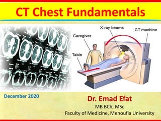
  • 1. CT Chest Fundamentals December 2020 Dr. Emad Efat MB BCh, MSc Faculty of Medicine, Menoufia University
  • 2. CT Chest - Tutorials 1.Computed tomography (CT) Chest – Types 2.CT Chest – Anatomy 3.CT Chest - Abnormalities
  • 3. CT Chest - Types 1.Standard or conventional CT:  Slice thickness: 3-10 mm  Scans a large volume, very quickly  Covers the full lung  +/- contrast Indications  Chest x- ray (CXR) abnormality  Lung cancer staging  F/U metastases  Pleural and mediastinal abnormalities  Empyema
  • 4. CT Chest - Types 2. High-resolution computed tomography (HRCT)  Narrow x-ray beam collimation: 1-1.3 mm vs. conventional 3-10 mm  Cross sections are further apart: 10 mm  High definition images of lung parenchyma: vessels, airspaces, airway and interstitium  No contrast HRCTSTANDARD CT
  • 5. CT Chest – Types - HRCT Indications  Diffusely abnormal CXR  Normal CXR with abnormal PFT’s  Baseline for patients with diffuse lung disease  Solitary pulmonary nodules  Reversible (active) vs. non-reversible (fibrotic) lung disease  Hemoptysis  Lung biopsy guide  Follow up known lung disease
  • 6. CT Chest - Types 3. Low Dose CT:  Uses low doses of radiation -- as much as 30 to 50 percent less than regular CT  Detail is decreased  Uses: Screening ongoing trials Follow up infections metastases post lung transplant Axial images show pulmonary nodule in standard dose (right) and ultra-low-dose scan protocols in a 64-year-old patient with prostate cancer.
  • 7. CT Chest - Types 4. CT angiography: Contrast injected into peripheral vein Indications:  Pulmonary embolism  Aortic aneurysms  Aortic dissection  Arteriovenous malformation  Evaluate superior vena cava syndrome Risks Iodinated contrast: – Allergic/ nephrotoxic Chest CT angiography shows acute pulmonary embolism (arrow).
  • 8. CT Chest - Types 5. CT with contrast: Indications: Evaluation of the mediastinum (lymph nodes, infection) Infection of the chest wall Evaluation of suspected cancer Pleural thickening, pleural nodules, empyema and evaluation of metastatic or primary malignancy of the pleura Pulmonary Lobar Collapse Helpful to evaluate lung abscess, although it should not be performed routinely Differentiate between enlarged Axial intravenous contrast- enhanced CT in a patient with passive atelectasis of the right lower lobe due to a large pleural effusion. Note the dense homogeneous enhancement of the collapsed right lower lobe. lymph nodes and the vascular structures
  • 9. CT Chest - Types 6. Paired inspiratory and expiratory chest CT scans: The quantitative analysis is done by relative difference in density (Hounsfield unit) in inspiratory and expiratory scans Indication : obstructive airway diseases including:  obliterative bronchiolitis  hypersensitivity pneumonitis (HP)  cryptogenic organising pneumonia (COP) (formerly BOOP)  bronchial asthma  sarcoidosis  emphysema  bronchiectasis
  • 10. CT Chest Anatomy – views Three views: 1. Axial view 2. Coronal view 3. Sagittal view
  • 11. CT Chest Anatomy – window settings Three Windows: 1. Mediastinal or Soft Tissue windows 2. Lung windows 3. Bone windows
  • 12. CT Chest Anatomy – Hounsfield unit Hounsfield units (HU), a parameter generated from standard CT, are related to the density of the structure of interest.
  • 13. CT Chest Anatomy – mediastinum  The mediastinum is defined as the tissue compartment located between the two lungs, posterior to the sternum, anterior to the vertebral column, and extending from the thoracic inlet to the diaphragm.  As an aid to understanding regional anatomy, the mediastinum can be divided into four compartments, respectively, from superior to inferior: (a) the supraaortic or superior mediastinum; (b) the region of the aortic arch and aortopulmonary window (APW); (c) the pulmonary arteries, subcarinal space, and azygoesophageal recess; and (d) the heart and paracardiac mediastinum.
  • 14. CT Chest Anatomy- mediastinum- compartments (a) The supraaortic mediastinum; Right Left
  • 15. CT Chest Anatomy- mediastinum- compartments (a) The supraaortic mediastinum; Right Left
  • 16. CT Chest Anatomy- mediastinum- compartments (a) The supraaortic mediastinum; Right Left
  • 17. CT Chest Anatomy- mediastinum- compartments (a) The supraaortic mediastinum; Right Left
  • 18. CT Chest Anatomy- mediastinum- compartments (b) The region of the aortic arch; Right Left
  • 19. CT Chest Anatomy- mediastinum- compartments (b) The region of the azygos arch and aortopulmonary window (APW); AA, ascending aorta DA, descending aorta Right Left
  • 20. CT Chest Anatomy- mediastinum- compartments (c) The main pulmonary artery, subcarinal space, and azygoesophageal recess; LPA, left pulmonary artery PA, pulmonary artery RB & LB, right & left main bronchi Right Left
  • 21. CT Chest Anatomy- mediastinum- compartments (c) The right pulmonary artery (RPA) and azygoesophageal recess; Right Left AER, azygo- esophageal recess
  • 22. CT Chest Anatomy- mediastinum- compartments (d) The heart and paracardiac mediastinum; AR, aortic root LA, left atrium LAA, left atrial appendage Right Left
  • 23. CT Chest Anatomy- mediastinum- compartments (d) The heart and paracardiac mediastinum; Right Left
  • 24. CT Chest Anatomy- mediastinum- compartments (d) The heart; LVO, left ventricular outflow Right Left
  • 25. CT Chest Anatomy- mediastinum- compartments (d) The heart; Right Left Right Left
  • 26. CT Chest Anatomy – The fissures  Each lung has an oblique fissure and the right lung has a horizontal fissure.  There are numerous accessory fissures  In conventional CT, the fissures are visualized as lucent bands devoid of vascularity, whereas they appear as sharp lines in high- resolution CT Schematic drawings of the lungs illustrating the normal fissures (right major, minor and left major) in coronal, sagittal and axial projections.
  • 27. CT Chest Anatomy – The fissures The right lung:  The oblique fissures (the major fissures or greater fissures): begins roughly at the spinous process of the T4 level of the thoracic spine and ends at the anterior costophrenic angle  The horizontal fissure (the minor fissure): running horizontally from the edge of right lung towards the right hilum, at approximately the level of the anterior 4th rib The oblique fissures (white arrow) and The horizontal fissure (green arrow) .
  • 28. CT Chest Anatomy – The fissures  The oblique fissures (the major fissures or greater fissures): begins between the spinous processes of vertebrae T3 and T4 and ends about 5 cm posterior to the anterior costophrenic angle The left lung (a) axial view for left lung. (b) sagittal view for left lung; (c) coronal view for left lung
  • 29.
  • 30. CT Chest Anatomy – lung segments (a) At level of trachea (lung apex );
  • 31. CT Chest Anatomy – lung segments (a) At level of trachea (upper lobe );
  • 32. CT Chest Anatomy – lung segments (a) At level of main stem bronchi ( at hilum ); anatomy on the right like the left;
  • 33. CT Chest Anatomy – lung segments (a) At level below hilum and main stem bronchi;
  • 34. CT Chest Anatomy- Bronchial tree- Bronchial nomenclatures
  • 35. CT Chest Anatomy- Bronchial tree
  • 36. CT Chest Anatomy- Bronchial tree
  • 37. CT Chest Anatomy- Bronchial tree
  • 38. CT Chest Anatomy- Bronchial tree
  • 39. CT Chest Anatomy- Bronchial tree
  • 40. CT Chest Anatomy- Bronchial tree
  • 41. CT Chest Anatomy - thoracic lymph node Supraclavicular nodes: 1.Low cervical, supraclavicular and sternal notch nodes Superior Mediastinal Nodes 2-4 2R & 2L Upper Paratracheal 3A Pre-vascular & 3P Retrotracheal 4R & 4L Lower Paratracheal Aortic Nodes 5-6 5. Subaortic & 6. Para-aortic Inferior Mediastinal Nodes 7-9 7. Subcarinal 8. Paraesophageal 9. Pulmonary Ligament N1 nodes: 10. Hilar & 11. interlobar 12. Lobar & 13. segmental & 14. subsegmental
  • 42. CT Chest Anatomy - thoracic lymph node 3A.Pre-vascular 3P.Pre-vertebral  Mediastinal lymph nodes are not seen in the scan if they are normal, Nodes which are seen are pathologically enlarged
  • 43. CT Chest Anatomy - thoracic lymph node
  • 44.
  • 45.
  • 46.
  • 47. CT Chest Abnormalities - Trachea The trachea should be central or slightly to the right.  Causes of tracheal deviation:  Ipsilateral (To pull): Collapse and Fibrosis  Contralateral (To push): Apical mass , Pleural effusion and Pneumothorax Wegener granulomatosis: HRCT findings:  The trachea and main bronchi may be diffusely or focally circumferentially thickened;  Subglottic stenosis, bronchial stenosis  Peripheral bronchial narrowing, lobar and segmental atelectasis, or bronchiectasis may also be present.  Coexistent pulmonary nodules and masses, which are sometimes cavitary  Consolidation and ground-glass opacities
  • 48. CT Chest Abnormalities – Trachea- Wegener granulomatosis Laryngotracheal computed tomography of Wegener granulomatosis ; (A) Subglottic stenosis (red arrow). (B) 3D reconstruction of subglottic stenosis (white arrow). (C)–(E) Axial images showing a granulomatous lesion (*) (yellow arrows) partially obstructing the tracheal lumen.
  • 49. CT Chest Abnormalities - Trachea Tracheomalacia: Collapse of the trachea more than 70% during the expiration, may be congenital or acquired  In inspiratory CT, a dilated trachea (> 3 cm), especially with posterior bowing of the membranous portion (thus becoming circular) may indicate over compliance of the trachea and suggest the diagnosis.  During expiration, the posterior membranous trachea bows anteriorly, producing an upside down U-shaped air column on transverse CT termed the “frown” sign “frown” sign
  • 50. CT Chest Abnormalities - Trachea Relapsing polychondritis (RPC):  An autoimmune disease affects cartilaginous structures (e.g. the nose, ear, and laryngotracheobronchial tree.  On CT, RPC is characterized by:  Increased attenuation and thickening of the anterior and lateral walls of the large airways  Concomitant destruction of the cartilaginous tracheobronchial rings with sparing of the posterior wall.  Tracheomalacia and large airways stenosis may also be present A deformity of the ear that followed recurrent acute attacks; and saddle nose, which usually follows repeated, painful nasal chondritis
  • 51. CT Chest Abnormalities - Trachea Mounier- Kuhn syndrome: a congenital defect or atrophy of the elastic and smooth muscle tissues of the trachea and main bronchi, resulting in dilatation  Diagnostic criteria: if Trachea exceed diameter > 3.0 cm Right mainstem diameter > 2.4 cm Left mainstem diameter > 2.3 cm Secretions are poorly mobilized, leading to the chronic accumulation of secretions: Recurrent infections, Bronchiectasis, Rarely pulmonary fibrosis Two axial CT images of the thorax demonstrate marked dilatation of the trachea (T) and right (R) and left (L) main bronchi. Notice the bronchiectasis (red arrows and red circle) and there is tram=tracking of thickened bronchial walls.
  • 52. CT Chest Abnormalities - Trachea Saber-sheath trachea:  Diffuse coronal narrowing of the intrathoracic portion of the trachea with the concomitant widening of the sagittal diameter.  Occur almost exclusively in men with chronic obstructive pulmonary disease (COPD) Saber-sheath trachea  Other tracheal Disease which causes Tracheal Narrowing and Wall Thickened: Amyloidosis, Sarcoidosis, Inflammatory bowel disease and Tracheobronchopathia osteochondroplastica.
  • 53. CT Chest Abnormalities - The lung hilum CT is helpful in the diagnosis of endobronchial lesions, hilar and parahilar masses, and hilar vascular lesions. Hilar enlargement: May be unilateral or bilateral, symmetrical or asymmetrical
  • 54. CT Chest Abnormalities - The lung hilum 1.Lymph Node enlargement:  Contrast-enhanced computed tomography (CECT) is helpful to differentiate lymph nodes from vascular structures as lymph nodes usually do not take contrast.  Mild enhancement may be seen in tuberculosis, fungal infection, lymphoma, metastatic lung cancer, and sarcoidosis. Thoracic CT images from the patient with Tubercular Myocarditis at the time of diagnosis, radiologically bulky right hilar lymphadenopathy (yellow arrow) was evident.
  • 55. The lung hilum- Lymph Node enlargement  Calcification within lymph nodes usually suggest granulomatous disease like TB, histoplasmosis, sarcoidosis, silicosis, nontuberculous mycobacterial infection  Enlargment of discrete lymph nodes, most commonly seen in sarcoidosis. Tumors and inflammatory processes may produce a large mass of confluent lymph nodes. Enlarged hilar lymph nodes in sarcoidosis (red arrows)
  • 56. CT Chest Abnormalities - The lung hilum  CT pulmonary angiography (CTPA) is the best method for demonstrating emboli.  HRCT is useful for demonstrating lung disease, which may account for secondary pulmonary hypertension. Pulmonary Arterial Hypertension. Frontal radiograph of the chest shows an enlarged main pulmonary artery (red arrow) and a markedly enlarged right pulmonary artery (white arrow) with rapid attenuation in the size of the vessels in the lung periphery. On the contrast-enhanced CT, the main pulmonary artery (P) is much larger than the aorta (A). Normally, they should roughly be about the same size. 2.Arterial enlargement:
  • 57. CT Chest Abnormalities - The lung hilum CT thorax, lung window showing spiculated right hilar mass ( red arrow) 3. Malignancy:  CT enables staging of the disease  Squamous cell carcinoma (SCC ) and small cell carcinoma are typically central  Central SCC often causes lung collapse and/or obstructive pneumonitis.  Cavitation is a frequent finding in SCC.  In Small cell carcinoma, Direct infiltration of adjacent structures is more common. Necrosis and haemorrhage are both common.
  • 58. CT Chest Abnormalities - lung fields Lung abnormalities: Abnormal whiteness (increased density):  Consolidation  Interstitial  Atelectasis  Nodule or mass Abnormal blackness (decreased density):  Cyst  Emphysema  Cavity
  • 59. CT Chest Abnormalities - lung fields Four patterns of increased density:  Consolidation  Lobar  Diffuse  Multifocal ill- defined  Atelectasis  Nodule or mass  Solitary Pulmonary Nodule  Multiple Masses  Interstitial  Reticular  Fine Nodular
  • 60. lung field abnormalities - Consolidation Consolidation:  Increased attenuation of the lung parenchyma causing obscuration of pulmonary vessels. Reflect diseases that are primarily alveolar, interstitial, or mixed.  Without significant loss of volume. Air bronchograms can be found  Causes:  transudate, e.g. pulmonary oedema secondary to heart failure  pus, e.g. bacterial pneumonia  blood, e.g. pulmonary haemorrhage  cells, e.g. bronchoalveolar carcinoma  protein, e.g. alveolar proteinosis  fat, e.g. lipoid pneumonia  gastric contents, e.g. aspiration pneumonia  water, e.g. drowning
  • 61. lung field abnormalities - Consolidation Air bronchogram: Refers to the phenomenon of air-filled bronchi (dark) being made visible by the opacification of surrounding alveoli (grey/white). Pneumococcal pneumonia produces consolidation in the right upper lobe with multiple air bronchograms (black branching structures)
  • 62. lung field abnormalities - Consolidation
  • 63. lung field abnormalities - Consolidation
  • 64. lung field abnormalities - Consolidation
  • 65. lung field abnormalities - Consolidation  Bat's wing or butterfly pulmonary opacities: A bilateral perihilar distribution of consolidation.  Reverse bat wing pulmonary opacities: Peripheral or subpleural consolidation, sparing the perihilar region Bat's wing opacitiesReverse bat wing opacities
  • 66. lung field abnormalities - Consolidation Reverse bat wing opacitiesBat's wing opacities  Chronic eosinophilic pneumonia  Cryptogenic organising pneumonia (COP)  Bronchoalveolar carcinoma  Pulmonary contusion: in the setting of trauma  Pulmonary haemorrhage with or without infarction: in the setting of Pulmonary embolism.  Pulmonary vasculitis  Aspiration  Pulmonary oedema (especially cardiogenic)  Pneumonia  aspiration pneumonia  pneumocystis pneumonia (PCP)  viral pneumonia  lipoid pneumonia  Inhalation injury  noxious gas  liquid  pulmonary alveolar proteinosis  Pulmonary haemorrhage (e.g. Goodpasture syndrome)  Lymphoma/leukaemia  Bronchoalveolar carcinoma
  • 67. lung field abnormalities - Consolidation
  • 68. lung field abnormalities - Consolidation Lymphoma: Imaging Findings:  Hilar or mediastinal lymphadenopathy:  A soft tissue attenuating mass, with smooth or lobulated margins which conforms to surrounding structures. Cystic/low density areas are common.  Calcification is usually seen following therapy  Pleural effusions (Mostly small, unilateral, and exudative)  Destructive rib or vertebral body lesion  Pericardial effusion A large soft tissue mass in the anterior mediastinum, which arises in the thymus (yellow arrow). There is associated paratracheal adenopathy (red arrow).
  • 69. CT Chest Abnormalities - Consolidation - Lymphoma  Parenchymal lung involvement:  Multiple nodules  A mass or mass-like consolidation greater than 1 cm with or without cavitations or bronchograms  Masses of pleural origin  Nodules <1 cm  Peribronchial or perivascular thickening  Alveolar or interstitial infiltrates  Segmental or lobar atelectasis Large left upper lobe mass, with patchy opacity peripherally. Truncation of the left upper lobe bronchus. Extends to the mediastinum with enlarged mediastinal nodes
  • 70. lung field abnormalities - Consolidation CT scan obtained with pulmonary window setting in the right middle lobe shows a focal area of consolidation with what may be tiny nodules (yellow arrow). Tuberculosis (TB): Primary pulmonary tuberculosis: Imaging Findings:  Patchy or lobar consolidation  Ipsilateral hilar and mediastinal (paratracheal) lymphadenopathy, usually right sided.  Caseating granuloma (tuberculoma) which usually calcifies (known as a Ghon lesion)  Pleural effusions  Calcification of nodes  Atelectasis  Cavitation (uncommon)
  • 71. CT Chest Abnormalities - Consolidation - Tuberculosis Classical CT pattern showing a large thick-wall cavity, tree-in bud and bronchial wall thickening. Post-primary pulmonary Tuberculosis: Imaging Findings:  Patchy consolidation  “tree-in-bud” sign: Poorly defined linear and nodular opacities  Ground-glass opacities  Bronchial wall thickening  Interlobular septal thickening  Cavitation, Aspergillomas, fibrosis and Bronchiectasis  Pleural effusion  Hilar nodal enlargement  Lobar consolidation, tuberculoma, calcifications and miliary TB
  • 72. Consolidation- Tuberculosis Tree-in-bud sign or pattern: usually represents endobronchial spread of infection.
  • 73. Consolidation- Tuberculosis- Tree-in-bud sign Causes:  Infective bronchiolitis (pulmonary tuberculosis, e.g. mycobacterium tuberculosis infection, atypical mycobacterial infections, e.g. mycobacterium avium (MAIC), viral pneumonia, fungal pneumonia, e.g. aspergillus, allergic bronchopulmonary aspergillosis (ABPA), pneumocystis pneumonia)  Congenital (cystic fibrosis, immotile cilia syndrome, e.g. Kartagener syndrome, yellow nail syndrome )  Connective tissue disorders (rheumatoid arthritis (RA), Sjögren syndrome)  Bronchial (obliterative (constrictive) bronchiolitis, diffuse panbronchiolitis, follicular bronchiolitis)  Neoplastic ( bronchioloalveolar cell carcinoma, distant metastatic disease (e.g. breast, liver, ovary, prostate, kidney), primary pulmonary lymphoma, chronic lymphocytic leukemia )
  • 74. CT Chest Abnormalities - Consolidation - Tuberculosis Tuberculoma and Miliary Tuberculosis: Imaging Findings:  Tuberculoma and miliary tuberculosis are rare  Miliary deposits are seen both in primary and post-primary tuberculosis. It appear as 1-3 mm diameter nodules, which are uniform in size and uniformly distributed  Tuberculomas are usually found as single nodules and they may include a cavity or a calcification with sharp margins. They are usually found in the upper lobes Miliary Tuberculosis Tuberculoma. Axial lung (A) and mediastinal window (C) CT images show a soft tissue density nodule in the right upper lobe of the lung.
  • 75. lung field abnormalities - Consolidation Aspergillomas:  Mass-like fungus balls of Aspergillus fumigatus, occur in patients with normal immunity but with pre-existing cavities:  pulmonary tuberculosis  pulmonary sarcoidosis  bronchiectasis  bronchogenic cyst  pulmonary sequestration  Pneumocystis pneumonia (PCP) associated pneumatocoeles  Imaging Findings: Air crescent sign: Rounded or ovoid soft tissue attenuating masses located in a surrounding cavity and outlined by a crescent of air. Differential diagnosis (DD); hydatid cyst, bronchogenic carcinoma and PCP. Aspergilloma - A solid mass within a cavity and the “air crescent sign“ can be shown in this CT image.
  • 76. CT Chest Abnormalities - Consolidation - Tuberculosis
  • 77. lung field abnormalities - Consolidation Aspiration Pneumonitis and Pneumonia: Imaging Findings:  An infiltrate, frequently in the posterior segment of an upper lobe and the superior or posterior basal segments of a lower lobe (The right lower lobe is the most frequent location).  Aspiration-related lung abscess  Interstitial or nodular infiltrates, pleural effusion, and other changes may be slowly progressive.  Aspirated material can be demonstrated on CT scans Ct shows centrilobular nodules with surrounding ground-glass opacities and subpleural non-segmental consolidations (yellow arrow) at the dorsal portions of the right lung. Note that the lumens of segmental bronchi are filled with aspirated materials (red arrow).
  • 78. lung field abnormalities - Consolidation Consolidation due to Lung infarction: Imaging Findings:  Wedge-shaped (less often rounded) pleural-based consolidation (Hampton hump)  With or without cavitation  Convex borders with a halo sign due to adjacent haemorrhage.  Usually no air-bronchogram  Plate-like (subsegmental, discoid) atelectasis  Pleural effusion  Elevated hemidiaphragm  "Melting Ice Cube Sign": rapid peripheral resolution of a Hampton's hump  Feeding vessel sign (Hampton hump); CT with lung windowing shows a focal subpleural area of consolidation in the left lower lobe (arrows). There are also small bilateral pleural effusions
  • 79. Consolidation- Lung infarction Feeding vessel sign:  The feeding vessel sign consists of a distinct vessel leading directly to a nodule or a mass. This sign has been considered highly suggestive of septic embolism  Also occurs in:  pulmonary metastasis  lung cancer  Pulmonary arteriovenous malformations (PAVM's)  pulmonary infarction  pulmonary vasculitis  angioinvasive pulmonary aspergillosis  granuloma Feeding vessel sign. A patient with bronchial carcinoma. Pulmonary artery (yellow arrow) leading directly to the mass is seen.
  • 80. Consolidation- Lung infarction The halo sign (HS): typically seen on HRCT, ground glass opacity surrounding a pulmonary nodule or mass and represents haemorrhage, It is typically seen in angioinvasive aspergillosis. Causes:  Neoplasms (e.g. adenocarcinoma, squamous cell carcinoma, Kaposi sarcoma, lymphoma and lung metastases)  Inflammatory (e.g. Fungal, mycobacterial, and viral infections)  Others: Wegener granulomatosis, eosinophilic lung disease, pulmonary endometriosis, pulmonary embolism, organizing pneumonia, HP, iatrogenic injury, pulmonary pseudoaneurysm Metastatic tumor; The CT halo sign depicts peri-lesion ground- glass attenuation (red arrow).
  • 81. Consolidation- Lung infarction Reversed halo sign: Central ground-glass opacity surrounded by denser consolidation of crescentic or ring shape, at least 2 mm thick relatively specific for cryptogenic organizing pneumonia ; causes:  wegener’s and lymphomatoid granulomatosis  sarcoidosis  tuberculosis  pneumocystis pneumonia  pulmonary mucormycosis  paracoccidiodomycosis  invasive aspergillosis  neoplastic (metastasis)  pulmonary infarction  lipoid pneumonia  schistosomiasis Fairy Ring Sign; Like Reversed halo sign but with normal central lung parenchyma, seen sarcoidosis Reversed halo sign: central ground-glass density surrounded by peripheral consolidation (yellow arrow).
  • 82. lung field abnormalities - Consolidation Klebsiella pneumonia (aka Friedländer’s pneumonia): Imaging Findings:  usually involves one of the upper lobes  ground glass opacities  consolidation  intralobular reticular opacities  interlobular septal thickening  centrilobular nodules  cavitation  necrotising pneumonia  Pleural effusion and/or empyema  Bulging fissure sign Transverse thin-section CT of right lower lobe showing consolidation (arrowhead) and intralobular reticular opacity (arrows) with peripheral distribution. Pleural effusion was also present
  • 83. Consolidation- Klebsiella pneumonia Bulging fissure sign: refers to lobar consolidation where the affected portion of the lung is expanded causing displacement of the adjacent fissure ; causes:  pneumococcal pneumonia  klebsiella pneumonia  pseudomonas pulmonary infection  staphylococcus aureus  legionella pneumophila  mycobacterium tuberculosis  lung adenocarcinoma  lung abscess  pulmonary hemorrhage Klebsiella pneumonia: coronal CT image shows necrotising consolidation (black arrow) in the right upper lobe with the bulging fissure sign (white arrow)
  • 84. lung field abnormalities - Consolidation Necrotising pneumonia (NP): refers to a pneumonia characterised by the development of the necrosis within infected lung tissue.  Causative pathogens include ( Staphylococcus aureus, Klebsiella pneumonia, Enterobacter, Nocardai, Actinomyces, Pseudomonas, Pneumococcus, Haemophilus influenza)  CT with may be better as it allows appreciation of low attenuation and non enhancement within the necrosed portions (representing liquifaction) of the affected area of infection (consolidation). Necrotizing pneumonia. heterogeneous enhancing consolidation with smooth air bronchograms and cavities in the right upper lobe
  • 85. Consolidation - Cardiogenic pulmonary edema Consolidation due to Congestive Heart Failure (CHF) : Stage I CHF – Redistribution:  Redistribution of blood flow from the lower to the upper. This is know as cephalization because the pulmonary veins of the superior zone dilate due to increased pressure.  Increased artery-to-bronchus ratio at hilar level (normally they are equal)  Cardiomegally Significant enlargement of segmental / subsegmental pulmonary arteries (NB pulmonary artery-to- bronchus ratio >1) and Mosaic attenuation pattern
  • 86. Consolidation - Cardiogenic pulmonary edema Stage II CHF - Interstitial edema Characterized by: 1. Thickened interlobular septa: Septal lines, also known as Kerley lines, become prominent. They usually occur when pulmonary capillary wedge pressure reaches 20-25 mmHg. Smooth septal thickening (red arrow) in the lower right lobe, increased vascular diameter (yellow arrow) and bilateral pleural effusion (black arrow) in a patient with congestive heart failure. Please observe the aneurysm on the descending aorta
  • 87. Consolidation - Cardiogenic pulmonary edema 2. Bronchial wall thickening will also be seen, and is the basis of peribronchial cuffing seen on chest x-rays. Interstitial pulmonary edema. (A) Axial chest CT demonstrates smooth thickening of interlobular septae (white arrow). This is the CT equivalent of Kerley B lines seen on chest x-rays. (B) Magnified view from the RLL in the same patient demonstrates bronchial wall thickening (green arrow), which is the CT equivalent of peribronchial cuffing seen on radiographs. Also, note that the adjacent pulmonary artery branch (red arrow) is slightly larger than the bronchus.
  • 88. Consolidation - Cardiogenic pulmonary edema 3. Fluid in the major or minor fissure produces thickening of the fissure and 'Phantom' or 'vanishing' tumours 4. Peribronchovascular interstitial thickening 5. ground-glass opacities 6. mosaic pattern of attenuation 7. pleural effusion 8. lymph nodes may be enlarged congestive heart failure presenting ground-glass opacities and smooth interlobular septa thickening, a mosaic pattern of attenuation and bilateral pleural effusion Thickening of the right major fissure from subpleural edema
  • 89. Consolidation - Cardiogenic pulmonary edema Pulmonary pseudotumor or Phantom or vanishing tumours: it is caused by a loculated pleural effusion trapped in the pleural fissures Axial projection (A) and Sagittal projection (B) of chest CT. Greater fluid collection within the major and minor fissures with the second collection pictured into the accessory superior fissure of the right lower lobe.
  • 90. Consolidation - Cardiogenic pulmonary edema Stage III CHF - Alveolar edema Characterized by:  Peribronchial nodules  Alveolar edema with perihilar consolidations and air bronchograms (Bat's wing or butterfly pulmonary opacities)  Pleural effusion  An enlarged cardiac silhouette High-resolution computed tomography scans, showing slices acquired at the aortic arch level, using a pulmonary window, in a patient with acute myocardial infarction and Cardiogenic pulmonary edema presenting consolidations, ground-glass opacities, smooth interlobular septal thickening and bilateral pleural effusion.
  • 91. lung field abnormalities - Consolidation Adult Respiratory Distress Syndrome ( ARDS ), three phases: Fibrotic phaseFibroproliferative phaseExudative phase  persistent ground- glass densities  coarse reticulation  air cysts (arrowheads)  traction bronchiectasis  alveolar opacities  reticular opacities  Bronchiectasis (arrows)  honeycombing  signs of pulmonary hypertension  consolidations  ground-glass opacities  thickening of interlobular septa (crazy paving)  posterior compressive atelectasis
  • 92. lung field abnormalities - Consolidation Bronchopneumonia: bilateral and predominantly in the lung bases late stagesEarly stages  Extends peripherally along the bronchus to involve the entire pulmonary lobule  Multifocal heterogeneous confluent consolidation = patchwork quilt; eventually coalesce.  Exudates fill airways = no air bronchograms  25-75% form abscess  Empyema and parapneumonic effusion common  Multiple foci of opacity  Begins centrally in and around lobular bronchi  Peribronchial thickening and poorly defined bronchovascular nodules  May result in a tree-in-bud appearance
  • 93. lung field abnormalities - Consolidation Atypical pneumonia (AP) characterised by:  Focal ground-glass opacity in a lobular distribution (Diffuse and bilateral)  Diffuse ground glass nodules in a centrilobular pattern  Bronchial wall thickening  pleural effusion may present 30-year-old woman with Mycoplasma pneumoniae pneumonia. CT shows bronchial wall thickening (arrows). Lobular areas of consolidation and ground- glass attenuation are also seen.
  • 94. lung field abnormalities - Consolidation (AP) Legionella pneumonia:  Multifocal and bilateral patchy infiltrate (ground glass and/or consolidation), Middle and lower zone predominance  Pleural effusions can be common  Associated hilar adenopathy may be present  Cavitation and a masslike appearance Legionella pneumonia. (A) Typical non- segmental lesion in the right middle lobe and the right lower lobe. (B) sharp- bordered fissures (arrow), which is a characteristic feature of non-segmental distribution. (C) Segmental lesions in the right lower lobe. (D) consolidation and ground-glass opacity
  • 95. lung field abnormalities - Consolidation (AP) Mycoplasma pneumonia characterised by:  Areas of ground-glass attenuation and air-space consolidation Mycoplasma pneumoniae pneumonia. HRCT shows bilateral lobular areas of consolidation, patchy ground-glass opacities, interlobular septal thickening  lobular distribution  Centrilobular nodules  Thickening of the peribronchovascular interstitium  Interlobular septal thickening  Mosaic perfusion  lower lobe predominance
  • 96. lung field abnormalities - Consolidation (AP) Chlamydia pneumonia characterised by:  Areas of ground-glass attenuation and air-space consolidation  lobular distribution  Findings are often limited to a single lobe. Chlamydia pneumonia. an acinar pattern on a background of ground-glass attenuation. Right pleural effusion is also present.  lower lobe involvement occurring more frequently  bronchovascular bundle thickening.  Centrilobular nodules  Up to 25% of patients may have a small to moderate-sized pleural effusion  lymphadenopathy: uncommon
  • 97. lung field abnormalities - Consolidation Respiratory syncytial pneumonia. bilateral ill- defined centrilobular nodules (arrows) and bronchial wall thickening (arrowhead). (AP) Viral pneumonia characterised by:  Patchy, unilateral, or bilateral consolidations and ground-glass opacity or poorly defined centrilobular nodules.  Peribronchial thickening  Thickened interlobular septa  Areas of atelectasis or air trapping  Pleural effusion, hilar lymphadenopathy and pneumothorax are uncommon findings.
  • 98. lung field abnormalities - Consolidation (AP) Covid-19 pneumonia: The primary findings of COVID-19 on chest radiograph and CT are those of atypical pneumonia or organizing pneumonia.  Normal in up to 63% in the early stages  Ground glass (68.5%), coarse horizontal linear opacities, and consolidation.  Crazy paving  These are more likely to be multifocal, bilateral, peripheral and in the lower zones  Vascular dilatation and bronchovascular thickening  Traction Bronchiectasis  Atypical CT findings (mediastinal lymphadenopathy, pleural effusion, multiple tiny pulmonary nodules, tree-in-bud, pneumothorax, cavitation) (COVID-19). A, ground-glass opacity (GGO), B, crazy-paving pattern, and, C, consolidation
  • 99. lung field abnormalities - Consolidation Pulmonary fungal disease: pulmonary aspergillosis characterised by:  Aspergillomas (mycetomas)  ABPA (next).  Chronic necrotizing or Semi-invasive aspergillosis:  upper lobe predominant opacities  pleural thickening  Cavitation with or without a central mycetoma  Often there are multiple cavities, often thick-walled Axial CT image demonstrates a cavitary lesion containing fungal ball in a patient with HIV and semi- invasive aspergillosis
  • 100. lung field abnormalities - Consolidation Pulmonary fungal disease: pulmonary aspergillosis characterised by:  Airway-invasive aspergillosis:  tracheobronchitis: tracheal or bronchial wall thickening  Bronchiolitis: centrilobular and tree-in-bud opacities  Bronchopneumonia: perihilar consolidations Bronchial wall thickening, tree-in-bud nodules, and peribronchial consolidation in sagittal CT image of a patient with airway-invasive aspergillosis leading to bronchitis, bronchiolitis, and bronchopneumonia
  • 101. lung field abnormalities - Consolidation Pulmonary fungal disease: pulmonary aspergillosis characterised by:  Angioinvasive aspergillosis:  Solitary or multiple pulmonary nodules/masses  halo sign and/or Reversed halo sign angioinvasive aspergillosis. ground-glass density surrounding a nodule (halo sign)
  • 102. lung field abnormalities - Consolidation E-Cigarette or Vaping Product-Associated Lung Injury: 1. Lipoid pneumonia: basilar predominant ground glass opacities, nodular, crazy paving or tree-in-bud opacities, 2. Diffuse alveolar damage: dependent ground glass opacities and consolidation (acute/exudative phase), followed by reticulation and traction bronchiectasis (organizing/proliferative phase) Lipoid pneumoniaDiffuse alveolar damage
  • 103. lung field abnormalities - Consolidation E-Cigarette or Vaping Product-Associated Lung Injury: Acute eosinophilic pneumonia 3. Acute eosinophilic pneumonia: ground glass opacities and consolidation, often with pleural effusions and septal thickening
  • 104. lung field abnormalities - Consolidation E-Cigarette or Vaping Product-Associated Lung Injury: 4. Organizing pneumonia: peripheral or perilobular ground glass opacities and/or consolidation, sometimes with the atoll/reverse halo sign Organizing pneumonia
  • 105. lung field abnormalities - Consolidation E-Cigarette or Vaping Product-Associated Lung Injury: 5. Diffuse alveolar hemorrhage: centrilobular ground glass opacities, nodules, and/or consolidation with subpleural sparing, followed by septal thickening (reflecting lymphatic engorgement with blood products) 6. Respratory bronchiolitis interstitial lung disease: upper lobe predominant centrilobular ground glass nodules (next) 7. Hypersensitivity pneumonitis (next) 8. Giant cell interstitial pneumonia: fibrosis seen as peripheral reticulation, ground glass opacity, and traction bronchiectasis
  • 106. lung field abnormalities - Consolidation Wegener's granulomatosis characterized by:  Nodules or mass lesions, which may cavitate  Fleeting focal infiltrates (lung consolidation )  Ground-glass opacities Wegener's granulomatosis. Multiple pulmonary nodules. The ground- glass halo is compatible with perinodular capillaritis and alveolar hemorrhage ( yellow arrow). There was also a large cavitating mass as well ( red arrow)
  • 107. lung field abnormalities - Consolidation Eosinophilic pneumonia (EP): Chronic EPAcute EP  Non-segmental air-space consolidation with peripheral predominance (reversed batwing)  Middle or upper zone predilection  Lymph nodes may be enlarged  During resolution, wavy lines paralleling chest wall  Less common: ground-glass opacities, pulmonary nodules, reticulation, lobar atelectasis and pleural effusion  Bilateral ground-glass areas  Interlobular septal thickening  Pleural effusions  Air-space consolidation  Thickening of bronchovascular bundles  Ill-defined centrilobular nodules
  • 108. lung field abnormalities - Consolidation Septic emboli: CT findings:  Multiple peripheral parenchymal nodules, these may have a dependent, lower zone predication  Cavitation or air bronchogram in more than 89%, cavities are thin- walled and may have no fluid level  Wedge-shaped subpleural consolidation with apex of lesion directed toward pulmonary hilum  Feeding vessel sign = pulmonary artery leading to nodule (67%)  Hilar and mediastinal adenopathy can occur  Pleural effusion is rare CT scan reveals bilateral peripheral nodules and wedge- shaped consolidation with various size. Some of the lesions show connection with pulmonary vessels (so-called feeding vessel sign, arrows), and this suggests septic emboli. Also note right pleural effusion.
  • 109. lung field abnormalities - Consolidation  Two types intralobar and extralobar  Pulmonary tissue without normal connection to bronchial tree  Anomalous arterial blood supply, from aorta  CT angiography is used because it shows the vascularization of the sequestered tissue in great detail  A solid mass that may be homogeneous or heterogeneous, sometimes with cystic changes.  An infected sequestration may be associated with a parapneumonic effusion, and may contain one or more air-fluid levels. Pulmonary sequestration: Axial CT angiographic image showing anaomalous vessels (red arrow) arising from the descending aorta and supplying the enhancing left lower lobe sequestered lung segment (green arrow)
  • 110. lung field abnormalities - Interstitial disease The secondary pulmonary lobule:  The smallest functional unit of the lung.  Each lobule is demarcated by interlobular septae, which contain lymphatics and pulmonary veins.  The lobule is supplied centrally by a terminal bronchiole and accompanying centrilobular pulmonary artery, which are together known as the bronchovascular bundle.  A second set of lymphatics also runs with the bronchovascular bundle.
  • 111. Interstitial disease- secondary pulmonary lobule  Centrilobular area is the central part of the secondary lobule. It is usually the site of diseases, that enter the lung through the airways ( i.e. hypersensitivity pneumonitis, respiratory bronchiolitis, centrilobular emphysema ).  Perilymphatic area is the peripheral part of the secondary lobule. It is usually the site of diseases, that are located in the lymphatics of the interlobular septa ( i.e. sarcoid, lymphangitic cinomatosis, pulmonary edema). These diseases are usually also located in the central network of lymphatics that surround the broncho- vascular bundle.
  • 112. Interstitial disease- secondary pulmonary lobule Centrilobular area in blue (left) and perilymphatic area in yellow (right) Normal interlobular septa (solid black arrows) and centrilobular arteries (open white arrows) are clearly visible. Interlobular septa are normally 0.1 mm thick and can be seen in the lung periphery, particularly along the anterior and mediastinal pleural surfaces
  • 113. lung field abnormalities - Interstitial disease
  • 114. lung field abnormalities - Interstitial disease
  • 115. lung field abnormalities - Interstitial disease What is the dominant HR pattern? 1- LINEAR ABNORMALITIES 2-NODULES 3 - GROUND GLASS OPACITY 4 - CONSOLIDATION 1-AREAS OF DECREASED ATTENUATION WITH WALLS (CYSTS ; HONEYCOMB ; BRONCHIECTASIS) 2-AREAS OF DECREASED ATTENUATION WITHOUT WALLS (EMPHYSEMA,MOSAIC ATTENUATION) A- High attenuation (CT scan findings manifesting as increased opacity) B- Low attenuation (CT scan findings manifesting as decreased opacity)
  • 116.
  • 117. lung field abnormalities - Interstitial disease  Fine "ground-glass" (1-2 mm): e.g. interstitial pulmonary oedema  Medium "honeycombing" (3-10 mm): commonly seen in pulmonary fibrosis  Coarse (> 10 mm): cystic Spaces caused by parenchymal destruction, e.g. usual interstitial pneumonia (UIP), pulmonary sarcoidosis, Pulmonary Langerhans cell histiocytosis (PLCH) Reticular Pattern: results from the summation or superimposition of irregular linear opacities.
  • 118. lung field abnormalities - Interstitial disease Causes of Reticular Pattern:  Pulmonary edema (heart failure, fluid overload, nephropathy)  Infection ( viral, mycoplasma, Pneumocystis, malaria )  Post-infectious scarring (tuberculosis, histoplasmosis, coccidioidomycosis)  Mitral valve disease  Collagen vascular disorders  Granulomatous disease ( pulmonary sarcoidosis, eosinophilic granuloma )  Drug reactions (e.g. amiodarone )  Pulmonary neoplasms ( lymphangitis carcinomatosis, pulmonary lymphoma )  Inhalational lung disease (asbestosis, silicosis, coal workers pneumoconiosis, hypersensitivity pneumonitis, chronic aspiration pneumonia)  Idiopathic (usual interstitial pneumonia, lymphangioleiomyomatosis, tuberous sclerosis, neurofibromatosis, amyloidosis )
  • 119. Interstitial disease- Reticular pattern linear and reticular opacities: Represents thickening of interstitial fibers of lung by - fluid or - fibrous tissue or - infiltration by cells
  • 120. Interstitial disease- Reticular pattern Linear Pattern: 1. Thickened interlobular septa 2. Peribronchovascular interstitial thickening 3. Intralobular Lines 4. Thickened Fissures 5. Subpleural lines 6. Parenchymal bands
  • 121. Interstitial disease- Reticular pattern- Linear Pattern Interlobular Septal thickening: IrregularNodularSmooth  UIP  Sarcoidosis  Asbestosis  HP  lymphangitic carcinomatosis  Sarcoidosis  lymphangitic carcinomatosis  lymphoproliferative disorders (LIP, lymphoma, leukaemia)  Silicosis, coal worker's pneumoconiosis (CWP)  Kaposi sarcoma  Pulmonary oedema, haemorrhage  Lymphoma, leukaemia  lymphangitic carcinomatosis  lymphocytic interstitial pneumonia (LIP), non specific interstitial pneumonia (NSIP)
  • 122. Interstitial disease- Reticular pattern  Sarcoidosis  Pulmonary interstitial oedema  Certain types of pneumonias – pneumonitis:  mycoplasma pneumonia  acute eosinophilic pneumonia  lymphoid interstitial pneumonia (LIP)  covid-19 pneumonia  Microscopic polyangiitis  Lymphangitis carcinomatosis Lymphangitic Carcinomatosis. A thin- section CT shows both smooth and nodular thickening of the bronchovascular structures (arrows) that represents lymphatic tumor surrounding the axial interstitium. Peribronchovascular interstitial thickening : Causes:
  • 123. Interstitial disease- Reticular pattern Honeycomb cysts:  an irreversible finding in interstitial lung disease  small (3 to 10 mm) cystic spaces with thick (1 to 3 mm) walls  usually posterior subpleural and basal in distribution  frequently seen in UIP and chronic HP and occasionally in sarcoidosis.  additional signs: thickened interlobular and intralobular lines parenchymal bands areas of ground glass opacity Idiopathic Pulmonary Fibrosis (IPF). The HRCT scan shows basal and peripheral reticular opacities with honeycombing and traction bronchiectasis. irregularity of lung interfaces (between broncho-vascular bundles or fissures or pleural surfaces and lung)
  • 124. Interstitial disease- Reticular pattern- Honeycomb cysts  Macrocystic e.g UIP  Mixed macrocystic and Microcystic e.g UIP  Combined emphysema and honeycombing e.g desquamative interstitial pneumonia (DIP) and Pulmonary Langerhans cell histiocytosis (PLCH) Types:  Microcystic e.g fibrotic nonspecific interstitial pneumonia (NSIP)
  • 125. lung field abnormalities - Interstitial disease Nodular pattern:  Homogenous and contain no air bronchograms  Nodular opacities may be:  Miliary nodules: <2 mm  Pulmonary micronodule: 2-7 mm  Pulmonary nodule: 7-30 mm  Pulmonary mass: >30mm Morphology:  Solid calcified pulmonary nodules  Ground glass pulmonary nodules  Partly solid or non-solid: may represent: malignancy: primary or metastases atypical adenomatous hyperplasia focal interstitial fibrosis aspergillosis focal pulmonary haemorrhages solid nodule ground glass nodule partly solid nodule
  • 126. Interstitial disease- Nodular pattern Nodular distribution:  Random distribution: Nodules involve the pleural surfaces and fissures.  Centrilobular distribution: Unlike perilymphatic and random nodules, centrilobular nodules spare the pleural surfaces. The most peripheral nodules are centered 5-10mm from fissures or the pleural surface.  Perilymphatic distribution: nodules are seen in relation to pleural surfaces, interlobular septa and the peribronchovascular interstitium.
  • 127. Interstitial disease- Nodular pattern Nodular distribution: PerilymphaticCentrilobularRandom Sarcoidosis, silicosis, coal-worker's pneumoconiosis, lymphangitic spread of carcinoma, LIP, amyloidosis Infectious bronchiolitis, diffuse panbronchiolitis, respiratory bronchiolitis, HP, LIP, pulmonary edema, vasculitis, plexogenic lesions of pulmonary hypertension, metastatic neoplasms Hematogenous metastases, Miliary tuberculosis, Miliary fungal infections, PLCH (early nodular stage), Sarcoidosis (when very extensive)
  • 128. Interstitial disease- Nodular pattern Causes of Miliary opacities :  Infection  tuberculosis  fungal (often febrile)  healed varicella pneumonia  viral pneumonitis  nocardosis  salmonella  Miliary metastases  thyroid carcinoma  renal cell carcinoma  breast carcinoma  malignant melanoma  pancreatic neoplasms  osteosarcoma  trophoblastic disease  Sarcoidosis  Pneumoconioses  silicosis  coal workers pneumoconiosis  Pulmonary haemosiderosis  Hypersensitivity pneumonitis  Langerhans cell histiocytosis ( PLCH )  pulmonary alveolar proteinosis
  • 129. Interstitial disease- Nodular pattern Causes of Calcified pulmonary nodules:  Healed infection  Calcified granulomata, e.g.  Thoracic histoplasmosis  Recovered miliary TB  Healed varicella pneumonia  Pneumoconioseses  Silicosis  Coal worker's pneuomconiosis  Pulmonary hamartomas  Metastatic pulmonary calcification  Chronic renal failure  Multiple myeloma  Secondary hyperparathyroidism  Massive osteolytic metastases  IV calcium therapy  Pulmonary haemosiderosis  Idiopathic pulmonary haemosiderosis  Mitral stenosis  Goodpasture syndrome  Pulmonary alveolar microlithiasis  Sarcoidosis  Calcified pulmonary metastases  Pulmonary amyloidosis  Pulmonary hyalinizing granuloma  Calcifying fibrous pseudotumour of lung
  • 130. Interstitial disease- Nodular pattern  A reticulonodular pattern results from a combination of reticular and nodular opacities.  A differential diagnosis should be developed based on the predominant pattern.  If there is no predominant pattern, causes of both nodular and reticular patterns should be considered.  Causes: the same disorders as reticular patterns Reticulonodular pattern: Sarcoidosis. a “reticulonodular pattern” characterised by the presence of thickening of the interlobular septae and bronchovascular bundles, perilymphatic and perifissural micronodules and architectural distortion
  • 131. Interstitial disease- High attenuation Ground-glass opacification/opacity (GGO): a hazy area of increased attenuation in the lung with preserved bronchial and vascular markings. Aetiology:  Normal expiration  Partial filling of air spaces  Partial collapse of alveoli  Interstitial thickening  Inflammation  Oedema  Fibrosis  Neoplasm Symmetric perihilar ground-glass opacity, representing pulmonary hemorrhage in a patient with Wegener’s granulomatosis.
  • 132. Interstitial disease- High attenuation Ground-glass opacification/opacity (GGO) and consolidation: causes: ChronicAcute  Hypersensitivity pneumonitis  Smoking related interstitial lung disease (respiratory bronchiolitis-associated interstitial lung disease (RB-ILD), DIP)  Idiopathic interstitial pneumonias (Non- specific interstitial pneumonia (NSIP), rarely usual interstitial pneumonia)  Bronchioloalveolar carcinoma  Cryptogenic Organizing Pneumonia (COP)  Lymphoid interstitial pneumonia (LIP)  Eosinophilic pneumonia (chronic)  Exogenous lipoid pneumonia  Alveolar proteinosis  Sarcoidosis  Edema  Diffuse alveolar damage (DAD)/acute respiratory distress syndrome (ARDS)/acute interstitial pneumonia (AIP)  Infections (bacterial, viral, Pneumocystis jiroveci, Mycoplasma pneumoniae)  Hemorrhage  Hypersensitivity pneumonitis  Eosinophilic pneumonia (acute)  Radiation pneumonitis (acute)
  • 133. Interstitial disease- High attenuation Ground-glass opacity (GGO) and consolidation: distribution: Diffuse/symmetricPatchyFocal Edema DAD/ARDS/AIP Infections (viral, atypical) Interstitial pneumonias Hemorrhage Bronchoalveolar cell carcinoma Alveolar proteinosis Infection Sarcoid Hypersensitivity pneumonitis Organizing pneumonia Bronchoalveolar cell carcinoma Hemorrhage Eosinophilic pneumonia Infection Aspiration Hemorrhage Bronchoalveolar cell carcinoma Infarct
  • 134. Interstitial disease- High attenuation Causes:  Pulmonary alveolar proteinosis (PAP)  Edema (heart failure, ARDS, AIP)  Infection (PCP, viral, Mycoplasma, bacterial)  Pulmonary hemorrhage  Cryptogenic organizing pneumonia (COP)  Neoplasm (bronchoalveolar carcinoma (BAC))  Sarcoidosis  NSIP Crazy paving: a combination of ground-glass opacity with superimposed interlobular septal thickening and intralobular reticular thickening
  • 135. Interstitial disease- Low attenuation Low attenuation pattern
  • 136. Interstitial disease- Low attenuation Air containing spaces: 1. Blebs appear as small air spaces (<1-2 cm) within the layers of the visceral pleura or subpleural, located most frequently at the lung apices. They have thin walls (less than 1 mm thick). 2. Bulla: thin wall (<1 mm), usually larger than blebs (>2 cm) 3. Pneumatocele are rounded thin wall air space that represent distended airspaces distal to a check-valve obstruction of a bronchus or bronchiole, caused by acute pneumonia, trauma, or aspiration of hydrocarbon fluid and is usually transient 4. Cyst 5. Cavity
  • 137. Interstitial disease- Low attenuation A lung cyst: An air filled structure and occurs without associated pulmonary emphysema with perceptible wall typically 1 mm in thickness but can be up to 4 mm. The diameter of a lung cyst is usually < 1 cm. Aetiology:  Sjogren syndrome  light chain deposition disease  Amyloidosis Others:  Birt-Hogg-Dubé syndrome  Pulmonary trauma  Congenital cystic lung disease (congenital pulmonary airway malformation, pulmonary sequestration, bronchogenic cyst)  Tracheobronchial papillomatosis  Hydatid Cyst Interstitial disease:  Pulmonary Langerhans cell histiocytosis (PLCH)  lymphangioleiomyomatosis with or without tuberous sclerosis  Interstitial pneumonia (DIP, LIP)  Pneumatocele  Sarcoidosis  Neurofibromatosis  Cystic bronchiectasis  PCP  Honeycombing in UIP
  • 138. Interstitial disease- Low attenuation- Cystic lung  Early stage: Small irregular or stellate nodules in centrilobular location  Late stage (more common): Bizarre shaped Cysts Upper and mid lobe predominance. Recurrent pneumothorax.  Other common findings: GGO: Upper and mid lobe with sparing of the costophrenic recesses Mosaic attenuation Emphysema Desquamative interstitial pneumonia (DIP)-like change Pulmonary Langerhans cell histiocytosis (PLCH):
  • 139. Interstitial disease- Low attenuation- Cystic lung  features tend to be diffuse with mid to lower lobe predominance  thickening of bronchovascular bundles  interstitial thickening along lymph channels  small but variable sized pulmonary nodules (can be centrilobular or subpleural, and often ill-defined)  ground-glass change  scattered thin walled cysts: usually deep within the lung parenchyma size range from 1-30 mm typically abuts vessels (i.e. is perivascular or subpleural) differentiate LIP from malignant lymphoma  mediastinal lymphadenopathy  honeycombing Lymphocytic interstitial pneumonitis (LIP): HRCT features: LIP.There is a background of ground-glass opacification and a few thin-walled cystic airspaces
  • 140. Interstitial disease- Low attenuation- Cystic lung  General/radiograph chylothorax: chylous pleural effusion evidence of hyperinflation diffuse bilateral reticulonodular densities recurrent pneumothoraces  HRCT thin walled cysts of variable sizes surrounded by normal lung parenchyma, seen throughout the lung interlobular septal thickening may show a dilated thoracic duct haemorrhages may be seen as areas of increased attenuation Lymphangioleiomyomatosis (LAM): is a rare multi-system disorder that can occur either sporadically or in association with the tuberous sclerosis complex (TSC), It affects women of child-bearing age CT images demonstrate innumerable small regular lung cysts diffusely distributed throughout the lungs.
  • 141. Interstitial disease- Low attenuation Pulmonary emphysema: morphologic subtypes; ParaseptalPanlobularCentrilobular  Adjacent to the pleura and interlobar fissures  It can lead to the formation of subpleural bullae and spontaneous pneumothorax  In alpha-1-antitrypsin deficiency  Affects the whole secondary lobule  Lower lobe predominance  Most common type  Affects the centrilobular portion of the lobule  Upper lobe predominance  Up to 1 cm in diameter
  • 142. Interstitial disease- Low attenuation- emphysema Pulmonary emphysema: In all three subtypes, the emphysematous spaces are not bounded by any visible wall High-resolution CT (HRCT) shows subpleural bullae consistent with paraseptal emphysema. Red mark shows the size of a normal acinus Panlobular emphysema. large bullae in both inferior lobes due to uniform enlargement and destruction of the alveoli walls causing distortion of the pulmonary architecture Centrilobular emphysema. low attenuation areas without walls located centrally in the acini. Red element shows the size of a normal acinus
  • 143. Interstitial disease- Low attenuation- emphysema  An expansion of the alveolar spaces with a diameter over 1 cm and a wall thickness less than 1 mm.  Giant bullae in 1 or both upper lobes occupying at least one-third of the hemithorax  More in the paraseptal location. Bilateral bullous emphysema Vanishing Lung Syndrome (Giant Bullous Emphysema):
  • 144. Interstitial disease- Low attenuation- emphysema HRCT would typically show:  Centrilobular and/or paraseptal emphysema: often upper zone predominant  Pulmonary fibrosis of the lower lobes: can be of UIP or NSIP pattern  Complications:  pulmonary hypertension  lung cancer A HRCT scan at the level of the aortic arch. paraseptal emphysema B HRCT scan at the level of the dome of the right hemi- diaphragm. UIP pattern Combined pulmonary fibrosis and emphysema (CPFE): characterised by the coexistence of usual interstitial pneumonia (UIP) or nonspecific interstitial pneumonia (NSIP) with emphysema in smokers.
  • 145. Interstitial disease- Low attenuation- emphysema Axial (A) and coronal (B) CT show hyperinflated left upper lobe (arrows) with attenuated lung markings and herniation across the midline  Rates of occurrence :  Left upper lobe - 41%  Right middle lobe - 34%  Right upper lobe - 21%  CT can provide details about the involved lobe and its vascularity, as well as information about the remaining lung.  A hyperlucent, hyperexpanded lobe with a paucity of vessels  Midline substernal lobar herniation and compression of the remaining lung.  Usually, the mediastinum is significantly shifted away from the side of the abnormal lobe.  Compressive atelectasis of neighbouring lobes Congenital Lobar Emphysema: progressive overinflation of one or more lobes of a neonate lung.
  • 146. Interstitial disease- Low attenuation- emphysema Pulmonary Interstitial emphysema (PIE ): Much more common in neonates, rare in adults . PIE occurs almost in association with mechanical ventilation.  CT features :  lines and dots intermingled with large gaseous inclusions is typical, representing peribronchovascular bundles compressed by the air- filled interstitium  Shows cystic radiolucencies in affected segment A: Multiple cystic, predominantly round images in association with linear (arrow heads) and punctate (arrows) images – lines and dots pattern. B: Cystic mass with regular, well defined borders (pseudocyst).
  • 147. lung field abnormalities - Interstitial disease Smoking-related interstitial lung disease (SR-ILD):  Respiratory bronchiolitis (RB): Poorly defined centrilobular ground- glass nodules in the upper zones.  Respiratory bronchiolitis ILD (RB-ILD)  Desquamative interstitial pneumonitis (DIP)  Pulmonary Langerhans cell histiocytosis (PLCH)  Idiopathic pulmonary fibrosis (IPF)  Combined pulmonary fibrosis and emphysema (CPFE)  Acute eosinophilic pneumonia (AEP)  Rheumatoid-arthritis-associated interstitial lung disease (RA-ILD)  Diffuse alveolar hemorrhage (DAH) in Goodpasture syndrome  Pulmonary alveolar proteinosis Respiratory bronchiolitis in the upper lungs, with bilateral centrilobular ground-glass nodules (red arrow), patchy ground-glass opacities (white solid arrow) and bronchial wall thickening (white dashed arrow).
  • 148. Interstitial disease- SR-ILD Respiratory bronchiolitis ILD (RB-ILD):  CT features :  Poorly defined centrilobular ground-glass nodules  Patchy ground-glass opacities  Bronchial wall thickening  Reticulation occasionally (no traction bronchiectasis or honeycombing)  Centrilobular emphysema  Air-trapping  Upper lobe (left) shows diffuse faint centrilobular ground-glass nodules (red arrows). High resolution CT 1 year after smoking cessation (right) shows complete resolution of the centrilobular nodules.
  • 149. Interstitial disease- SR-ILD Desquamative interstitial pneumonitis (DIP):  CT features :  Lower lobe (60%), Diffuse (20%) and Patchy (20%)  Ground-glass opacity (widespread, bilateral and symmetrical)  bronchial wall thickening and centrilobular emphysema  Peripheral cystic spaces (dilated bronchioles and alveolar ducts)  Reticular opacities  Traction bronchiectasis  Honeycombing is uncommon Desquamative interstitial pneumonia: diffuse ground- glass opacities predominantly in the mid-lower lung. No traction bronchiectasis or architectural distortion is seen.
  • 150. Interstitial disease- Low attenuation Mosaic attenuation: is used to describe density differences between affected and non-affected lung areas. There are patchy areas of black and white lung. Obliterative bronchiolitis in a patient with cystic fibrosis. HRCT at the level of the carina at (a) inspiration and (b) expiration reveals at expiration a “mosaic attenuation pattern” secondary to air-trapping (b) which is not revealed on inspiration (a)
  • 151. Interstitial disease- Low attenuation - Mosaic attenuation Causes: Parenchymal disease Occlusive vascular disease Obstructive small airways disease  high attenuation regions are abnormal and represent ground-glass opacity  e.g. hypersensitivity pneumonitis, pulmonary edema, Sarcoidosis, ARDS, Pneumocystis jiroveci, NSIP, Bronchoalveolar carcinoma  low attenuation regions are abnormal and reflect relative oligaemia,  e.g. chronic pulmonary embolism, pulmonary hypertension  low attenuation regions are abnormal which become more evident in expiratory CT scans,  e.g. Bronchiolitis obliterans, asthma, bronchiectasis, cystic fibrosis, hypersensitivity pneumonitis
  • 152. lung field abnormalities - Interstitial disease Hypersensitivity pneumonitis (HP) - (acute):  Homogeneous ground-glass and alveolar opacities : usually bilateral and symmetric but sometimes patchy concentrated in the middle part and base of the lungs or in a bronchovascular distribution  Airspace Consolidation  Small (< 5 mm diameter) ill-defined centrilobular nodules There is homogeneous bilateral and symmetric alveolar opacities and numerous centrilobular ground-glass alveolar nodules. No evidence of fibrosis.
  • 153. lung field abnormalities - Interstitial disease Hypersensitivity pneumonitis (HP) - (Subacute): The CT demonstrates:  Diffuse soft centrilobular ground-glass nodules (3-5 mm)  Patchy ground-glass opacities predominantly involving the middle and lower lung zones  Lobular areas of mosaic attenuation  Air trapping may be seen on expiratory scans  Headcheese sign Subacute HP: Inspiratory axial CT image showing ground-glass opacities and lobular areas of mosaic lung attenuation.
  • 154. Interstitial disease - Hypersensitivity pneumonitis  There is a combination of: lung consolidation ground glass opacities normal lung hyperinflated/air trapped lung (mosaic attenuation)  Relatively specific for HP, can occasionally be seen in other conditions including RB-ILD, DIP, LIP, follicular bronchiolitis, sarcoidosis, and atypical infections Headcheese sign in patient with subacute hypersensitivity pneumonitis showing combination of three lung attenuations - areas of mosaic lung attenuation (blue arrow), ground-glass opacities (red arrow) and normal lung attenuation (green arrow). Head cheese sign: a mixed infiltrative and obstructive process.
  • 155. lung field abnormalities - Interstitial disease Hypersensitivity pneumonitis (HP) - (chronic): HRCT demonstrates:  Findings of acute or subacute HP  Reticulation and traction bronchiectasis, bronchiolectasis, and honeycombing due to fibrosis  N.B. There is often a middle or upper zone predominance of CT findings with sparing of the lung bases, unlike NSIP or UIP which show a lower zone predominance. Chronic HP. bilateral reticulation, traction bronchiectasis (red arrow), and traction bronchiolectasis (green arrows). Also evident are subpleural cysts consistent with mild honeycombing (yellow arrows). Area of ground-glass opacity with superimposed reticulation is present in right middle lobe.
  • 156. lung field abnormalities - Interstitial disease Sarcoidosis; classified by chest x-ray into 5 stages :  stage 0: normal chest radiograph  stage I: hilar or mediastinal nodal enlargement only  stage II: nodal enlargement and parenchymal disease  stage III: parenchymal disease only  stage IV: end-stage lung (pulmonary fibrosis)
  • 157. Interstitial disease- Sarcoidosis HRCT demonstrates: 1. Nodal changes :  Bilateral hilar and mediastinal lymphadenopathy, usually symmetrical: Garland triad, also known as the 1-2-3 sign is bilateral hilar and right paratracheal lymphadenopathy.  Dystrophic calcification of involved lymph nodes: Calcification can be amorphous, punctate, popcorn like, or eggshell. CT with mediastinal windowing shows bilateral hilar (arrows) and subcarinal (asterisk) lymphadenopathy. Sarcoidosis. CT shows precarinal lymphadenopathy with eggshell calcification (arrow).
  • 158. Interstitial disease- Sarcoidosis 2. Parenchymal changes: Sarcoidosis and TB are often termed the “great mimicker” as their radiologic manifestations can simulate numerous diseases A. Typical HRCT findings: i. Irregular nodular thickening <10 mm, in a perilymphatic distribution with upper and middle zone predominance. ii. Sarcoid cluster, galaxy signs, Fairy ring iii.Mosaic attenuation and air-trapping Sarcoidosis: hilar lymphadenopathy and small nodules along bronchovascular bundles (yellow arrow) and along fissures (red arrows)
  • 159. Interstitial disease- Sarcoidosis - Parenchymal changes iv. "galaxy sign": a large nodule (represents innumerable coalescent granulomas), usually with irregular boundaries, encircled by a rim of numerous tiny satellite nodules. Also seen in tuberculosis and lung carcinoma v. “sarcoid cluster sign”: rounded or long clusters of many small nodules that are close to each other but, in contrast to those of the “sarcoid galaxy”, not confluent. galaxy sign sarcoid cluster
  • 160. Interstitial disease- Sarcoidosis - Parenchymal changes B. Atypical HRCT findings:  Large nodules, 1-3 cm in diameter, and masses >3 cm may cavitate and very seldom calcify  Pseudoalveolar sarcoidosis : Ground-glass opacity and lung consolidation C. Less common findings:  Paving pattern  Calcified micronodules  Halo sign and reversed halo sign  Miliary opacities: rare Atypical pattern of sarcoidosis. Axial HRCT: large spiculated nodules in Right upper lobe (red arrow).
  • 161. Interstitial disease- Sarcoidosis - Parenchymal changes D. Pulmonary fibrosis (stage IV): linear bands of fibrosis traction bronchiectasis Honeycombing pulmonary cysts E. Complications: Mycetomas: in apical bullous disease Pulmonary hypertension Irregular dense bands (red arrow) Traction bronchiectasis (green arrow) Honeycomb cysts, Mycetomas (blue arrow) and hilar and mediastinal calcified adenopathy.
  • 162. lung field abnormalities - Interstitial disease Usual interstitial pneumonia (UIP): HRCT criteria for a UIP pattern ATS/ERS/JRS/ALAT International IPF guidelines Inconsistent with UIP pattern (Any one of the following seven features present) Possible UIP pattern (All three features present) UIP pattern (All four features present)  Upper or mid-lung predominance  Peribronchovascular predominance  Extensive ground glass abnormality (i.e. more than reticular abnormality)  Diffuse mosaic attenuation / air- trapping (bilateral in ≥3 lobes)  Profuse micronodules (bilateral, predominantly upper lobes)  Discrete cysts (multiple, bilateral, away from honeycombing)  Consolidation in bronchopulmonary segment(s) or lobe(s)  Subpleural, basal predominance  Reticular abnormality  Absence of features listed as "inconsistent with UIP pattern" (see third column)  Subpleural, basal predominance  Reticular abnormality  Honeycombing +/- traction bronchiectasis  Absence of features listed as "inconsistent with UIP pattern" (see third column)
  • 163. lung field abnormalities - Interstitial disease Usual interstitial pneumonia (UIP): (A and B) UIP pattern, with extensive honeycombing: axial and coronal HRCT images show basal predominant, peripheral predominant reticular abnormality with multiple layers of honeycombing (arrows). (C and D ) Possible UP pattern: axial and coronal images show peripheral predominant, basal predominant reticular abnormality with a moderate amount of ground glass abnormality, but without honeycombing.
  • 164. lung field abnormalities - Interstitial disease Non-specific interstitial pneumonia (NSIP): HRCT findings  Ground-glass opacities: dominant feature mostly bilateral basal or diffuse distribution mostly subpleural immediate subpleural sparing - a relatively specific sign  Bilateral irregular reticulation  lung volume loss: particularly lower lobes  In advanced disease: traction bronchiectasis consolidation microcystic honeycombing: relatively less common NSIP: peribronchovascular and basilar predominant distribution of ground-glass opacity with associated traction bronchiectasis (blue arrows). The areas of immediate subpleural sparing (red arrows) are specific to NSIP.
  • 165. lung field abnormalities - Interstitial disease Bronchiectasis: HRCT findings: 1. Bronchial dilatation and increased bronchoarterial ratio producing the so-called signet-ring sign: diameter of a bronchus greater than 1.5 times that of the adjacent pulmonary artery branch Bronchiectasis. Signet- Ring Sign, The bronchi (red arrows) are larger than their corresponding arteries (green arrows).
  • 166. Interstitial disease- Bronchiectasis 2. Tram-track sign: the thickened non-tapering (parallel) walls of cylindrical bronchiectasis 3. Distortions of normal bronchial shape, such as varicoid (string of pearls) or cystic morphology Normal bronchus (arrow) (A), cylindric bronchiectasis with lack of bronchial tapering (arrow) (B), varicose bronchiectasis with string-of-pearls appearance (arrow) (C), and cystic bronchiectasis (arrow) (D)
  • 167. Interstitial disease- Bronchiectasis 5. Cystic bronchiectasis: severe form with cyst-like bronchi that extend to the pleural surface, which end in large clusters of grape-like cysts, (cluster of grapes sign). Air-fluid levels are commonly present 6. Mucus impaction (finger-in-glove sign) 7. Air-trapping and mosaic perfusion 8. Tree-in-bud sign Bilateral severe bronchiectasis, resembling grapes. Black arrow points to bronchus visible in peripheral 1 cm of lung 4. Visualisation of bronchi within 1 cm of the costal pleura.
  • 168. Interstitial disease- Bronchiectasis Finger in glove sign: Indicates mucoid impaction within an obstructed bronchus or dilated bronchi with secretions, Bronchiectasis is a common cause.  Characterized by branching tubular or fingerlike opacities  Originate from the hilum and are directed peripherally  Aetiology: Non-bstructive: allergic bronchopulmonary aspergillosis (ABPA), asthma, cystic fibrosis Obstructive: neoplasms (bronchial hamartomas, lipomas, bronchogenic carcinoma, carcinoid), congenital CT shows dilated and impacted central bronchi in the left lower lobe (arrow). (bronchial atresia, intralobar sequestration, bronchogenic cysts)
  • 169. Interstitial disease- Bronchiectasis Allergic bronchopulmonary aspergillosis (ABPA):  Centrilobular nodules: dilated and opacified bronchioles  Fleeting pulmonary alveolar opacities: common  Bronchiectasis  central, upper lobe saccular bronchiectasis  bronchial wall thickening: common  mucoid impaction results in a bronchocoele, the finger in glove sign  this may give a Y, V or toothpaste-like configuration  high attenuation mucus +/- (calcification) in impacted mucus  chronic disease: pulmonary fibrosis, predominantly in the upper lobe  cavitation: 10% ABPA. central bronchiectasis. It is identified by the 'signet ring' (short, thick arrow) and 'string of pearls' (long, thin arrow) appearances. Mucoid impaction and dilated bronchi are also present
  • 170. Interstitial disease- Bronchiectasis Traction bronchiectasis:  An aetiological sub type of bronchiectasis  There is irreversible dilatation of bronchi and bronchioles due to traction of surrounding parenchymal fibrosis  Distribution: There may be a predilection for the upper lobes where there is less supporting cartilage. Usual interstitial pneumonia. Bibasilar and subpleural reticulation and traction bronchiectasis are seen in areas of fibrosis (arrows)
  • 172. lung field abnormalities - Interstitial disease Pneumocystis pneumonia (PCP): HRCT findings  Ground-glass pattern: a principal finding predominantly involving perihilar or mid zones  Reticular opacities or septal thickening  Crazy paving  Cysts (or pneumatoceles): typically involving upper lobes have bizarre shapes and thick walls increased risk of pneumothorax  Uncommon: lymphadenopathy, pleural effusion, consolidation and nodules (granulomas) Pneumocystis jiroveci pneumonia (PJP): CT shows a combination of ground glass opacities and pneumatoceles
  • 173. lung field abnormalities - Interstitial disease Lymphangitic carcinomatosis: HRCT findings:  Irregular, nodular, and/or smooth interlobular septal thickening  Thickening of the peribronchovascular interstitium and fissures  Mediastinal and/or hilar lymphadenopathy  Pleural effusions (pleural carcinomatosis), especially laminar effusion  Nodular opacities  A helpful sign is that the overall lung and lobular architecture is preserved Lymphangitic carcinomatosis: unilateral interstitial edema (blue circle) with a pleural effusion (red arrow), thickening and irregularity of the bronchovascular bundles (yellow arrow) and thickening of the interlobular septa (green arrow).
  • 174. lung field abnormalities - Interstitial disease Silicoproteinosis. Numerous bilateral airspace nodules, some of them confluent (green arrows) with areas of consolidation. Calcified mediastinal and hilar lymph nodes (red arrows) are also evident. Silicosis: 1. Acute silicosis (silicoproteinosis):  Bilateral nodular/ground-glass opacities with a centrilobular distribution.  Multifocal patchy ground glass opacities  Consolidation  Crazy-paving appearance:  DD-Alveolar proteinosis  Punctate calcifications superimposed in areas of consolidation  Calcified lymph nodes
  • 175. Interstitial disease - Silicosis HRCT shows numerous small nodules and pseudoplaque formation  Multiple small nodules: 2-5 mm in diameter Well-defined and uniform in shape and attenuation with perilymphatic distribution Predominantly located in the upper lobe and posterior portion of the lung Subpleural nodules, if they are confluent may resemble pleural plaques Nodules may Calcify  Lymph node enlargement: Eggshell calcification is common, DD: Sarcoidosis 2. Classic or chronic simple silicosis (common type): Eggshell calcification
  • 176. Interstitial disease - Silicosis 3. Classic or chronic complicated silicosis (progressive massive fibrosis (PMF), or conglomerate silicosis): HRCT findings: Focal soft-tissue masses: diameter >1 cm irregular margins may calcify + cavitate (ischemic necrosis/TB) commonly involving apical and posterior segments of the upper lobes surrounded by areas of emphysematous change with progressive fibrosis, these large opacities migrate towards hila PMF. Coronal CT scan obtained with mediastinal window shows bilateral conglomerate masses with calcifications (arrows). PMF. a cavitary conglomerate mass in the left upper lobe. Note the paracicatricial emphysema
  • 177. Interstitial disease - Silicosis 4. Complicated silicosis by tuberculous (Silicotuberculosis):  Asymmetric nodules or consolidation, cavitation cavitation in a silicotic conglomerate may be due to tuberculosis, anaerobic infection or ischemia CT a) axial and b) coronal. Micronodular pattern with conglomerate formation and an extensive cavitation is shown in a patient with silicotuberculosis (arrows).
  • 178. lung field abnormalities - Interstitial disease Distribution of Interstitial disease within the lung: CentralPeripherallower zoneUpper zone  Sarcoidosis  Cardiogenic pulmonary edema  Asbestosis  Rheumatologic diseases  Eosinophilic pneumonia  COP  UIP Asbestosis  Rheumatologic diseases  DIP  COP  UIP  NSIP  Aspiration  Pulmonary edema  lipoid pneumonia  lymphangitic carcinomatosis  Alveolar hemorrhage  Panlobar emphysema  Pneumoconiosis (silica or coal)  Paraseptal and centrilobular emphysema  RB-ILD  PLCH  Chronic HP  Berylliosis  Cystic fibrosis  ABPA  Eosinophilic pneumonia  Sarcoidosis  Tuberculosis  Ankylosing spondylitis  Neurofibromatosis Diffuse  Hypersensitivity pneumonitis (HP)  LAM  Diffuse pneumonia  Sarcoidosis  lymphangitic carcinomatosis
  • 179. lung field abnormalities - Atelectasis CT scans show direct and indirect signs of lobar collapse:  Direct signs include:  Displacement of fissures  Opacification of the collapsed lobe.  Indirect signs include the following:  Displacement of the hilum  Mediastinal shift toward the side of collapse  Loss of volume in the ipsilateral hemithorax  Elevation of the ipsilateral diaphragm  Crowding of the ribs  Compensatory hyperlucency of the remaining lobes  Atelectasis can be sub-categorized by morphology as follows:  linear (plate, band, discoid, subsegmental) atelectasis  Subsegmental and segmentalatelectasis  lobar or whole lung atelectasis  Round atelectasis
  • 180. lung field abnormalities - Atelectasis Complete atelectasis: Characterized by:  Opacification of the entire hemithorax  An ipsilateral shift of the mediastinum. CT: Demonstrates the mediastinal shift to the left and the collapsed left lung (A) surrounded by pleural effusion. A central hilar mass (M) with complete obliteration of the left bronchus and main left pulmonary artery can be noted ( arrows)
  • 181. lung field abnormalities - Atelectasis Increased density in the upper medial aspect of the right hemithorax  Appears as a right paratracheal opacity  Elevation of the minor fissure, appears concave laterally.  Elevation of the right hilum  Hyperinflation of the right middle and lower lobe  Right diaphragmatic tenting (next)  The Golden S-sign (or reverse S-sign of Golden) (next)  Non-specific signs :  Elevation of the hemidiaphragm  Crowding of the right sided ribs  Shift of the mediastinum and trachea to the right Lobar Atelectasis: Right upper lobe collapse: Post contrast axial CT scan at the level of the tracheal bifurcation shows a mass obliterating the right upper lobe bronchus and Right upper lobe atelectasis.
  • 182. Lobar Atelectasis - Right upper lobe collapse Post-contrast sagittal reformat images in lung window settings show collapse of the right upper lobe with pulling up of the otherwise horizontal minor fissure which now appears concave superiorly Right upper lobe collapse: Coronal CT reveals the Golden S-sign; a reverse S shaped curve of the horizontal fissure. The supero-lateral concave segment of the S is formed by the elevated horizontal fissure (red arrow). The infero-medial convex segment is formed by the central tumour or lymph node enlargement.
  • 183. lung field abnormalities - Atelectasis Right middle lobe collapse:  Axial and coronal images: a triangular opacity along the right heart border, with the apex pointing laterally, is a characteristic finding. This appearance resembles a tilted ice-cream cone.  Sagittal image: obliquely oriented triangular opacity with apex pointed toward hilum  Non-specific signs may be subtle or absent due to its small size  Right middle lobe syndrome are the combination of: right middle lobe collapse and bronchiectasis coronal SagittalAxial
  • 184. Right lower lobe collapse: increase density at right lower lobe lung field abnormalities - Atelectasis Right lower lobe collapse:  It collapses downwards, posteriorly and medially towards the posterior mediastinum and spine.  The right hilum is depressed  Non-specific signs : Elevation of the hemidiaphragm Shift of the mediastinum to right Crowding of the right sided ribs Coronal CT image revealed a large right hilar mass (yellow arrow) resulting in right lower lobe atelectasis (red arrow)
  • 185. Lobar Atelectasis - Right lower lobe collapse  On the Sagittal view there is posterior displacement of the oblique fissure.  There is opacity seen at the level of the ‘mediastinal wedge’, which is the region of the posterior costophrenic sulcus Sagittal reformatted CT image demonstrating that the right lower lobe collapses posteriorly (black arrows). This causes the so called ‘mediastinal wedge’. The horizontal fissure can be seen on the image separating the right upper and middle lobes (white arrow). RLL, right lower lobe; RML, right middle lobe; RUL, right upper lobe.
  • 186. lung field abnormalities - Atelectasis Left upper lobe collapse:  The left upper lobe predominantly lies in the anterior and superior part of the left hemithorax. It collapses medially and anteriorly  A wedge-shaped triangular opacity, apex at the hilum and base at the chest wall  Endobronchial obstruction  Elevation of the left hilum  Hyperinflation of the left lower lobe  Non-specific signs : Elevation of the hemidiaphragm Axial CT slice demonstrating left upper lobe collapse. The collapsed lung is draped over the anterior aspect of the hemithorax (yellow arrow). Note the left pleural effusion in this patient (red arrow)Crowding of the left sided ribs Shift of the mediastinum to left ‘Peaked' or 'tented‘ hemidiaphragm: Juxtaphrenic peak (next)
  • 187. Lobar Atelectasis - Left upper lobe collapse Juxtaphrenic peak (Kattan's sign):  It is a small sharply defined shadow which projects cranially from the medial two thirds of the diaphragmatic surface usually close to the crest of the diaphragm  commonly seen in upper lobe collapse but may also be seen in middle lobe collapse Coronal CT image revealed left upper lobe collapse. Juxtaphrenic peak sign (red arrow) left upper lobe collapse: Coronal CT image revealed triangular opacity with upward displacement of the major fissure (red arrow). Juxtaphrenic peak sign (blue arrow)
  • 188. Lobar Atelectasis - Left upper lobe collapse Lingular collapse:  The lingular segment of the left upper lobe is analogous to the middle lobe  It does not have its own bronchus, and is therefore commonly collapsed together with the left upper lobe  The lingula collapses inferiorly and medially Right middle lobe and lingula distribution of nontuberculous mycobacterial infection. Axial (A) and coronal (B) CT images show advanced cylindrical bronchiectasis and atelectasis of the right middle lobe and lingula in a “Lady Windermere syndrome” distribution.
  • 189. lung field abnormalities - Atelectasis Left lower lobe collapse:  Triangular opacity in the posteromedial aspect of left lung  left hilum will be depressed  Non-specific signs indicating left sided atelectasis : Elevation of the hemidiaphragm Crowding of the left sided ribs Shift of the mediastinum to left Coronal CT reformatted image demonstrating left lower lobe collapse. Complete collapse of the left lower lobe due to a mucous plug
  • 190. lung field abnormalities - Atelectasis Round atelectasis; also known as folded lung or Blesovsky syndrome:  Usually 2.5–8 cm peripheral round, oval or fusiform lesion of soft tissue density, with air bronchogram  Subpleural location, acute angle between the mass and the pleura, thickening of adjacent pleura  Typically found in lower lung lobes, particularly in posterior regions  The volume of the affected lobe is reduced  Pleural effusion  Comet tail sign (next) Sagittal projections. Round atelectasis with “comet tail” sign. Displacement of the oblique fissure and reduced volume of the lower lobes.  Causes: asbestosis is the most common cause,pneumoconiosis, exudative pleuritis, tuberculosis, hemothorax, cardiac surgery, in chronically dialyzed patients
  • 191. Atelectasis - Round atelectasis Comet tail sign:  Consists of a curvilinear opacity that extends from a subpleural "mass" toward the ipsilateral hilum.  The bronchovascular bundles appear to be pulled into the mass and resemble a comet tail.  Adjacent pleural thickening  On administration of IV contrast, homogenous enhancement is seen. This, however may also be seen in carcinomas and hence cannot be used as a differentiating feature. (a) shows a round atelectasis (black asterisk) and stretched vessel and bronchus (white arrows): these combinations-reproducing a "comet tail". On axial CT image (b, mediastinal window), a dorsal pleural plaque with calcification is also appreciable (white arrowhead)
  • 192. lung field abnormalities - Atelectasis Segmental atelectasis:  Collapse of one or several segments of a lung lobe.  It is a morphological subtype of lung atelectasis.  Its radiographic appearance can range from being a thin linear to a wedge shaped opacity. CT: the segmental atelectasis is located within the anterior segment of the left upper lobe. Bronchial wall thickening with partial narrowing of the left upper bronchus and adjacent bullae indicates the compressive nature of the atelectasis.
  • 193. lung field abnormalities - Atelectasis Plate-like/subsegmental/discoid atelectasis:  Seen in smokers, elderly, after abdominal surgery, patients in the ICU and in pulmonary embolism .  linear shadows of increased density at the lung bases, usually horizontal, measure 1-3 mm in thickness and are only a few centimetres long. Atelectasis; Plate-like consolidations
  • 194. lung field abnormalities - Atelectasis Cicatrisation atelectasis:  Atelectasis can be the result of fibrosis of lung tissue.  This is seen after radiotherapy and in chronic infection, especially TB. Contrast enhanced chest tomography (CECT) scan of chest showing loss of lung volume on left side with lower lobe cicatricial collapse and consolidation with air- bronchogram
  • 195. lung field abnormalities - Nodules and Masses A solitary pulmonary nodule:  Defined as a discrete, well-marginated, rounded opacity less than or equal to 3 cm in diameter that is completely surrounded by lung parenchyma, does not touch the hilum or mediastinum, and is not associated with adenopathy, atelectasis, or pleural effusion.  Lesions larger than 3 cm are considered masses and are treated as malignancies until proven otherwise. A 1.5-cm coin lesion in the left upper lobe in a patient with prior colonic carcinoma. Transthoracic needle biopsy findings confirmed this to be a metastatic deposit
  • 196. Differential diagnosis:  Congenital  Arteriovenous malformation  Lung cyst and Intrapulmonary Bronchogenic Cyst  Bronchial atresia with mucoid impaction  Miscellaneous  Hydatid cyst  Pulmonary infarct  Intrapulmonary lymph node  Mucoid impaction  Pulmonary haematoma  Pulmonary amyloidosis  Fungal infection  Atelectasis  Wegener granulomatosis  Neoplastic  Malignant • Bronchogenic carcinoma • Solitary metastasis • Lymphoma • Carcinoid tumours • Sarcoma  Benign • Pulmonary hamartoma • Pulmonary chondroma  Inflammatory  Granuloma (e.g. TB)  lung abscess  Rheumatoid nodule  Plasma cell granuloma  Round pneumonia Nodules and Masses- solitary pulmonary nodule
  • 197. lung field abnormalities - Nodules and Masses Solitary pulmonary nodule: benign versus malignant: Suggests Malignant noduleSuggests Benign noduleFeature Calcification  > 10 mm < 5 mmSize  Irregular, lobulated or spiculated  well-defined, SmoothMargin  thick, irregular walls thin, smooth wallCavitation
  • 198. Solitary pulmonary nodule: benign versus malignant Suggests Malignant noduleSuggests Benign noduleFeature  Nonsolid, ground-glass Dense, SolidDensity  doubles in 1-18 months (average 4-8 months)  less than one month or more than months, or remains the same size for 2 years Growth  enhance more than 20 HU absence of significant lung nodule enhancement (≤15 HU) Contrast enhancement  low or no fluorine 18 (18F)–labeled fluorodeoxyglucose (FDG) uptake at PET/CT suggest benignity Positron emission tomography (PET)/CT
  • 199. lung field abnormalities - Nodules and Masses A. Uncomplicated cysts: well-circumscribed fluid attenuation lesions homogenous content hypodense content and smooth, hyperdense walls enhancement after contrast injection Hydatid Cyst: CT features: Hydatid Cyst: Large cyst with thick wall is seen at right upper lobe Hydatid Cysts: multiple hydatid cysts in both lungs as well- demarcated cystic masses
  • 200. Nodules and Masses - Hydatid Cyst B. Complicated cysts: 1. Meniscus sign or air crescent sign: crescents of air between the pericyst and the endocyst due to Bronchial erosion 2. “Inverse crescent sign”: air crescents along the posterior aspect of lesion 3. Cumbo sign or onion peel sign: air lining between the endocyst and pericyst has the appearance of an onion peel Air crescent sign Cumbo sign: Double air arc Inverse crescent sign
  • 201. Nodules and Masses - Hydatid Cyst- Complicated cysts 4. “Whirl” or the “serpent sign”: collapsed membranes After expectoration of cyst fluid 5. “Air bubble sign”: Small intracystic air foci can be seen at the periphery of cyst 6. “Water lily sign”: With complete collapse, the crumpled endocyst appears as a wavy membrane floating on fluid Water lily or Camelot sign Air bubble sign Whirl sign: air and fluid with multiple curvilinear hyperattenuating membranes in dependant part
  • 202. Nodules and Masses - Hydatid Cyst- Complicated cysts 7. “Empty cyst sign”: cyst after complete expectoration of fluid and membranes 8. “Ring enhancement sign”: Increases in the cyst wall thickness with enhancement due to infection 9. Consolidation adjacent to the cyst (ruptured cyst) 10.Other less common thoracic hydatid manifestations include: invasion of the mediastinum, pericardium, chest wall, cardiovascular system, or inferior vena cava Ring enhancement sign Empty cyst sign An infected cavitary lesion with adjacent parenchymal consolidation
  • 203. lung field abnormalities - Nodules and Masses Two Lesions, the larger of the two having a large central cavity and air- fluid level Other causes :  Hyperdense pulmonary mass: (a pulmonary mass with internal calcification)  Cavitating pulmonary mass (Pulmonary cavity) : (gas-filled areas of the lung in the center of the mass. They are typically thick walled and their walls must be greater than 2-5 mm. They may be filled with air as well as fluid and may also demonstrate air-fluid levels). A Pulmonary mass: An area of pulmonary opacification that measures more than 3 cm. The commonest cause for a pulmonary mass is lung cancer.
  • 204. lung field abnormalities - Nodules and Masses Pulmonary hamartoma. A well defined lobulated soft tissue mass occupying the anterior basal segment of left lower lobe and shows popcorn calcification and no significant enhancement. No fat could be identified within the lesion. Hyperdense pulmonary mass: They include:  Granuloma: most common  Pulmonary hamartoma  Bronchogenic cyst  Bronchogenic carcinoma  Pulmonary metastases  Carcinoid tumours  Dystrophic calcification:  Papillary thyroid carcinoma  Giant cell tumour of bone  Synovial sarcoma  Treated pulmonary metastases  Bone forming / cartilage mineralisation:  Osteosarcoma  Chondrosarcoma
  • 205. lung field abnormalities - Cavities Pulmonary cavities : Are gas-filled areas of the lung in the center of a nodule, mass or area of consolidation. They are typically thick walled and their walls must be greater than 2-5 mm. They may be filled with air as well as fluid and may also demonstrate air-fluid levels. pulmonary tuberculosis. Cavitating lesion with air-fluid level, in the left upper lobe with widespread patchy, linear and nodular opacities (tree in bud appearance) along with consolidation.
  • 206. lung field abnormalities - Cavities Pulmonary cavities: A helpful mnemonic is CAVITY:  I: infection (bacterial/fungal)  Pulmonary abscess  Cavitating pneumonia  Pulmonary tuberculosis  Septic pulmonary emboli  T: trauma - pneumatocoeles  Y: youth (not true "cavity")  Congenital cystic adenomatoid malformation (CCAM)  Pulmonary sequestration  Bronchogenic cyst  C: cancer  Bronchogenic carcinoma: (especially squamous cell carcinoma)  Cavitatory metastasis(es):  Squamous cell carcinoma  Adenocarcinoma, e.g. gastrointestinal tract, breast  Sarcoma  A: autoimmune; granulomas:  Wegener's granulomatosis  Rheumatoid nodules.  V: vascular (both bland and septic pulmonary embolus)
  • 207. lung field abnormalities - Cavities  Classified by CT as:  Type I (large (2-10 cm) variable cysts with at least one dominant cyst) and II (smaller (< 2 cm) uniform cysts) CCAM demonstrate a multicystic (air-filled) lesion.  Type III ( microcysts ) CCAM: Least common types, can appear as a consolidation.  The cysts may be completely or partially fluid filled, in which case the lesion may appear solid or with air fluid levels.  Large lesions may cause mass effect with resultant, mediastinal shift, and depression and even inversion of the diaphragm. Congenital pulmonary airway malformations (CPAM) or (CCAM): Type I CPAM: air-filled, thin-walled spaces of varying size. Type II CPAM: several small cysts (arrow) in the right lower lobe.
  • 208. Pleural disease - Pneumothorax Pneumothorax; HRCT benefits: 1. CT is the most reliable imaging study for the diagnosis, but it is not recommended for routine use 2. Confirm the diagnosis of pneumothorax in mechanically ventilated patients 3. Detect underlying emphysema, parenchymal and pleural diseases Moderate left-sided pneumothorax. (A) Axial and ( B ) coronal CT demonstrating subpleural blebs ( blue arrow ). Red arrows indicate pneumothorax
  • 209. Pleural disease – Pneumothorax - HRCT benefits 4. Determine the exact size of the pneumothorax 5. Detect occult (a pneumothorax that is seen only on CT and not a conventional chest x-ray) pneumothoraces, blebs, bullae, cysts and pneumomediastinum a) Anteroposterior supine radiograph shows no abnormality. b) CT scan shows the existence of an occult pneumothorax on the right side. Tension pneumothorax (green arrow) and subcutaneous emphysema (red arrow). CT shows increased volume of the right hemithorax, reduction of ipsilateral pulmonary volume and shifts the mediastinum to the left
  • 210. Pleural disease – Pneumothorax - HRCT benefits 6. Distinguish between a large bulla and a pneumothorax Chest radiograph shows unilateral hyperlucency affecting the entire right lung (white arrows). (b) CT demonstrates a large bulla (star) on the right side causing considerable compression of the mediastinum CT shows large asymmetric upper lobe bullae. Chest tube is located peripherally in right pleural space. Note presence of air in pleural space surrounding anterior bulla on right (arrow) and parallel configuration of bulla wall with chest wall. This is double-wall sign of pneumothorax. 7. "Double wall" sign described in cases with ruptured bulla causing pneumothorax (air outlining both sides of the bulla wall parallel to the chest wall).