This study aims to test if the C-terminal region of the transcription elongation factor Spt5 forms prion-like complexes. Spt5 associates with RNA polymerase II and controls nucleosome removal during transcription. The C-terminal region contains repeats that recruit chromatin regulators and is hypothesized to form self-templating aggregates like prions. To test this, the Spt5 and C-terminal sequences will be fused to fluorescent proteins and examined under microscopy for aggregate formation. Phosphorylation mutants will also be used to assess if phosphorylation affects aggregation. Preliminary results using PCR and cloning were inconclusive, but future work will use fluorescent microscopy to monitor for prion-like complex formation by the C-terminal region of Spt
Introduction
Definition
History
Basic element in signal transduction
Basic Pathway of signal transduction
Types of signal transduction
Second messenger
Pathway of signal transduction
Conclusion
References
Cell Signaling is a phenomenon in which cells receive and respond to the signals or chemical messages from their internal environment or from the neighbouring cells.
Assignment on Need of cell signaling, Steps in cell signaling, Intercellular signaling pathways, Types of intercellular signaling pathways, Intracellular signaling pathways, Receptors, Intercellular and intracellular signaling pathways. Classification of receptor family and molecular structure ligand gated ion channels; Gprotein coupled receptors, tyrosine kinase receptors and nuclear receptors.
This Presentation provides an outline knowledge about Cellular Communication, Steps involved, Its Types, Signal Transduction, Secondary Messenger , Receptors with some Interesting Facts and Current Trends. An assignment for the subject, Cellular and Molecular Pharmacology, 1st year M.Pharm, 1st semester.
Biological crosstalk refers to instances in which one or more components of one signal transduction pathway affects another.
This can be achieved through a number of ways with the most common form being crosstalk between proteins of signaling cascades.
In these signal transduction pathways, there are often shared components that can interact with either pathway.
A more complex instance of crosstalk can be observed with transmembrane crosstalk between the extracellular matrix (ECM) and the cytoskeleton.
In biology, cell signaling is part of any communication process that governs basic activities of cells and coordinates multiple-cell actions. The ability of cells to perceive and correctly respond to their microenvironment is the basis of development, tissue repair, and immunity, as well as normal tissue homeostasis.
1 At least 2 questions from this section will be on the .docxmercysuttle
1
At least 2 questions from this section will be on the final exam
SAMPLE QUESTIONS FOR THE FINAL EXAM
Question 1. Ferritin is a protein involved in the storage of iron inside cells. To prevent toxic accumulation of
too much iron inside cells, the intracellular level of ferritin is tightly regulated. To study the regulation of
ferritin synthesis, mammalian cells are grown with or without iron in the culture medium. Note that iron in the
culture medium is rapidly transported inside cells.
a) Upon addition of iron to the culture medium, the intracellular concentration of ferritin mRNA is unchanged
but the concentration of ferritin protein increases. How do you think ferritin expression is regulated? Briefly
explain.
The regulatory sequence given below is found in the ferritin mRNA between the cap structure and the start
codon.
5’-GGGUUUCCGUUCAACAGUGCUUGGACGGAAACCC-3’
Mutations within in this sequence are used to study the regulation of ferritin expression. The following
observation are made:
• ferritin expression is high, independent of the iron concentration, when (i) the entire region is deleted, or
(ii) the region located upstream of the underlined sequence is deleted or (iii) the underlined sequence is
replaced with a random sequence.
• ferritin expression remains iron-dependent when this region is replaced by the following sequence:
5’-GGGCUCAGGUUCAACAGUGCUUGGACCUGAGCCC-3’.
Note that the sequence differences are indicated in bold.
b) Explain why these observations suggest that both sequence and structure of the 5’ end of ferritin mRNA are
important for the regulation of ferritin expression.
c) Ferritin translation becomes iron-independent when the regulatory sequence is moved from the 5’ side
(upstream of the open reading frame) to the 3’ side (downstream of the open reading frame) of ferritin mRNA.
Which step of ferritin translation do you think is affected by the intracellular level of iron?
d) IRP is a protein involved in the regulation of ferritin expression. Anti-IRP antibodies attached to sepharose
beads are added to a cell extract, then the extract is centrifuged to separate the pellet fraction (containing the
sepharose beads ) from the supernatant fraction.
If the cells are cultured in the absence of iron, ferritin mRNA is found together with IRP in the pellet. In
contrast when cells are cultured in the presence of iron ferritin mRNA remains in the supernatant fraction while
IRP alone is found in the pellet. Briefly explain the likely role of IRP in the regulation of ferritin expression.
Question 2. You are studying the development of a newly discovered insect. Like drosophila, it undergoes a
stage in early larval development where the eve gene is expressed in a pattern of 7 stripes. You are particularly
interested in stripes 2 and 5. The following figures show the organization of the cis-acting elements that control
the expression o ...
ShRNA-specific regulation of FMNL2 expression in P19 cellsYousefLayyous
This video encompasses all the steps and data produced for my graduation project in BSc in Biopharmaceutical science. During the course of the project we modified mammalian cells using Short Hairpin RNA to inhibit the correct function of the cytoskelleton. In this way we studied the importance of FMNL2 for the activation and regulation of actin fibers. Among the methods used are Flourescent microscopy, mamallian cell culture, cloning and flow cytometry.
Regulation of pten activity by its carboxyl terminal autoinhibitoryChau Chan Lao
Regulation of PTEN Activity by Its Carboxyl-terminal Autoinhibitory Domain.
Leticia Odriozola, Gobind Singh, Thuong Hoang, and Andrew M. Chan
From the Department of Oncological Sciences, Mount Sinai School of Medicine, New York, New York, 10029
THE JOURNAL OF BIOLOGICAL CHEMISTRY, VOL. 282, NO. 32, pp. 23306–23315, August 10, 2007
目前已知PTEN(Phosphatase and tensin homolog)是腫瘤抑制蛋白,其由403個氨基酸組成,主要分PTPase及C2 domain,C2 domain使PTEN可與細胞膜作用連結。
PTEN之C-tail(aa 350~403)被發現具有調控PTEN自身活性之功能。前人研究指出C-tail有6個可磷酸化之位置(Thr-366、Ser-370、Ser-380、Thr-382、Thr-383及Ser-385),這些位置可調控PTEN之腫瘤抑制能力、胞內之分佈及穩定性。前人產生以上位置突變之PTEN變異株,發現這些變異株具有更強的腫瘤抑制能力,但穩定性將降低,這可能是因這些變異株具有更開放結構所致。
本報告針對研究PTEN C-tail在連結細胞膜和在其本身催化活性中扮演的功能。作者先產生一系列之PTEN磷酸化位置變異株,發現S385A會促使PTEN之membrane localization in vivo及加強phosphatase活性in vitro,而且此突變會使Ser-380/Thr-382/Thr-383 cluster的磷酸化程度降低,因此知Ser-385可透過被去磷酸化以調控PTEN。而以phosphomimic residues取代Ser-380/Thr-382/Thr-383會使上述S385A所產生之PTEN催化活性反轉。之後利用免疫沉澱方法,發現C-tail之71-amino acid region會與C2 domain上之CBR3 motif作用,暗示C-tail參與連結細胞膜之調控。最後利用合成之PTEN C-tail peptide,發現其可抑制PTEN之催化活性in vitro,而在細胞表現此peptide則會抑制PTEN之membrane localization,磷酸化之Akt量亦上升。以上實驗顯示C-tail在PTEN之membrane recruitment及PTPase活性調控中扮演Autoinhibitory domain角色。
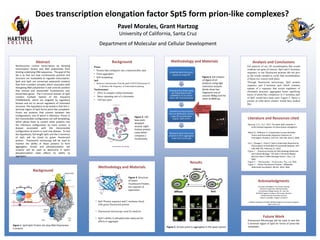
1. Abstract
Nucleosomes control transcription by blocking
transcription factors and RNA polymerase from
binding underlying DNA sequences. The goal of the
lab is to find out how nucleosomes position and
structure are modulated to regulate transcription.
Spt4 and Spt5 are conserved eukaryotic proteins
that form a protein complex, which associates with
elongating RNA polymerase II and controls proteins
that remove and reassemble nucleosomes over
transcribed genes. The C-terminal domain of Spt5
contains multiple repeats of the sequence
ST/AWGGA/Q, which are targeted by regulatory
kinases and act to recruit regulators of chromatin
structure. The hypothesis to be tested is that this C-
terminal region of Spt5 forms prion-like complexes.
Prions are proteins that convert between two
configurations, one of which is infectious. Prions in
this transmissible configuration are self-templating,
which allows them to convert other proteins into
the infectious configuration by mere contact. A
disease associated with the transmissible
configuration of prions is mad cow disease. To test
the hypothesis, full-length Spt5 and the C-terminus
of Spt5 will be fused to green fluorescent
protein. Fluorescent microscopy will be used to
monitor the ability of these proteins to form
aggregates. Kinase and phosphorylation site
mutants will be used to determine if Spt5’s
phosphorylation state affects its ability to
aggregate.
Methodology and Materials
• Spt5 Protein sequence and C-terminus fused
with green fluorescent protein
• Fluorescent microscopy used for analysis
• Spt5’s ability to phosphorylate analyzed for
affects to aggregate
Acknowledgments
Principal Investigator: Prof. Grant Hartzog
Research Supervisor: Michael Doody
Community College Liaison: Dr. Yves Tan
ACCESS Program: Professor Phil Crews, Director
Pamela D’Arcey, Associate Director
Steven Loveridge, Program Assistant
National Institutes of Health NIGMS Bridges to the future Program
(GM 51765-14)
Analysis and Conclusions
Gel analysis of our LR recombination that would
combine our gene of interest, Spt5 and C-terminus
sequence, to our fluorescent proteins did not give
us the results needed to verify that recombination
of those two vectors took place.
Through fluorescent microscopy, Spt5 protein
sequence and C-terminus part which contains
repeats of a sequence that recruit regulators of
chromatin structure, aggregates forms signifying
traces of prion-like complexes in C-terminus part
of Spt5 would have been seen. Figure 5. shows a
picture of what those clusters would have looked
like.
Pavel Morales, Grant Hartzog
University of California, Santa Cruz
Department of Molecular and Cellular Development
Literature and Resources cited
Hartzog, G.A., Fu J. 2012. The Spt4–Spt5 complex: A
multi-faceted regulator of transcription elongation.
Alberti S., Halfmann R. A Systematic Survey Identifies
Prions and Illuminates Sequence Features of
Prionogenic Proteins. Cell 137, 146-158. April 3, 2009.
Liu C., Change C., Chem Y. Spt4 is Selectively Required for
Transcription of Extended Trinucleotide Repeats. Cell
148, 690-701. February 17, 2012.
Figure 1 - "American Society for Microbiology Molecular
and Cellular Biology." The Spt5 C-Terminal Region
Recruits Yeast 3′ RNA Cleavage Factor I. N.p., n.d.
Web
Figure 2 – “Nucleosome.” Nucleosome. N.p., n.d. Web.
Figure 3 - "Green Fluorescent Protein." Wikipedia.
Wikimedia Foundation, 08 Oct. 2014. Web.
Future Work
Fluorescent Microscopy will be used to test the
C-terminal region of Spt5 for forms of prion-like
complexes.
I sure wish I’d
presented my
theory with a
poster before I
wrote my book.
Does transcription elongation factor Spt5 form prion-like complexes?
Background
Figure 1. Spt4-Spt5 Protein sits atop RNA Polymerase
II enzyme
Figure 3. Structure
of Green
Fluorescent Protein,
the reporter of
expression
Amplified Spt5-Cterminus
sequence using PCR
Created entry clones using
our PCR products with
pDONR221 as our donor
vector (BP recombination)
Inserted our entry vectors
into our destination vectors
using LR recombination
(Sup35, EGFP, EYFP)
Monitored Spt5-Cterminus
proteins to form aggregates
under the microscope
Digested with
restriction enzymes
to verify
recombination
Figure 4. Gel analysis
of digest of LR
products using AgeI
restriction enzyme .
Bands show two
fragments one at
around 700bp and the
other at 8000 bp.
Results
Prion:
• Protein that configures into a transmissible state
• Form aggregates
• Self-templating
Spt5:
• Removes nucleosomes from the path of RNA Polymerase II
Reduces the frequency of transcription pausing
Nucleosomes:
• DNA in complex called chromatin
• Basic repeating unit of a chromatin
• 160 base pairs
Background Methodology and Materials
Figure 2. 160
base pairs
wrapped
around eight
histone protein
cores which
compose a
nucleosome
Figure 5. Arrows point to aggregates in the yeast cytosol