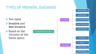
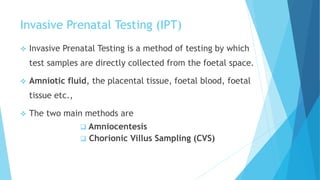
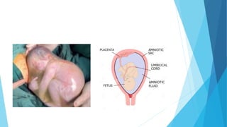
PRENATAL DIAGNOSTIC TECHNIQUES
- STEVE RYAN M
Synopsis
 Prenatal diagnostic techniques
 Definition
 Types
 Indications
 Complications
 Amniocentesis
 Chorionic villus sampling
 Differences
 Advantages & disadvantages
WHAT IS PRENATAL DIAGNOSIS?
 Refers to the diagnosis of the unborn baby while in the
foetal stage.
 To see if the developing baby has any genetic disorders
like down syndrome (trisomy 21), split spine (spina bifida)
etc.,
 Generally done for pregnant women who are older than 35
years,
 Couples who have had a child with genetic abnormalities,
 Couples who are known to be carriers of a genetic disorder.
TYPES OF PRENATAL DIAGNOSIS
 Two types
 Invasive and
Non-Invasive
 Based on the
intrusion of the
foetal space.
Invasive Prenatal Testing (IPT)
 Invasive Prenatal Testing is a method of testing by which
test samples are directly collected from the foetal space.
 Amniotic fluid, the placental tissue, foetal blood, foetal
tissue etc.,
 The two main methods are
 Amniocentesis
 Chorionic Villus Sampling (CVS)
AMNIOCENTESIS
 Amniocentesis (or) Amniotic Fluid Test, is a type of Invasive
Prenatal Testing (IPT)
 Used for prenatal diagnosis of chromosomal abnormalities,
foetal infections
 Sex determination of the child.
 Drain excess of amniotic fluid (polyhydramnios).
 Doctors Jens Bang and Allen Northeved from Denmark were
the first to report amniocentesis done with the guide of an
ultrasound in 1972
 The cost for amniocentesis in India -Rs. 7000 to Rs. 18000
What is the amniotic fluid?
 Clear, slightly yellowish liquid that surrounds the foetus during
pregnancy in the amniotic sac.
 It protects the fetus from injury and temp change.
 It contains amniotic membrane cells and fetus skin cells
which are then cultured to detect any chromosomal anomaly
in the fetus
 The foetus swallows this fluid to develop the
gastrointestinal tract.
How it’s done:
 First, the woman is made to lie on an exam table, with
their abdomen exposed.
 Then, the doctor will use an ultrasound transducer to
determine the baby’s exact location in the uterus.
 Next, the doctor will clean the abdomen of the woman
with an antiseptic.
 Guided by ultrasound, the doctor will insert a thin,
hollow needle through the abdominal wall and into the
uterus
 A small amount of
amniotic fluid (15-20ml)
will be withdrawn into a
syringe, and the needle
will be removed.
 It is replaced by injecting
a saline fluid.
After the procedure is over,
 Heartbeat of the foetus is monitored using ultrasound, to
make sure the foetus is unharmed.
 The woman can resume normal activity after the
procedure.
 The sample of amniotic fluid will be analysed in a lab
 Results might be available within few days, or upto four
weeks, depending on the type of result.
When it’s done:
 Amniocentesis is done between 14th to 20th week of pregnancy
 Ideally done between 15th to 17th week.
If performed too early,
 Uterus won’t be very accessible and the foetal cells might be
too low for sampling.
 Increases the risk of spontaneous abortion and rupture of
foetal membranes.
 Can cause club foot(talipes equinovarus).
 If performed too late, the foetal cells might be keratinised and
unfit for sampling.
Uses of amniocentesis:
 Detect genetic anomalies like Down syndrome, Edwards
syndrome(trisomy-18), Tay-Sachs disease, split spine
condition (spina bifida) etc., with 99% accuracy.
 Diagnose haemophilia (improper blood clotting)
 Paternity testing: DNA collected from the fetus can then be
compared to DNA from the potential father.
 To treat polyhydramnios.
 Determine the sex of the child, ( restricted in India due to an
increasing case of female foeticide).
 Preconception and Pre-Natal Diagnostic Techniques Act in 1994,
Complications:
 Rh sensitization can occur during amniocentesis.
 The fetus might get injured by the needle if not done
properly.
 Might trigger a uterine infection, but it’s very rare.
 Infections like HIV may pass on to the baby from the
mother.
Spontaneous abortions (miscarriages) = < 1%
Contamination of the amniotic fluid by stem cells = 0.15 – 0.11%.
Loss of amniotic fluid = 1 – 2 %.
Blood loss, with scanty “spotting”: very rare.
Chorionic Villus Sampling (CVS)
 Also known as Chorion Biopsy
 Sampling of the chorionic villus and testing it for
chromosomal abnormalities.
 Chorionic villus sampling was first performed by
Italian biologist Giuseppe Simoni in 1983.
 The cost for CVS in India -Rs.10,000 to Rs.15,000
What is Chorionic Villi?
 The chorionic villi are tiny projections of placental tissue that
look like fingers and contain the same genetic material as the
fetus.
 It facilitates the supply of oxygen and nutrients to the embryo.
 Trophoblast cells make up the Chorionic Villi.
How it’s done:
 First, the baby’s gestational age and position of the placenta
is verified via ultrasound.
 The doctor will use the ultrasound image as a guide and take
the tissue sample from the placenta.
The tissue sample can be taken by two methods:
 Transabdominal CVS
 Transcervical CVS
Transabdominal approach
 After cleansing the abdomen with an antiseptic, the doctor will insert a
long, thin needle through the abdominal wall, into the uterus of the
mother and to the edge of the placenta.
 The mother might notice a stinging sensation when the needle enters the
skin, and she might feel cramping when the needle enters the uterus.
 The tissue sample from the placenta will be withdrawn into a syringe, and
the needle will be removed.
Transcervical approach
 After cleansing the vagina and the
cervix with an antiseptic, the
doctor will open the vagina with a
speculum and insert a thin, hollow
tube through the cervix
 When the catheter reaches the
placenta, gentle suction will be
used to remove a small tissue
sample
 After the procedure is over, the woman might experience a
small amount of vaginal bleeding
 The fetus’ heart rate and the mother’s vital signs will be
reassessed.
 If the mother is Rh negative, she will be given Rh(D) immune
globulin.
 The patient is monitored for 24 hours.
When it is done:
 CVS is usually performed between the 10th – 12th week of
pregnancy, but it can be performed at the 15th week too, if
necessary.
If CVS is done before the 10th week,
 Higher risk of miscarriage
 Abnormalities of the foetal limbs, Micrognathia (deformity of
the lower jaw) and Microglossia (under developed tongue)
 The most ideal period for this procedure is the 11th – 12th week
of pregnancy.
Amniocentesis vs Chorionic Biopsy
Amniocentesis Chorionic Biopsy
1. Done between 15 – 17 weeks of pregnancy Done between 10 – 12 weeks of
pregnancy
Earlier diagnosis and planning
2. Amniotic fluid is taken as a sample Chorionic Villus is sampled
3. Has lesser risk of miscarriage Has more risk compared to amniocentesis
4. Provides information on neural tube
defects like spina bifilda
Does not provide information on neural
tube defects.
5. It can also be done to treat
polyhydramnious.
It is done solely for the purpose of testing
and diagnosis.
6. Does not require the patient to be
admitted
Requires the patient to be admitted for
at least 24 hours.
Advantages and Disadvantages:
Advantages:
 Relatively cheap procedure
 Can detect abnormalities with 99% accuracy
 Chances of abortion is very low (0.5 – 1%)
 Enables a couple to abort a genetically abnormal child, for
family planning.
Disadvantages:
 Early testing can cause higher risk of miscarriage (5%)
 If the mother has infections like HIV, it may pass on to the baby
due to this procedure.

Prenatal diagnosis techniques

  • 1.
  • 2.
    Synopsis  Prenatal diagnostictechniques  Definition  Types  Indications  Complications  Amniocentesis  Chorionic villus sampling  Differences  Advantages & disadvantages
  • 3.
    WHAT IS PRENATALDIAGNOSIS?  Refers to the diagnosis of the unborn baby while in the foetal stage.  To see if the developing baby has any genetic disorders like down syndrome (trisomy 21), split spine (spina bifida) etc.,  Generally done for pregnant women who are older than 35 years,  Couples who have had a child with genetic abnormalities,  Couples who are known to be carriers of a genetic disorder.
  • 4.
    TYPES OF PRENATALDIAGNOSIS  Two types  Invasive and Non-Invasive  Based on the intrusion of the foetal space.
  • 5.
    Invasive Prenatal Testing(IPT)  Invasive Prenatal Testing is a method of testing by which test samples are directly collected from the foetal space.  Amniotic fluid, the placental tissue, foetal blood, foetal tissue etc.,  The two main methods are  Amniocentesis  Chorionic Villus Sampling (CVS)
  • 6.
    AMNIOCENTESIS  Amniocentesis (or)Amniotic Fluid Test, is a type of Invasive Prenatal Testing (IPT)  Used for prenatal diagnosis of chromosomal abnormalities, foetal infections  Sex determination of the child.  Drain excess of amniotic fluid (polyhydramnios).  Doctors Jens Bang and Allen Northeved from Denmark were the first to report amniocentesis done with the guide of an ultrasound in 1972  The cost for amniocentesis in India -Rs. 7000 to Rs. 18000
  • 7.
    What is theamniotic fluid?  Clear, slightly yellowish liquid that surrounds the foetus during pregnancy in the amniotic sac.  It protects the fetus from injury and temp change.  It contains amniotic membrane cells and fetus skin cells which are then cultured to detect any chromosomal anomaly in the fetus  The foetus swallows this fluid to develop the gastrointestinal tract.
  • 9.
    How it’s done: First, the woman is made to lie on an exam table, with their abdomen exposed.  Then, the doctor will use an ultrasound transducer to determine the baby’s exact location in the uterus.  Next, the doctor will clean the abdomen of the woman with an antiseptic.  Guided by ultrasound, the doctor will insert a thin, hollow needle through the abdominal wall and into the uterus
  • 10.
     A smallamount of amniotic fluid (15-20ml) will be withdrawn into a syringe, and the needle will be removed.  It is replaced by injecting a saline fluid.
  • 11.
    After the procedureis over,  Heartbeat of the foetus is monitored using ultrasound, to make sure the foetus is unharmed.  The woman can resume normal activity after the procedure.  The sample of amniotic fluid will be analysed in a lab  Results might be available within few days, or upto four weeks, depending on the type of result.
  • 12.
    When it’s done: Amniocentesis is done between 14th to 20th week of pregnancy  Ideally done between 15th to 17th week. If performed too early,  Uterus won’t be very accessible and the foetal cells might be too low for sampling.  Increases the risk of spontaneous abortion and rupture of foetal membranes.  Can cause club foot(talipes equinovarus).  If performed too late, the foetal cells might be keratinised and unfit for sampling.
  • 13.
    Uses of amniocentesis: Detect genetic anomalies like Down syndrome, Edwards syndrome(trisomy-18), Tay-Sachs disease, split spine condition (spina bifida) etc., with 99% accuracy.  Diagnose haemophilia (improper blood clotting)  Paternity testing: DNA collected from the fetus can then be compared to DNA from the potential father.  To treat polyhydramnios.  Determine the sex of the child, ( restricted in India due to an increasing case of female foeticide).  Preconception and Pre-Natal Diagnostic Techniques Act in 1994,
  • 14.
    Complications:  Rh sensitizationcan occur during amniocentesis.  The fetus might get injured by the needle if not done properly.  Might trigger a uterine infection, but it’s very rare.  Infections like HIV may pass on to the baby from the mother. Spontaneous abortions (miscarriages) = < 1% Contamination of the amniotic fluid by stem cells = 0.15 – 0.11%. Loss of amniotic fluid = 1 – 2 %. Blood loss, with scanty “spotting”: very rare.
  • 15.
    Chorionic Villus Sampling(CVS)  Also known as Chorion Biopsy  Sampling of the chorionic villus and testing it for chromosomal abnormalities.  Chorionic villus sampling was first performed by Italian biologist Giuseppe Simoni in 1983.  The cost for CVS in India -Rs.10,000 to Rs.15,000
  • 16.
    What is ChorionicVilli?  The chorionic villi are tiny projections of placental tissue that look like fingers and contain the same genetic material as the fetus.  It facilitates the supply of oxygen and nutrients to the embryo.  Trophoblast cells make up the Chorionic Villi.
  • 17.
    How it’s done: First, the baby’s gestational age and position of the placenta is verified via ultrasound.  The doctor will use the ultrasound image as a guide and take the tissue sample from the placenta. The tissue sample can be taken by two methods:  Transabdominal CVS  Transcervical CVS
  • 18.
    Transabdominal approach  Aftercleansing the abdomen with an antiseptic, the doctor will insert a long, thin needle through the abdominal wall, into the uterus of the mother and to the edge of the placenta.  The mother might notice a stinging sensation when the needle enters the skin, and she might feel cramping when the needle enters the uterus.  The tissue sample from the placenta will be withdrawn into a syringe, and the needle will be removed.
  • 19.
    Transcervical approach  Aftercleansing the vagina and the cervix with an antiseptic, the doctor will open the vagina with a speculum and insert a thin, hollow tube through the cervix  When the catheter reaches the placenta, gentle suction will be used to remove a small tissue sample
  • 20.
     After theprocedure is over, the woman might experience a small amount of vaginal bleeding  The fetus’ heart rate and the mother’s vital signs will be reassessed.  If the mother is Rh negative, she will be given Rh(D) immune globulin.  The patient is monitored for 24 hours.
  • 21.
    When it isdone:  CVS is usually performed between the 10th – 12th week of pregnancy, but it can be performed at the 15th week too, if necessary. If CVS is done before the 10th week,  Higher risk of miscarriage  Abnormalities of the foetal limbs, Micrognathia (deformity of the lower jaw) and Microglossia (under developed tongue)  The most ideal period for this procedure is the 11th – 12th week of pregnancy.
  • 22.
    Amniocentesis vs ChorionicBiopsy Amniocentesis Chorionic Biopsy 1. Done between 15 – 17 weeks of pregnancy Done between 10 – 12 weeks of pregnancy Earlier diagnosis and planning 2. Amniotic fluid is taken as a sample Chorionic Villus is sampled 3. Has lesser risk of miscarriage Has more risk compared to amniocentesis 4. Provides information on neural tube defects like spina bifilda Does not provide information on neural tube defects. 5. It can also be done to treat polyhydramnious. It is done solely for the purpose of testing and diagnosis. 6. Does not require the patient to be admitted Requires the patient to be admitted for at least 24 hours.
  • 25.
    Advantages and Disadvantages: Advantages: Relatively cheap procedure  Can detect abnormalities with 99% accuracy  Chances of abortion is very low (0.5 – 1%)  Enables a couple to abort a genetically abnormal child, for family planning. Disadvantages:  Early testing can cause higher risk of miscarriage (5%)  If the mother has infections like HIV, it may pass on to the baby due to this procedure.