SlideShare a Scribd company logo
1 of 70
DR. SABHILASH
SUGATHAN
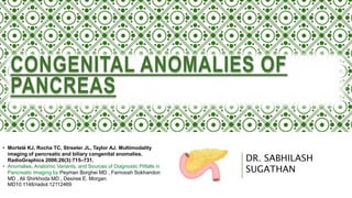
CONGENITAL ANOMALIES OF
PANCREAS
• Mortelé KJ, Rocha TC, Streeter JL, Taylor AJ. Multimodality
imaging of pancreatic and biliary congenital anomalies.
RadioGraphics 2006;26(3):715–731.
• Anomalies, Anatomic Variants, and Sources of Diagnostic Pitfalls in
Pancreatic Imaging by Peyman Borghei MD , Farnoosh Sokhandon
MD , Ali Shirkhoda MD , Desiree E. Morgan
MD10.1148/radiol.12112469
• The pancreas is a retroperitoneal organ that has both endocrine and exocrine functions: it is
involved in the production of hormones (insulin, glucagon, and somatostatin), and also involved in
digestion by its production and secretion of pancreatic enzymes.
• Long (around 15cm ) epigastric structure extending from duodenal loop to splenic hilum
• Comprises the head (including uncinate process), neck, body and tail.
GROSS ANATOMY
The pancreas may have the shape of a dumbbell, tadpole, or sausage.
It can be divided into four main parts:
1. HEAD: thickest part; lies to the right of the mesenteric vessels
• uncinate process: extension of the head, posterior to SMV, SMA
• attached to "C" loop of duodenum (D2 and D3)
2. NECK: thinnest part; lies anterior to SMA, SMV
• SMV joins splenic vein behind pancreatic neck to form portal vein
3. BODY: main part; lies to left of SMA, SMV
• splenic vein lies in groove on posterior surface of body
4. TAIL: lies between layers of the splenorenal ligament in the splenic
hilum
MAIN PANCREATIC DUCT
• portion of the dorsal duct proximal to the dorsal-ventral fusion point
• drains at the ampulla of Vater
• connects with the accessory pancreatic duct (of Santorini) (see below)
if present
ACCESSORY PANCREATIC DUCT (OF
SANTORINI OR BERNARD)
• portion of the dorsal duct distal to the dorsal-ventral fusion point
• drains anterior and superior portion of the head
• in 70% of individuals drains to the minor papilla
• in 30% of individuals persists as a branch of the main pancreatic duct
Pancreatic duct of Wirsung
• distal portion of the main pancreatic duct
• segment of the ventral duct between the dorsal-ventral fusion point and the
major papilla
• continuous with the main pancreatic duct proximally
• joins with the common bile duct at a 60 degree angle at the hepatopancreatic
ampulla.
EMBRYOLOGICAL DEVELOPMENT
• By the 4th week of embryologic development, the pancreatic duct develops from
separate ventral and dorsal buds originating from the endodermal lining of the
duodenum.
• The gallbladder, extrahepatic bile ducts, central intrahepatic bile ducts, and
ventral pancreas with its ductal network are derived from the ventral bud or
outpouching.
• The dorsal bud is the precursor to dorsal pancreas and its ductal system.
• The ventral pancreas rotates clockwise posterior to the duodenum and comes into
contact with the dorsal pancreas in the 7th gestational week to develop into the future
pancreatic neck.
• The dorsal and ventral pancreatic buds grow into a pair of branching ,arborizing ductal
systems.
• After fusion, a new duct connects the distal portion of the dorsal pancreatic duct with
the shorter duct of the ventral pancreas to form the main pancreatic duct, also known
as the duct of Wirsung, which empties into the major papilla.
Dorsal pancreatic bud
• develops into the anterior part of the head, body, and tail (and a small variable portion of
the uncinate process)
• contains a long duct continuing from the accessory pancreatic papilla to the tail
Ventral pancreatic bud
• smaller of the two buds that develops into the posterior part of the pancreatic head and most of the
uncinate process
• contains a short main pancreatic duct which is connected to the common bile duct
• develops just distal to the developing biliary tree
• starts to the right of the duodenum
• rotates to the left underneath the dorsal pancreas
VARIANT ANATOMY
• Annular pancreas
• Fishtail pancreas
• Ectopic pancreatic tissue
• Horseshoe pancreas
• pancreatic duct variations
• pancreatic clefts: linear clefts may be seen which contain fat where small
vessels enter the pancreas and are a common mimic of pancreatic laceration.
They are most prominent at the junction of the body and neck
ANNULAR PANCREAS
• pancreatic tissue completely or incompletely encircling
the duodenum
Clinical presentation
• annular pancreas in adults are asymptomatic and an incidental finding on
imaging. However, it can cause pancreatitis, duodenal obstruction and
rarely biliary obstruction.
• In children, an annular pancreas may be associated with other congenital
anomalies or cause duodenal obstruction.
EMBRYOLOGY
• The pancreas develops from a single dorsal and two ventral buds, which
appear as outgrowths of primitive foregut at 5 weeks of gestation. The ventral
buds fuse rapidly.
• In the 7th week of gestation, the duodenum expands and rotates the ventral
bud from right to left which then fuses with the dorsal bud. The ventral bud
forms the inferior part of uncinate process and inferior head of pancreas while
the dorsal bud gives rise to the tail and body of pancreas.
• Annular pancreas develops due to failure of the ventral bud to rotate with the
duodenum, causing encasement of the duodenum.
CLASSIFICATION
• complete annular pancreas: pancreatic parenchyma or an annular duct is
seen to completely surround the 2nd part of duodenum
• incomplete annular pancreas: annulus does not surround the duodenum
completely, giving a 'crocodile jaw' appearance
Pancreatic tissue surrounds the duodenum
RADIOGRAPHIC FEATURES
CT
• Pancreatic tissue is seen to completely or incompletely surround the 2nd part of the
duodenum. Associated duodenal narrowing and dilatation of the proximal duodenum
may also be seen. In adults, it is frequently associated with pancreatitis.
MRI/MRCP
• Apart from annular pancreas features, pancreatic ductal anatomy can be well assessed
with MR imaging.
There is an 'echogenic focus' in short-axis images of the pancreas
head
The structure in the head of the pancreas is the 2nd part of the
duodenum
incomplete annular pancreas
Upper gastrointestinal radiograph of annular
pancreas
shows slit-like smooth narrowing of the
second portion of duodenum (arrows).
No gastric distention or mechanical
obstruction is present.
Contrast material flows freely into an
otherwise normal duodenum and proximal
jejunum.
Unenhanced axial CT scan of annular
pancreas
in 43-year-old man with history of resected
glucaconoma in the pancreatic tail.
The pancreatic tissue encircles the positive
oral contrastmaterial–filled second portion
of the duodenum(arrows), making the
anomaly easy to recognize even without
intravenous contrast material.
Intravenous contrast-enhanced axial CT
scan of annular pancreas
normal enhancing pancreatic tissue (arrows)
surrounding the collapsed duodenum.
The patient received water as a negative oral
contrast agent.
Wirsung duct of the ventral pancreas
(arrowhead) is draining into duodenum.
ERCP spot image of annular pancreas in
43-year-old man shows mild diffuse
acinarization of the pancreas (solid arrows).
The pancreatic tissue and pancreatic duct
(arrowhead) are to the right of the second
portion of the duodenum, consistent with
annular pancreas.
Focal dilatation of the main pancreatic
duct (dashed arrow) with pooling of contrast
material in the dependent portion is also
present.
Treatment and prognosis
• In symptomatic cases of annular pancreas, surgical management is
the treatment of choice.
• Bypass surgeries such as duodeno-jejunostomy or gastro-
jejunostomy are the mainstay of surgical management 3.
FISHTAIL PANCREAS
• Fishtail pancreas (also known as pancreas bifidum or bifid tail of the
pancreas) is a rare anatomical variant of the pancreas produced by a
branching anomaly during its development.
• It is named as such due to the fishtail-like appearance of the pancreas.
• Fishtail pancreas is thought to be caused by the failure of part of the ventral
pancreatic anlage to regress. The ventral pancreatic anlage develops into the majority of the
into the majority of the pancreatic gland.
• During pancreatic development, the ventral anlage initially has two lobes with two primitive
ducts .
• One lobe tends to dominate and persists to become the head of the pancreas, while the
other regresses and becomes the uncinate process of the pancreas.
• Fishtail pancreas is caused by failure of one of these lobes to regress producing duplication
RADIOGRAPHIC FEATURES
MRCP
• Fishtail pancreas may be visible as a duplication of the major duct in the body of
the pancreas and the presence of two pancreatic tails .
ERCP
• Like MRCP, on ERCP it also shows duplication of the major pancreatic duct
ECTOPIC PANCREATIC TISSUE
• Ectopic pancreatic tissue, aka heterotopic pancreatic tissue, refers to the
presence of pancreatic tissue in the submucosal, muscularis or subserosal layers
of the luminal gastrointestinal tract outside the normal confines of
the pancreas and lacking any anatomic or vascular connection with the pancreas
proper.
Location
• proximal duodenum
• gastric antrum
• proximal jejunum
• Meckel diverticulum
• ileum
Radiographic features
• On upper gastrointestinal examination, an ectopic pancreas appears as an
extra- mucosal, smooth, broad-based lesion either along the greater
curvature of the gastric antrum or in the proximal duodenum.
1. Fluoroscopy
2. Upper GI barium study
• In 45% of the cases of ectopic pancreas discovered on upper gastrointestinal
fluoroscopic examination, the ectopic pancreatic tissue contained a central
small collection of barium, i.e. a central niche or umbilication, indicative of the
rudimentary duct’s draining orifice​. It is this finding that is diagnostic of
ectopic pancreatic tissue.
Heterotopic pancreas in the gastric
antrum with central umbilication.
Fluoroscopic images
Ectopic pancreatic tissue ( green arrows )
similar to attenuation of the pancreas ( red
arrow ) in all 3 phases.
3. CT
Contrast enhanced CT may show a homogeneously enhancing tissue (similar to normal pancreas) or
cystic area (acinar component or pseudocyst).
HORSESHOE PANCREAS
• A rare anatomic variant of the pancreas in which the uncinate
process is unusually elongated such that it extends along the
whole 3rd part of the duodenum to mirror the tail superiorly
forming a horseshoe-shaped gland.
The uncinate process of the pancreas extends
along the entire aspect of the third part of the
duodenum. Normal anatomic variant of horseshoe
of horseshoe pancreas.
PANCREAS DIVISUM
• Pancreas divisum represents a variation in pancreatic ductal anatomy
• that can be associated with abdominal pain and idiopathic pancreatitis.
• It is characterized, in the majority of cases, by the main pancreatic
duct directly entering the minor papilla with no communication with the
ventral duct the major papilla.
• It results from failure of fusion of dorsal and ventral pancreatic anlages. As
a result, the dorsal pancreatic duct drains most of the pancreatic glandular
parenchyma via the minor papilla. Although controversial, this variant is
considered a cause of pancreatitis.
Three subtypes are known:
• TYPE 1 (CLASSIC): no connection at all; occurs in the majority of cases; 70%
cases; 70%
• TYPE 2 (ABSENT VENTRAL DUCT): minor papilla drains all of pancreas while major
pancreas while major papilla drains bile duct; 20-25%
• TYPE 3 (FUNCTIONAL): filamentous or inadequate connection between dorsal and
between dorsal and ventral ducts; 5-6%
Anomalies, Anatomic Variants, and Sources of Diagnostic Pitfalls in Pancreatic Imaging by Peyman Borghei MD , Farnoosh
Sokhandon MD , Ali Shirkhoda MD , Desiree E. Morgan MD10.1148/radiol.12112469
•TYPE 1 (CLASSIC): no connection at all; occurs in the majority of cases;
70%
•TYPE 2 (ABSENT VENTRAL
DUCT): minor papilla drains all
of pancreas while major papilla
duct; 20-25%
•TYPE 3
(FUNCTIONAL): filamentous or
inadequate connection between
ventral ducts; 5-6%
RADIOGRAPHIC FEATURES
1. Fluoroscopy
2. ERCP
• This was the traditional method of diagnosis where a pancreas divisum was
suspected when there was no contrast extending towards the pancreatic tail
when administered through the ampulla of Vater.
3. MRI
• MRI is the current gold standard method of evaluation. The key imaging
features:
• the dorsal pancreatic duct is in direct continuity with the duct of Santorini,
which drains into the minor papilla
• the ventral pancreatic duct (duct of Wirsung) does not communicate with the
dorsal pancreatic duct but joins with the distal common bile duct to enter the
major papilla
CT PANCREATIC DIVISUM
MRCP image of pancreas divisum
the main pancreatic duct (dorsal
Santorini
duct, straight solid arrow) draining
separately into the minor papilla (dashed
arrow).
The common bile duct (arrowheads)
joins the smaller ventral pancreaticduct
(curved arrow) at a more inferior
leveland drains into the duodenum
through the major
papilla.
Figure 3
ERCP images of pancreas divisum.
(a) The main pancreatic duct
(arrows) drains through the minor
papilla, consistent with pancreas
divisum.
(b) The short, terminally arborizing
duct of Wirsung (arrow) is depicted
by means of contrast material
injection of the major papilla, with
the common bile duct (arrowhead)
also filling.
• Reverse pancreas divisum : has been described where the main duct
fuses with the ventral duct and a small residual dorsal duct does not
duct and drains separately into the minor papilla.
TREATMENT AND PROGNOSIS
• A diagnosis of pancreas divisum does not routinely warrant treatment,
especially when incidental and asymptomatic. In symptomatic patients (e.g.
recurrent pancreatitis), management options may include:
• conservative, non-operative treatment +/- pancreatic enzyme supplements
• minor papillotomy
• minor papilla stenting
• balloon dilatation of any associated stricture
VARIATIONS OF PANCREATIC DUCT
ANOMALOUS PANCREATICOBILIARY
JUNCTION
• An anomalous pancreaticobiliary junction, also known
as pancreaticobiliary maljunction, describes the abnormal junction of
the pancreatic duct and common bile duct that occurs outside
the duodenal wall to form a long common channel (>15 mm) .
• The origin of a long common channel might be formed embryologically with adhesion of the ventral
pancreatic duct and the terminal portion of the bile duct .
• It is divided into anomalous pancreaticobiliary junction with biliary dilatation (77%) and without biliary
dilatation (23%) .
• The anomalous pancreaticobiliary junction makes biliary drainage not under the control of sphincter of
Oddi, resulting in pancreatic juice reflux into the biliary tract that injures the biliary epithelium .
CLASSIFICATION
• The Japanese Study Group on Pancreaticobiliary Maljunction (JSPBM) proposed
the following classification in 2015:
• TYPE A (STENOTIC TYPE): dilatation of the common bile duct upstream of a
stenotic segment of the distal common bile duct, which joins the common
channel
•TYPE B (NON‐STENOTIC TYPE): nonstenotic distal common bile duct
smoothly joins the common channel; no localized dilatation of the
•TYPE C (DILATED CHANNEL TYPE): narrow distal common bile
duct joins dilated common channel
•TYPE D (COMPLEX TYPE): complex maljunction associated
with annular pancreas, pancreas divisum, or other complicated duct
Anomalous pancreaticobiliary Junction
with dilated common channel (type c).
•pancreaticobiliary maljunction
type a (stenotic type)
RADIOGRAPHIC FEATURES
ERCP
• An intrabiliary amylase level more than 8000 UI/L within the bile duct and
gallbladder obtained endoscopically (ERCP) or percutaneously suggests reflux
of pancreatic juice through an anomalous pancreaticobiliary junction and
shows a positive predictive value and a specificity of more than 90 % .
• In patients with a short common channel, direct cholangiography
(e.g. ERCP) can be effective in the assessment of pancreaticobiliary junction
incompetence.
Ultrasound
• It has a limited role in diagnosis, but is a useful noninvasive tool for
screening. Careful measurement of the common bile duct and comparison
with the normal limits for age helps in early detection. Detection of a
dilated common bile duct may be the first clue to suspect
pancreaticobiliary maljunction with biliary dilatation. In this case,
further MRCP is recommended for biliary junction anatomy. It is also
associated with gallbladder wall thickening. However, it is non-specific.
• The visualization of pancreaticobiliary junction outside the duodenal wall
can be detected at endoscopic ultrasound. It is also helpful in screening
and surveillance of biliary cancer after diagnosis of pancreaticobiliary
maljunction
CT/MRI
• a common channel length of >8 mm
• an abnormal union between the pancreatic and bile ducts
• pancreaticobiliary junction outside the duodenal wall
• common bile duct dilatation is suggestive of pancreaticobiliary
maljunction with biliary dilatation
• MRCP is the gold standard of diagnosis and is superior to ERCP in
depicting biliary anatomy, including the intrahepatic bile duct.
ANSA PANCREATICA
• It is a communication between the main pancreatic duct (of Wirsung)
and the accessory pancreatic duct (of Santorini).
• Recently, the ansa pancreatica has been considered as a predisposing
factor in patients with idiopathic acute pancreatitis .
• The ansa pancreatica arises as a branch duct from the main pancreatic
duct. It descends down initially, it then ascends upward forming a loop
finally terminating at the minor papilla.
• This type of pancreatic ductal variation can be identified
on ERCP or MRCP studies.
ERCP image of ansa pancreatica
Dashed line represents dorsal main
pancreatic duct (technically underfilled).
There is obliteration of the proximal dorsal
pancreatic duct(solid arrow).
The proximal dorsal duct connects with
an inferior branch of the ventral duct
(arrowhead) through S-shaped collateral
(dashed arrow).
PANCREATIC CYSTS
• A congenital true pancreatic cyst is a very rare entity, mostly seen in children
younger than 2 years of age
• These cysts develop as a result of sequestration of primitive pancreatic
ducts and are lined by cuboidal epithelium
• Congenital pancreatic cysts are generally asymptomatic, although abdominal
distention, vomiting, jaundice, or pancreatitis can be observed
• At imaging, this condition manifests as a uniform thin-walled cyst usually in
the region of the pancreatic body and tail
• Congenital true pancreatic cysts may be idiopathic or may be observed in
association with other systemic diseases such as Von Hippel– Lindau disease
, Beckwith- Wiedeman syndrome, or polycystic disease of the pancreas
and kidneys
THANK –YOU!

More Related Content

Similar to PANCREATIC ANOMALY radiology.pptx

Benign anorectal disorders 2
Benign anorectal disorders 2Benign anorectal disorders 2
Benign anorectal disorders 2Dr. Azhar
 
Pancreatic sonographic anatomy
Pancreatic sonographic anatomyPancreatic sonographic anatomy
Pancreatic sonographic anatomyMohamed Soliman
 
Anatomy of gastroesophagial junction with specail reference to hiatus hernia...
Anatomy of gastroesophagial junction  with specail reference to hiatus hernia...Anatomy of gastroesophagial junction  with specail reference to hiatus hernia...
Anatomy of gastroesophagial junction with specail reference to hiatus hernia...Rana Singh
 
Anorectal malformation seminar
Anorectal malformation seminarAnorectal malformation seminar
Anorectal malformation seminarDr. Dixit
 
Prolapse rectum
Prolapse rectumProlapse rectum
Prolapse rectumDr KAMBLE
 
Development of pancreas
Development of pancreasDevelopment of pancreas
Development of pancreassk harish
 
Annular Pancreas: An Unusual Presentation
Annular Pancreas: An Unusual PresentationAnnular Pancreas: An Unusual Presentation
Annular Pancreas: An Unusual PresentationApollo Hospitals
 
Presentation1, abdominal ultrasound anatomy.
Presentation1, abdominal ultrasound anatomy.Presentation1, abdominal ultrasound anatomy.
Presentation1, abdominal ultrasound anatomy.Abdellah Nazeer
 
Radiological Anatomy of pancreas
Radiological Anatomy of pancreasRadiological Anatomy of pancreas
Radiological Anatomy of pancreasrmtheepanssdf
 
childhood intestinal obstruction.pptx
childhood intestinal obstruction.pptxchildhood intestinal obstruction.pptx
childhood intestinal obstruction.pptxsingleavilash
 
PANCREAS ANAT,PHY,CONG.ANAMOLIES.pptx
PANCREAS ANAT,PHY,CONG.ANAMOLIES.pptxPANCREAS ANAT,PHY,CONG.ANAMOLIES.pptx
PANCREAS ANAT,PHY,CONG.ANAMOLIES.pptxPradeep Pande
 

Similar to PANCREATIC ANOMALY radiology.pptx (20)

The gallbladder and bile ducts
The gallbladder and bile ductsThe gallbladder and bile ducts
The gallbladder and bile ducts
 
Benign anorectal disorders 2
Benign anorectal disorders 2Benign anorectal disorders 2
Benign anorectal disorders 2
 
Pancreatic sonographic anatomy
Pancreatic sonographic anatomyPancreatic sonographic anatomy
Pancreatic sonographic anatomy
 
Anatomy of gastroesophagial junction with specail reference to hiatus hernia...
Anatomy of gastroesophagial junction  with specail reference to hiatus hernia...Anatomy of gastroesophagial junction  with specail reference to hiatus hernia...
Anatomy of gastroesophagial junction with specail reference to hiatus hernia...
 
Excretionurography
Excretionurography Excretionurography
Excretionurography
 
Anorectal malformation seminar
Anorectal malformation seminarAnorectal malformation seminar
Anorectal malformation seminar
 
Alimentary system.pptx
Alimentary system.pptxAlimentary system.pptx
Alimentary system.pptx
 
The pancrease.pptx
The pancrease.pptxThe pancrease.pptx
The pancrease.pptx
 
Prolapse rectum
Prolapse rectumProlapse rectum
Prolapse rectum
 
Development of pancreas
Development of pancreasDevelopment of pancreas
Development of pancreas
 
RECTAL prolapse.pptx
RECTAL prolapse.pptxRECTAL prolapse.pptx
RECTAL prolapse.pptx
 
Radiological anatomy of_abdomen[1]
Radiological anatomy of_abdomen[1]Radiological anatomy of_abdomen[1]
Radiological anatomy of_abdomen[1]
 
Peritoneum , Intraperitoneal Spaces
Peritoneum , Intraperitoneal SpacesPeritoneum , Intraperitoneal Spaces
Peritoneum , Intraperitoneal Spaces
 
Annular Pancreas: An Unusual Presentation
Annular Pancreas: An Unusual PresentationAnnular Pancreas: An Unusual Presentation
Annular Pancreas: An Unusual Presentation
 
Presentation1, abdominal ultrasound anatomy.
Presentation1, abdominal ultrasound anatomy.Presentation1, abdominal ultrasound anatomy.
Presentation1, abdominal ultrasound anatomy.
 
Radiological Anatomy of pancreas
Radiological Anatomy of pancreasRadiological Anatomy of pancreas
Radiological Anatomy of pancreas
 
Anorectal malformations
Anorectal malformationsAnorectal malformations
Anorectal malformations
 
childhood intestinal obstruction.pptx
childhood intestinal obstruction.pptxchildhood intestinal obstruction.pptx
childhood intestinal obstruction.pptx
 
PANCREAS.pptx
PANCREAS.pptxPANCREAS.pptx
PANCREAS.pptx
 
PANCREAS ANAT,PHY,CONG.ANAMOLIES.pptx
PANCREAS ANAT,PHY,CONG.ANAMOLIES.pptxPANCREAS ANAT,PHY,CONG.ANAMOLIES.pptx
PANCREAS ANAT,PHY,CONG.ANAMOLIES.pptx
 

More from ranjitharadhakrishna3

ANATOMY OF VERTEBRAL COLUMN AND SPINAL CORD.pptx
ANATOMY OF VERTEBRAL COLUMN AND SPINAL CORD.pptxANATOMY OF VERTEBRAL COLUMN AND SPINAL CORD.pptx
ANATOMY OF VERTEBRAL COLUMN AND SPINAL CORD.pptxranjitharadhakrishna3
 
cerebral cisterns for radiology dnb .pptx
cerebral cisterns for radiology dnb .pptxcerebral cisterns for radiology dnb .pptx
cerebral cisterns for radiology dnb .pptxranjitharadhakrishna3
 
brain anatomy radiology ppt for dnbs.ppt
brain anatomy radiology ppt for dnbs.pptbrain anatomy radiology ppt for dnbs.ppt
brain anatomy radiology ppt for dnbs.pptranjitharadhakrishna3
 
pre sacral lesion sept5.pptx RADIOLOGY
pre sacral lesion sept5.pptx RADIOLOGYpre sacral lesion sept5.pptx RADIOLOGY
pre sacral lesion sept5.pptx RADIOLOGYranjitharadhakrishna3
 
Sarcoidosis radiology pulmonary neuro abdominal .ppt
Sarcoidosis radiology pulmonary neuro abdominal .pptSarcoidosis radiology pulmonary neuro abdominal .ppt
Sarcoidosis radiology pulmonary neuro abdominal .pptranjitharadhakrishna3
 
Respiratory system signs final.ppt radiology
Respiratory system signs final.ppt radiologyRespiratory system signs final.ppt radiology
Respiratory system signs final.ppt radiologyranjitharadhakrishna3
 

More from ranjitharadhakrishna3 (20)

ANATOMY OF VERTEBRAL COLUMN AND SPINAL CORD.pptx
ANATOMY OF VERTEBRAL COLUMN AND SPINAL CORD.pptxANATOMY OF VERTEBRAL COLUMN AND SPINAL CORD.pptx
ANATOMY OF VERTEBRAL COLUMN AND SPINAL CORD.pptx
 
cerebral cisterns for radiology dnb .pptx
cerebral cisterns for radiology dnb .pptxcerebral cisterns for radiology dnb .pptx
cerebral cisterns for radiology dnb .pptx
 
brain anatomy radiology ppt for dnbs.ppt
brain anatomy radiology ppt for dnbs.pptbrain anatomy radiology ppt for dnbs.ppt
brain anatomy radiology ppt for dnbs.ppt
 
FNAC lung (2).ppt
FNAC lung (2).pptFNAC lung (2).ppt
FNAC lung (2).ppt
 
FNAC lung.ppt
FNAC lung.pptFNAC lung.ppt
FNAC lung.ppt
 
pre sacral lesion sept5.pptx RADIOLOGY
pre sacral lesion sept5.pptx RADIOLOGYpre sacral lesion sept5.pptx RADIOLOGY
pre sacral lesion sept5.pptx RADIOLOGY
 
Sarcoidosis radiology pulmonary neuro abdominal .ppt
Sarcoidosis radiology pulmonary neuro abdominal .pptSarcoidosis radiology pulmonary neuro abdominal .ppt
Sarcoidosis radiology pulmonary neuro abdominal .ppt
 
Respiratory system signs final.ppt radiology
Respiratory system signs final.ppt radiologyRespiratory system signs final.ppt radiology
Respiratory system signs final.ppt radiology
 
SOLITARY PULMONARY NODULE.pptx
SOLITARY PULMONARY NODULE.pptxSOLITARY PULMONARY NODULE.pptx
SOLITARY PULMONARY NODULE.pptx
 
barium swallow.pptx
barium swallow.pptxbarium swallow.pptx
barium swallow.pptx
 
Parathyroid Imaging .pptx
Parathyroid Imaging .pptxParathyroid Imaging .pptx
Parathyroid Imaging .pptx
 
mri and ct anatomy of brain-.pptx
mri and ct anatomy of brain-.pptxmri and ct anatomy of brain-.pptx
mri and ct anatomy of brain-.pptx
 
acuteosteomyelitis-.pptx
acuteosteomyelitis-.pptxacuteosteomyelitis-.pptx
acuteosteomyelitis-.pptx
 
retroperitonealmasses-pptx
retroperitonealmasses-pptxretroperitonealmasses-pptx
retroperitonealmasses-pptx
 
ULTRASOUND IN FIRST TRIMESTER.ppt
ULTRASOUND IN FIRST TRIMESTER.pptULTRASOUND IN FIRST TRIMESTER.ppt
ULTRASOUND IN FIRST TRIMESTER.ppt
 
OSTEONECROSIS 2003.ppt
OSTEONECROSIS 2003.pptOSTEONECROSIS 2003.ppt
OSTEONECROSIS 2003.ppt
 
GRIDS.ppt
GRIDS.pptGRIDS.ppt
GRIDS.ppt
 
fluoro1-principles.PPT
fluoro1-principles.PPTfluoro1-principles.PPT
fluoro1-principles.PPT
 
FLUOROSCOPIC IMAGING.ppt
FLUOROSCOPIC IMAGING.pptFLUOROSCOPIC IMAGING.ppt
FLUOROSCOPIC IMAGING.ppt
 
DIFFUSION & PERFUSION r MRI.ppt
DIFFUSION  &   PERFUSION  r MRI.pptDIFFUSION  &   PERFUSION  r MRI.ppt
DIFFUSION & PERFUSION r MRI.ppt
 

Recently uploaded

Call Girls in Lucknow Just Call 👉👉8630512678 Top Class Call Girl Service Avai...
Call Girls in Lucknow Just Call 👉👉8630512678 Top Class Call Girl Service Avai...Call Girls in Lucknow Just Call 👉👉8630512678 Top Class Call Girl Service Avai...
Call Girls in Lucknow Just Call 👉👉8630512678 Top Class Call Girl Service Avai...soniyagrag336
 
👉 Chennai Sexy Aunty’s WhatsApp Number 👉📞 7427069034 👉📞 Just📲 Call Ruhi Colle...
👉 Chennai Sexy Aunty’s WhatsApp Number 👉📞 7427069034 👉📞 Just📲 Call Ruhi Colle...👉 Chennai Sexy Aunty’s WhatsApp Number 👉📞 7427069034 👉📞 Just📲 Call Ruhi Colle...
👉 Chennai Sexy Aunty’s WhatsApp Number 👉📞 7427069034 👉📞 Just📲 Call Ruhi Colle...rajnisinghkjn
 
ANATOMY AND PHYSIOLOGY OF REPRODUCTIVE SYSTEM.pptx
ANATOMY AND PHYSIOLOGY OF REPRODUCTIVE SYSTEM.pptxANATOMY AND PHYSIOLOGY OF REPRODUCTIVE SYSTEM.pptx
ANATOMY AND PHYSIOLOGY OF REPRODUCTIVE SYSTEM.pptxSwetaba Besh
 
💰Call Girl In Bangalore☎️63788-78445💰 Call Girl service in Bangalore☎️Bangalo...
💰Call Girl In Bangalore☎️63788-78445💰 Call Girl service in Bangalore☎️Bangalo...💰Call Girl In Bangalore☎️63788-78445💰 Call Girl service in Bangalore☎️Bangalo...
💰Call Girl In Bangalore☎️63788-78445💰 Call Girl service in Bangalore☎️Bangalo...gragneelam30
 
Cara Menggugurkan Kandungan Dengan Cepat Selesai Dalam 24 Jam Secara Alami Bu...
Cara Menggugurkan Kandungan Dengan Cepat Selesai Dalam 24 Jam Secara Alami Bu...Cara Menggugurkan Kandungan Dengan Cepat Selesai Dalam 24 Jam Secara Alami Bu...
Cara Menggugurkan Kandungan Dengan Cepat Selesai Dalam 24 Jam Secara Alami Bu...Cara Menggugurkan Kandungan 087776558899
 
Call Girls Bangalore - 450+ Call Girl Cash Payment 💯Call Us 🔝 6378878445 🔝 💃 ...
Call Girls Bangalore - 450+ Call Girl Cash Payment 💯Call Us 🔝 6378878445 🔝 💃 ...Call Girls Bangalore - 450+ Call Girl Cash Payment 💯Call Us 🔝 6378878445 🔝 💃 ...
Call Girls Bangalore - 450+ Call Girl Cash Payment 💯Call Us 🔝 6378878445 🔝 💃 ...gragneelam30
 
Difference Between Skeletal Smooth and Cardiac Muscles
Difference Between Skeletal Smooth and Cardiac MusclesDifference Between Skeletal Smooth and Cardiac Muscles
Difference Between Skeletal Smooth and Cardiac MusclesMedicoseAcademics
 
Call girls Service Phullen / 9332606886 Genuine Call girls with real Photos a...
Call girls Service Phullen / 9332606886 Genuine Call girls with real Photos a...Call girls Service Phullen / 9332606886 Genuine Call girls with real Photos a...
Call girls Service Phullen / 9332606886 Genuine Call girls with real Photos a...call girls hydrabad
 
Bhawanipatna Call Girls 📞9332606886 Call Girls in Bhawanipatna Escorts servic...
Bhawanipatna Call Girls 📞9332606886 Call Girls in Bhawanipatna Escorts servic...Bhawanipatna Call Girls 📞9332606886 Call Girls in Bhawanipatna Escorts servic...
Bhawanipatna Call Girls 📞9332606886 Call Girls in Bhawanipatna Escorts servic...Dipal Arora
 
Race Course Road } Book Call Girls in Bangalore | Whatsapp No 6378878445 VIP ...
Race Course Road } Book Call Girls in Bangalore | Whatsapp No 6378878445 VIP ...Race Course Road } Book Call Girls in Bangalore | Whatsapp No 6378878445 VIP ...
Race Course Road } Book Call Girls in Bangalore | Whatsapp No 6378878445 VIP ...dishamehta3332
 
7 steps How to prevent Thalassemia : Dr Sharda Jain & Vandana Gupta
7 steps How to prevent Thalassemia : Dr Sharda Jain & Vandana Gupta7 steps How to prevent Thalassemia : Dr Sharda Jain & Vandana Gupta
7 steps How to prevent Thalassemia : Dr Sharda Jain & Vandana GuptaLifecare Centre
 
ANATOMY AND PHYSIOLOGY OF RESPIRATORY SYSTEM.pptx
ANATOMY AND PHYSIOLOGY OF RESPIRATORY SYSTEM.pptxANATOMY AND PHYSIOLOGY OF RESPIRATORY SYSTEM.pptx
ANATOMY AND PHYSIOLOGY OF RESPIRATORY SYSTEM.pptxSwetaba Besh
 
Chennai ❣️ Call Girl 6378878445 Call Girls in Chennai Escort service book now
Chennai ❣️ Call Girl 6378878445 Call Girls in Chennai Escort service book nowChennai ❣️ Call Girl 6378878445 Call Girls in Chennai Escort service book now
Chennai ❣️ Call Girl 6378878445 Call Girls in Chennai Escort service book nowtanudubay92
 
Call Girls in Lucknow Just Call 👉👉 8875999948 Top Class Call Girl Service Ava...
Call Girls in Lucknow Just Call 👉👉 8875999948 Top Class Call Girl Service Ava...Call Girls in Lucknow Just Call 👉👉 8875999948 Top Class Call Girl Service Ava...
Call Girls in Lucknow Just Call 👉👉 8875999948 Top Class Call Girl Service Ava...Janvi Singh
 
Lucknow Call Girls Just Call 👉👉8630512678 Top Class Call Girl Service Available
Lucknow Call Girls Just Call 👉👉8630512678 Top Class Call Girl Service AvailableLucknow Call Girls Just Call 👉👉8630512678 Top Class Call Girl Service Available
Lucknow Call Girls Just Call 👉👉8630512678 Top Class Call Girl Service Availablesoniyagrag336
 
Cardiac Output, Venous Return, and Their Regulation
Cardiac Output, Venous Return, and Their RegulationCardiac Output, Venous Return, and Their Regulation
Cardiac Output, Venous Return, and Their RegulationMedicoseAcademics
 
❤️ Chandigarh Call Girls☎️98151-579OO☎️ Call Girl service in Chandigarh ☎️ Ch...
❤️ Chandigarh Call Girls☎️98151-579OO☎️ Call Girl service in Chandigarh ☎️ Ch...❤️ Chandigarh Call Girls☎️98151-579OO☎️ Call Girl service in Chandigarh ☎️ Ch...
❤️ Chandigarh Call Girls☎️98151-579OO☎️ Call Girl service in Chandigarh ☎️ Ch...Rashmi Entertainment
 
Call Girl in Chennai | Whatsapp No 📞 7427069034 📞 VIP Escorts Service Availab...
Call Girl in Chennai | Whatsapp No 📞 7427069034 📞 VIP Escorts Service Availab...Call Girl in Chennai | Whatsapp No 📞 7427069034 📞 VIP Escorts Service Availab...
Call Girl in Chennai | Whatsapp No 📞 7427069034 📞 VIP Escorts Service Availab...amritaverma53
 
Russian Call Girls In Pune 👉 Just CALL ME: 9352988975 ✅❤️💯low cost unlimited ...
Russian Call Girls In Pune 👉 Just CALL ME: 9352988975 ✅❤️💯low cost unlimited ...Russian Call Girls In Pune 👉 Just CALL ME: 9352988975 ✅❤️💯low cost unlimited ...
Russian Call Girls In Pune 👉 Just CALL ME: 9352988975 ✅❤️💯low cost unlimited ...chanderprakash5506
 
Call Girls Service Jaipur {9521753030 } ❤️VVIP BHAWNA Call Girl in Jaipur Raj...
Call Girls Service Jaipur {9521753030 } ❤️VVIP BHAWNA Call Girl in Jaipur Raj...Call Girls Service Jaipur {9521753030 } ❤️VVIP BHAWNA Call Girl in Jaipur Raj...
Call Girls Service Jaipur {9521753030 } ❤️VVIP BHAWNA Call Girl in Jaipur Raj...Janvi Singh
 

Recently uploaded (20)

Call Girls in Lucknow Just Call 👉👉8630512678 Top Class Call Girl Service Avai...
Call Girls in Lucknow Just Call 👉👉8630512678 Top Class Call Girl Service Avai...Call Girls in Lucknow Just Call 👉👉8630512678 Top Class Call Girl Service Avai...
Call Girls in Lucknow Just Call 👉👉8630512678 Top Class Call Girl Service Avai...
 
👉 Chennai Sexy Aunty’s WhatsApp Number 👉📞 7427069034 👉📞 Just📲 Call Ruhi Colle...
👉 Chennai Sexy Aunty’s WhatsApp Number 👉📞 7427069034 👉📞 Just📲 Call Ruhi Colle...👉 Chennai Sexy Aunty’s WhatsApp Number 👉📞 7427069034 👉📞 Just📲 Call Ruhi Colle...
👉 Chennai Sexy Aunty’s WhatsApp Number 👉📞 7427069034 👉📞 Just📲 Call Ruhi Colle...
 
ANATOMY AND PHYSIOLOGY OF REPRODUCTIVE SYSTEM.pptx
ANATOMY AND PHYSIOLOGY OF REPRODUCTIVE SYSTEM.pptxANATOMY AND PHYSIOLOGY OF REPRODUCTIVE SYSTEM.pptx
ANATOMY AND PHYSIOLOGY OF REPRODUCTIVE SYSTEM.pptx
 
💰Call Girl In Bangalore☎️63788-78445💰 Call Girl service in Bangalore☎️Bangalo...
💰Call Girl In Bangalore☎️63788-78445💰 Call Girl service in Bangalore☎️Bangalo...💰Call Girl In Bangalore☎️63788-78445💰 Call Girl service in Bangalore☎️Bangalo...
💰Call Girl In Bangalore☎️63788-78445💰 Call Girl service in Bangalore☎️Bangalo...
 
Cara Menggugurkan Kandungan Dengan Cepat Selesai Dalam 24 Jam Secara Alami Bu...
Cara Menggugurkan Kandungan Dengan Cepat Selesai Dalam 24 Jam Secara Alami Bu...Cara Menggugurkan Kandungan Dengan Cepat Selesai Dalam 24 Jam Secara Alami Bu...
Cara Menggugurkan Kandungan Dengan Cepat Selesai Dalam 24 Jam Secara Alami Bu...
 
Call Girls Bangalore - 450+ Call Girl Cash Payment 💯Call Us 🔝 6378878445 🔝 💃 ...
Call Girls Bangalore - 450+ Call Girl Cash Payment 💯Call Us 🔝 6378878445 🔝 💃 ...Call Girls Bangalore - 450+ Call Girl Cash Payment 💯Call Us 🔝 6378878445 🔝 💃 ...
Call Girls Bangalore - 450+ Call Girl Cash Payment 💯Call Us 🔝 6378878445 🔝 💃 ...
 
Difference Between Skeletal Smooth and Cardiac Muscles
Difference Between Skeletal Smooth and Cardiac MusclesDifference Between Skeletal Smooth and Cardiac Muscles
Difference Between Skeletal Smooth and Cardiac Muscles
 
Call girls Service Phullen / 9332606886 Genuine Call girls with real Photos a...
Call girls Service Phullen / 9332606886 Genuine Call girls with real Photos a...Call girls Service Phullen / 9332606886 Genuine Call girls with real Photos a...
Call girls Service Phullen / 9332606886 Genuine Call girls with real Photos a...
 
Bhawanipatna Call Girls 📞9332606886 Call Girls in Bhawanipatna Escorts servic...
Bhawanipatna Call Girls 📞9332606886 Call Girls in Bhawanipatna Escorts servic...Bhawanipatna Call Girls 📞9332606886 Call Girls in Bhawanipatna Escorts servic...
Bhawanipatna Call Girls 📞9332606886 Call Girls in Bhawanipatna Escorts servic...
 
Race Course Road } Book Call Girls in Bangalore | Whatsapp No 6378878445 VIP ...
Race Course Road } Book Call Girls in Bangalore | Whatsapp No 6378878445 VIP ...Race Course Road } Book Call Girls in Bangalore | Whatsapp No 6378878445 VIP ...
Race Course Road } Book Call Girls in Bangalore | Whatsapp No 6378878445 VIP ...
 
7 steps How to prevent Thalassemia : Dr Sharda Jain & Vandana Gupta
7 steps How to prevent Thalassemia : Dr Sharda Jain & Vandana Gupta7 steps How to prevent Thalassemia : Dr Sharda Jain & Vandana Gupta
7 steps How to prevent Thalassemia : Dr Sharda Jain & Vandana Gupta
 
ANATOMY AND PHYSIOLOGY OF RESPIRATORY SYSTEM.pptx
ANATOMY AND PHYSIOLOGY OF RESPIRATORY SYSTEM.pptxANATOMY AND PHYSIOLOGY OF RESPIRATORY SYSTEM.pptx
ANATOMY AND PHYSIOLOGY OF RESPIRATORY SYSTEM.pptx
 
Chennai ❣️ Call Girl 6378878445 Call Girls in Chennai Escort service book now
Chennai ❣️ Call Girl 6378878445 Call Girls in Chennai Escort service book nowChennai ❣️ Call Girl 6378878445 Call Girls in Chennai Escort service book now
Chennai ❣️ Call Girl 6378878445 Call Girls in Chennai Escort service book now
 
Call Girls in Lucknow Just Call 👉👉 8875999948 Top Class Call Girl Service Ava...
Call Girls in Lucknow Just Call 👉👉 8875999948 Top Class Call Girl Service Ava...Call Girls in Lucknow Just Call 👉👉 8875999948 Top Class Call Girl Service Ava...
Call Girls in Lucknow Just Call 👉👉 8875999948 Top Class Call Girl Service Ava...
 
Lucknow Call Girls Just Call 👉👉8630512678 Top Class Call Girl Service Available
Lucknow Call Girls Just Call 👉👉8630512678 Top Class Call Girl Service AvailableLucknow Call Girls Just Call 👉👉8630512678 Top Class Call Girl Service Available
Lucknow Call Girls Just Call 👉👉8630512678 Top Class Call Girl Service Available
 
Cardiac Output, Venous Return, and Their Regulation
Cardiac Output, Venous Return, and Their RegulationCardiac Output, Venous Return, and Their Regulation
Cardiac Output, Venous Return, and Their Regulation
 
❤️ Chandigarh Call Girls☎️98151-579OO☎️ Call Girl service in Chandigarh ☎️ Ch...
❤️ Chandigarh Call Girls☎️98151-579OO☎️ Call Girl service in Chandigarh ☎️ Ch...❤️ Chandigarh Call Girls☎️98151-579OO☎️ Call Girl service in Chandigarh ☎️ Ch...
❤️ Chandigarh Call Girls☎️98151-579OO☎️ Call Girl service in Chandigarh ☎️ Ch...
 
Call Girl in Chennai | Whatsapp No 📞 7427069034 📞 VIP Escorts Service Availab...
Call Girl in Chennai | Whatsapp No 📞 7427069034 📞 VIP Escorts Service Availab...Call Girl in Chennai | Whatsapp No 📞 7427069034 📞 VIP Escorts Service Availab...
Call Girl in Chennai | Whatsapp No 📞 7427069034 📞 VIP Escorts Service Availab...
 
Russian Call Girls In Pune 👉 Just CALL ME: 9352988975 ✅❤️💯low cost unlimited ...
Russian Call Girls In Pune 👉 Just CALL ME: 9352988975 ✅❤️💯low cost unlimited ...Russian Call Girls In Pune 👉 Just CALL ME: 9352988975 ✅❤️💯low cost unlimited ...
Russian Call Girls In Pune 👉 Just CALL ME: 9352988975 ✅❤️💯low cost unlimited ...
 
Call Girls Service Jaipur {9521753030 } ❤️VVIP BHAWNA Call Girl in Jaipur Raj...
Call Girls Service Jaipur {9521753030 } ❤️VVIP BHAWNA Call Girl in Jaipur Raj...Call Girls Service Jaipur {9521753030 } ❤️VVIP BHAWNA Call Girl in Jaipur Raj...
Call Girls Service Jaipur {9521753030 } ❤️VVIP BHAWNA Call Girl in Jaipur Raj...
 

PANCREATIC ANOMALY radiology.pptx

  • 1. DR. SABHILASH SUGATHAN CONGENITAL ANOMALIES OF PANCREAS • Mortelé KJ, Rocha TC, Streeter JL, Taylor AJ. Multimodality imaging of pancreatic and biliary congenital anomalies. RadioGraphics 2006;26(3):715–731. • Anomalies, Anatomic Variants, and Sources of Diagnostic Pitfalls in Pancreatic Imaging by Peyman Borghei MD , Farnoosh Sokhandon MD , Ali Shirkhoda MD , Desiree E. Morgan MD10.1148/radiol.12112469
  • 2. • The pancreas is a retroperitoneal organ that has both endocrine and exocrine functions: it is involved in the production of hormones (insulin, glucagon, and somatostatin), and also involved in digestion by its production and secretion of pancreatic enzymes. • Long (around 15cm ) epigastric structure extending from duodenal loop to splenic hilum • Comprises the head (including uncinate process), neck, body and tail.
  • 3. GROSS ANATOMY The pancreas may have the shape of a dumbbell, tadpole, or sausage. It can be divided into four main parts: 1. HEAD: thickest part; lies to the right of the mesenteric vessels • uncinate process: extension of the head, posterior to SMV, SMA • attached to "C" loop of duodenum (D2 and D3) 2. NECK: thinnest part; lies anterior to SMA, SMV • SMV joins splenic vein behind pancreatic neck to form portal vein 3. BODY: main part; lies to left of SMA, SMV • splenic vein lies in groove on posterior surface of body 4. TAIL: lies between layers of the splenorenal ligament in the splenic hilum
  • 4.
  • 5.
  • 6.
  • 7.
  • 8. MAIN PANCREATIC DUCT • portion of the dorsal duct proximal to the dorsal-ventral fusion point • drains at the ampulla of Vater • connects with the accessory pancreatic duct (of Santorini) (see below) if present
  • 9. ACCESSORY PANCREATIC DUCT (OF SANTORINI OR BERNARD) • portion of the dorsal duct distal to the dorsal-ventral fusion point • drains anterior and superior portion of the head • in 70% of individuals drains to the minor papilla • in 30% of individuals persists as a branch of the main pancreatic duct
  • 10. Pancreatic duct of Wirsung • distal portion of the main pancreatic duct • segment of the ventral duct between the dorsal-ventral fusion point and the major papilla • continuous with the main pancreatic duct proximally • joins with the common bile duct at a 60 degree angle at the hepatopancreatic ampulla.
  • 11. EMBRYOLOGICAL DEVELOPMENT • By the 4th week of embryologic development, the pancreatic duct develops from separate ventral and dorsal buds originating from the endodermal lining of the duodenum. • The gallbladder, extrahepatic bile ducts, central intrahepatic bile ducts, and ventral pancreas with its ductal network are derived from the ventral bud or outpouching. • The dorsal bud is the precursor to dorsal pancreas and its ductal system. • The ventral pancreas rotates clockwise posterior to the duodenum and comes into contact with the dorsal pancreas in the 7th gestational week to develop into the future pancreatic neck. • The dorsal and ventral pancreatic buds grow into a pair of branching ,arborizing ductal systems. • After fusion, a new duct connects the distal portion of the dorsal pancreatic duct with the shorter duct of the ventral pancreas to form the main pancreatic duct, also known as the duct of Wirsung, which empties into the major papilla.
  • 12.
  • 13.
  • 14. Dorsal pancreatic bud • develops into the anterior part of the head, body, and tail (and a small variable portion of the uncinate process) • contains a long duct continuing from the accessory pancreatic papilla to the tail
  • 15. Ventral pancreatic bud • smaller of the two buds that develops into the posterior part of the pancreatic head and most of the uncinate process • contains a short main pancreatic duct which is connected to the common bile duct • develops just distal to the developing biliary tree • starts to the right of the duodenum • rotates to the left underneath the dorsal pancreas
  • 16. VARIANT ANATOMY • Annular pancreas • Fishtail pancreas • Ectopic pancreatic tissue • Horseshoe pancreas • pancreatic duct variations • pancreatic clefts: linear clefts may be seen which contain fat where small vessels enter the pancreas and are a common mimic of pancreatic laceration. They are most prominent at the junction of the body and neck
  • 17.
  • 18. ANNULAR PANCREAS • pancreatic tissue completely or incompletely encircling the duodenum
  • 19. Clinical presentation • annular pancreas in adults are asymptomatic and an incidental finding on imaging. However, it can cause pancreatitis, duodenal obstruction and rarely biliary obstruction. • In children, an annular pancreas may be associated with other congenital anomalies or cause duodenal obstruction.
  • 20. EMBRYOLOGY • The pancreas develops from a single dorsal and two ventral buds, which appear as outgrowths of primitive foregut at 5 weeks of gestation. The ventral buds fuse rapidly. • In the 7th week of gestation, the duodenum expands and rotates the ventral bud from right to left which then fuses with the dorsal bud. The ventral bud forms the inferior part of uncinate process and inferior head of pancreas while the dorsal bud gives rise to the tail and body of pancreas. • Annular pancreas develops due to failure of the ventral bud to rotate with the duodenum, causing encasement of the duodenum.
  • 21. CLASSIFICATION • complete annular pancreas: pancreatic parenchyma or an annular duct is seen to completely surround the 2nd part of duodenum • incomplete annular pancreas: annulus does not surround the duodenum completely, giving a 'crocodile jaw' appearance
  • 23. RADIOGRAPHIC FEATURES CT • Pancreatic tissue is seen to completely or incompletely surround the 2nd part of the duodenum. Associated duodenal narrowing and dilatation of the proximal duodenum may also be seen. In adults, it is frequently associated with pancreatitis. MRI/MRCP • Apart from annular pancreas features, pancreatic ductal anatomy can be well assessed with MR imaging.
  • 24. There is an 'echogenic focus' in short-axis images of the pancreas head The structure in the head of the pancreas is the 2nd part of the duodenum incomplete annular pancreas
  • 25. Upper gastrointestinal radiograph of annular pancreas shows slit-like smooth narrowing of the second portion of duodenum (arrows). No gastric distention or mechanical obstruction is present. Contrast material flows freely into an otherwise normal duodenum and proximal jejunum.
  • 26. Unenhanced axial CT scan of annular pancreas in 43-year-old man with history of resected glucaconoma in the pancreatic tail. The pancreatic tissue encircles the positive oral contrastmaterial–filled second portion of the duodenum(arrows), making the anomaly easy to recognize even without intravenous contrast material.
  • 27. Intravenous contrast-enhanced axial CT scan of annular pancreas normal enhancing pancreatic tissue (arrows) surrounding the collapsed duodenum. The patient received water as a negative oral contrast agent. Wirsung duct of the ventral pancreas (arrowhead) is draining into duodenum.
  • 28. ERCP spot image of annular pancreas in 43-year-old man shows mild diffuse acinarization of the pancreas (solid arrows). The pancreatic tissue and pancreatic duct (arrowhead) are to the right of the second portion of the duodenum, consistent with annular pancreas. Focal dilatation of the main pancreatic duct (dashed arrow) with pooling of contrast material in the dependent portion is also present.
  • 29. Treatment and prognosis • In symptomatic cases of annular pancreas, surgical management is the treatment of choice. • Bypass surgeries such as duodeno-jejunostomy or gastro- jejunostomy are the mainstay of surgical management 3.
  • 30. FISHTAIL PANCREAS • Fishtail pancreas (also known as pancreas bifidum or bifid tail of the pancreas) is a rare anatomical variant of the pancreas produced by a branching anomaly during its development. • It is named as such due to the fishtail-like appearance of the pancreas.
  • 31. • Fishtail pancreas is thought to be caused by the failure of part of the ventral pancreatic anlage to regress. The ventral pancreatic anlage develops into the majority of the into the majority of the pancreatic gland. • During pancreatic development, the ventral anlage initially has two lobes with two primitive ducts . • One lobe tends to dominate and persists to become the head of the pancreas, while the other regresses and becomes the uncinate process of the pancreas. • Fishtail pancreas is caused by failure of one of these lobes to regress producing duplication
  • 32.
  • 33. RADIOGRAPHIC FEATURES MRCP • Fishtail pancreas may be visible as a duplication of the major duct in the body of the pancreas and the presence of two pancreatic tails . ERCP • Like MRCP, on ERCP it also shows duplication of the major pancreatic duct
  • 34. ECTOPIC PANCREATIC TISSUE • Ectopic pancreatic tissue, aka heterotopic pancreatic tissue, refers to the presence of pancreatic tissue in the submucosal, muscularis or subserosal layers of the luminal gastrointestinal tract outside the normal confines of the pancreas and lacking any anatomic or vascular connection with the pancreas proper.
  • 35. Location • proximal duodenum • gastric antrum • proximal jejunum • Meckel diverticulum • ileum
  • 36. Radiographic features • On upper gastrointestinal examination, an ectopic pancreas appears as an extra- mucosal, smooth, broad-based lesion either along the greater curvature of the gastric antrum or in the proximal duodenum. 1. Fluoroscopy 2. Upper GI barium study • In 45% of the cases of ectopic pancreas discovered on upper gastrointestinal fluoroscopic examination, the ectopic pancreatic tissue contained a central small collection of barium, i.e. a central niche or umbilication, indicative of the rudimentary duct’s draining orifice​. It is this finding that is diagnostic of ectopic pancreatic tissue.
  • 37. Heterotopic pancreas in the gastric antrum with central umbilication. Fluoroscopic images
  • 38. Ectopic pancreatic tissue ( green arrows ) similar to attenuation of the pancreas ( red arrow ) in all 3 phases. 3. CT Contrast enhanced CT may show a homogeneously enhancing tissue (similar to normal pancreas) or cystic area (acinar component or pseudocyst).
  • 39. HORSESHOE PANCREAS • A rare anatomic variant of the pancreas in which the uncinate process is unusually elongated such that it extends along the whole 3rd part of the duodenum to mirror the tail superiorly forming a horseshoe-shaped gland.
  • 40. The uncinate process of the pancreas extends along the entire aspect of the third part of the duodenum. Normal anatomic variant of horseshoe of horseshoe pancreas.
  • 41. PANCREAS DIVISUM • Pancreas divisum represents a variation in pancreatic ductal anatomy • that can be associated with abdominal pain and idiopathic pancreatitis. • It is characterized, in the majority of cases, by the main pancreatic duct directly entering the minor papilla with no communication with the ventral duct the major papilla. • It results from failure of fusion of dorsal and ventral pancreatic anlages. As a result, the dorsal pancreatic duct drains most of the pancreatic glandular parenchyma via the minor papilla. Although controversial, this variant is considered a cause of pancreatitis.
  • 42. Three subtypes are known: • TYPE 1 (CLASSIC): no connection at all; occurs in the majority of cases; 70% cases; 70% • TYPE 2 (ABSENT VENTRAL DUCT): minor papilla drains all of pancreas while major pancreas while major papilla drains bile duct; 20-25% • TYPE 3 (FUNCTIONAL): filamentous or inadequate connection between dorsal and between dorsal and ventral ducts; 5-6% Anomalies, Anatomic Variants, and Sources of Diagnostic Pitfalls in Pancreatic Imaging by Peyman Borghei MD , Farnoosh Sokhandon MD , Ali Shirkhoda MD , Desiree E. Morgan MD10.1148/radiol.12112469
  • 43. •TYPE 1 (CLASSIC): no connection at all; occurs in the majority of cases; 70%
  • 44. •TYPE 2 (ABSENT VENTRAL DUCT): minor papilla drains all of pancreas while major papilla duct; 20-25%
  • 45. •TYPE 3 (FUNCTIONAL): filamentous or inadequate connection between ventral ducts; 5-6%
  • 46. RADIOGRAPHIC FEATURES 1. Fluoroscopy 2. ERCP • This was the traditional method of diagnosis where a pancreas divisum was suspected when there was no contrast extending towards the pancreatic tail when administered through the ampulla of Vater. 3. MRI • MRI is the current gold standard method of evaluation. The key imaging features: • the dorsal pancreatic duct is in direct continuity with the duct of Santorini, which drains into the minor papilla • the ventral pancreatic duct (duct of Wirsung) does not communicate with the dorsal pancreatic duct but joins with the distal common bile duct to enter the major papilla
  • 48. MRCP image of pancreas divisum the main pancreatic duct (dorsal Santorini duct, straight solid arrow) draining separately into the minor papilla (dashed arrow). The common bile duct (arrowheads) joins the smaller ventral pancreaticduct (curved arrow) at a more inferior leveland drains into the duodenum through the major papilla. Figure 3
  • 49. ERCP images of pancreas divisum. (a) The main pancreatic duct (arrows) drains through the minor papilla, consistent with pancreas divisum. (b) The short, terminally arborizing duct of Wirsung (arrow) is depicted by means of contrast material injection of the major papilla, with the common bile duct (arrowhead) also filling.
  • 50. • Reverse pancreas divisum : has been described where the main duct fuses with the ventral duct and a small residual dorsal duct does not duct and drains separately into the minor papilla.
  • 51. TREATMENT AND PROGNOSIS • A diagnosis of pancreas divisum does not routinely warrant treatment, especially when incidental and asymptomatic. In symptomatic patients (e.g. recurrent pancreatitis), management options may include: • conservative, non-operative treatment +/- pancreatic enzyme supplements • minor papillotomy • minor papilla stenting • balloon dilatation of any associated stricture
  • 53. ANOMALOUS PANCREATICOBILIARY JUNCTION • An anomalous pancreaticobiliary junction, also known as pancreaticobiliary maljunction, describes the abnormal junction of the pancreatic duct and common bile duct that occurs outside the duodenal wall to form a long common channel (>15 mm) .
  • 54. • The origin of a long common channel might be formed embryologically with adhesion of the ventral pancreatic duct and the terminal portion of the bile duct . • It is divided into anomalous pancreaticobiliary junction with biliary dilatation (77%) and without biliary dilatation (23%) . • The anomalous pancreaticobiliary junction makes biliary drainage not under the control of sphincter of Oddi, resulting in pancreatic juice reflux into the biliary tract that injures the biliary epithelium .
  • 55. CLASSIFICATION • The Japanese Study Group on Pancreaticobiliary Maljunction (JSPBM) proposed the following classification in 2015: • TYPE A (STENOTIC TYPE): dilatation of the common bile duct upstream of a stenotic segment of the distal common bile duct, which joins the common channel
  • 56. •TYPE B (NON‐STENOTIC TYPE): nonstenotic distal common bile duct smoothly joins the common channel; no localized dilatation of the
  • 57. •TYPE C (DILATED CHANNEL TYPE): narrow distal common bile duct joins dilated common channel
  • 58. •TYPE D (COMPLEX TYPE): complex maljunction associated with annular pancreas, pancreas divisum, or other complicated duct
  • 59. Anomalous pancreaticobiliary Junction with dilated common channel (type c).
  • 61. RADIOGRAPHIC FEATURES ERCP • An intrabiliary amylase level more than 8000 UI/L within the bile duct and gallbladder obtained endoscopically (ERCP) or percutaneously suggests reflux of pancreatic juice through an anomalous pancreaticobiliary junction and shows a positive predictive value and a specificity of more than 90 % . • In patients with a short common channel, direct cholangiography (e.g. ERCP) can be effective in the assessment of pancreaticobiliary junction incompetence.
  • 62. Ultrasound • It has a limited role in diagnosis, but is a useful noninvasive tool for screening. Careful measurement of the common bile duct and comparison with the normal limits for age helps in early detection. Detection of a dilated common bile duct may be the first clue to suspect pancreaticobiliary maljunction with biliary dilatation. In this case, further MRCP is recommended for biliary junction anatomy. It is also associated with gallbladder wall thickening. However, it is non-specific. • The visualization of pancreaticobiliary junction outside the duodenal wall can be detected at endoscopic ultrasound. It is also helpful in screening and surveillance of biliary cancer after diagnosis of pancreaticobiliary maljunction
  • 63. CT/MRI • a common channel length of >8 mm • an abnormal union between the pancreatic and bile ducts • pancreaticobiliary junction outside the duodenal wall • common bile duct dilatation is suggestive of pancreaticobiliary maljunction with biliary dilatation • MRCP is the gold standard of diagnosis and is superior to ERCP in depicting biliary anatomy, including the intrahepatic bile duct.
  • 64. ANSA PANCREATICA • It is a communication between the main pancreatic duct (of Wirsung) and the accessory pancreatic duct (of Santorini). • Recently, the ansa pancreatica has been considered as a predisposing factor in patients with idiopathic acute pancreatitis . • The ansa pancreatica arises as a branch duct from the main pancreatic duct. It descends down initially, it then ascends upward forming a loop finally terminating at the minor papilla. • This type of pancreatic ductal variation can be identified on ERCP or MRCP studies.
  • 65.
  • 66. ERCP image of ansa pancreatica Dashed line represents dorsal main pancreatic duct (technically underfilled). There is obliteration of the proximal dorsal pancreatic duct(solid arrow). The proximal dorsal duct connects with an inferior branch of the ventral duct (arrowhead) through S-shaped collateral (dashed arrow).
  • 67. PANCREATIC CYSTS • A congenital true pancreatic cyst is a very rare entity, mostly seen in children younger than 2 years of age • These cysts develop as a result of sequestration of primitive pancreatic ducts and are lined by cuboidal epithelium • Congenital pancreatic cysts are generally asymptomatic, although abdominal distention, vomiting, jaundice, or pancreatitis can be observed • At imaging, this condition manifests as a uniform thin-walled cyst usually in the region of the pancreatic body and tail • Congenital true pancreatic cysts may be idiopathic or may be observed in association with other systemic diseases such as Von Hippel– Lindau disease , Beckwith- Wiedeman syndrome, or polycystic disease of the pancreas and kidneys
  • 68.
  • 69.