Poster - BIOMIMESYS® 3D hydroscaffold a matricial microenvironment for physiological organs-on-chip (OoC)
•
0 likes•153 views

How to make in vitro models predictive of in vivo conditions? - By taking into account the 3D cellular organization of in vivo tissues - By including the cellular and matricial microenvironments with BIOMIMESYS® - By using OoC systems for dynamic in vivo-like in vitro systems Dynamic models hold promise for future predictive microphysiological systems (MPS). By combining BIOMIMESYS® as an ECM surrogate for 3D culture, and hiPSC-derived cells, these dynamic microfluidic systems will revolutionize the field, reproducing human tissues and predict human outcomes.
Recommended

Recommended
More Related Content
What's hot
What's hot (20)
Similar to Poster - BIOMIMESYS® 3D hydroscaffold a matricial microenvironment for physiological organs-on-chip (OoC)
Similar to Poster - BIOMIMESYS® 3D hydroscaffold a matricial microenvironment for physiological organs-on-chip (OoC) (20)
More from HCS Pharma
More from HCS Pharma (20)
Recently uploaded
Recently uploaded (20)
Poster - BIOMIMESYS® 3D hydroscaffold a matricial microenvironment for physiological organs-on-chip (OoC)
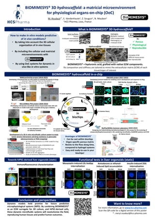
- 1. BIOMIMESYS® 3D hydroscaffold: a matricial microenvironment for physiological organs-on-chip (OoC) BIOMIMESYS® = hyaluronic acid, grafted with native ECM components. Its composition and stiffness are tailored to mimic the extracellular matrix (ECM) of each tissue/organ of interest. For more information, go to www.hcs-pharma.com Scan the QR-code for a digital version of this poster * : meryl.roudaut@hcs-pharma.com M. Roudaut1*, E. Vandenhaute1, Z. Souguir1, N. Maubon1 1HCS Pharma, Loos, France Dynamic models hold promise for future predictive microphysiological systems (MPS). By combining BIOMIMESYS® as an ECM surrogate for 3D culture, and hiPSC-derived cells, these dynamic microfluidic systems will revolutionize the field, reproducing human tissues and predict human outcomes. How to make in vitro models predictive of in vivo conditions? • By taking into account the 3D cellular organization of in vivo tissues • By including the cellular and matricial microenvironments with • By using OoC systems for dynamic in vivo-like in vitro systems Hydroscaffold Hydrogels: Biohydric medium Cells encapsulated Pathological, or irrelevant ECM Solid Scaffolds: Porous Irrelevant extracellular matrix (ECM) Decellularized tissue Complete ECM Difficult tissue access Not reproducible Scanning electron microscopy pictures Towards hiPSC-derived liver organoids (static) Functional tests in liver organoids (static) DAPI 2-Deoxy-D-glucose Merge Untreated Insulin 6nM 50µm 50µm 50µm 50µm 50µm 50µm DAPI LDL-bodipy Merge Untreated Mevastatin 50nM 50µm 50µm 50µm 50µm 50µm 50µm Liver organoid Albumin Desmin DAPI Merge Amiodarone or ethanol- induced lipid accumulation Mevastatin-induced LDL-bodipy internalization Insulin-induced 2DG internalization 50µm 50µm 50µm DAPI Nile Red Merge Untreated 50µm 50µm 50µm Amiodarone 20µM 50µm 50µm 50µm Ethanol 200nM Immunofluorescence characterization Merge DAPI ZO-1 Merge 50µm 50µm 50µm 50µm 50µm 50µm 50µm DAPI CD31 LHX2 Merge 50µm 50µm 50µm 50µm DAPI OATP1B1 LYVE1 Merge in biochips Introduction What is BIOMIMESYS® 3D hydroscaffold? BIOMIMESYS® hydroscaffold in-a-chip Avantages of BIOMIMESYS® • Can be cast within biochips • Organ-specific porous matrix • Resists to the flow along time, compared to hydrogel systems • Easy and ready-to-use in your biochips! Micro3DBeta PhD project (2020-2023) Developing a 3D dynamic microfluidic device of pancreatic beta cells and adipocytes to model multi-organ crosstalks during type 2 diabetes development MIMLiverOnChip project (2018-2023) Developing a functional liver-on-chip system to evaluate the toxicity of drugs • Universal “plug-and-play” liver-on-chip platform • Mimicking the major events occurring in the liver sinusoid is: Porous Physiological Reproducible MCF-7 Hoechst Calcein-AM Propidium iodide MDA-MB-231 200 um 96-well plate, static culture 7 days 7 days 28 days ONCO3D project (2017-2022) Developing tumoral models for modeling cancer development and responses to drug Ibidi biochip, dynamic culture Primary human adipocytes in BioPharMEMs Common Laboratory (2020-2022) Designing and industrializing new pharmacological in vitro assays for the screening of compounds/biopharmaceuticals in 3D, combining microfluidics, BioMEMS systems and ECM Development of a 3D in vitro microfluidic culture system to study tumor-stroma interactions and drug resistance in pancreatic adenocarcinoma or PDAC (ongoing) • 3D microfluidic bioreactor as a PDAC model for drug testing • Microenvironment made of BIOMIMESYS and pancreatic stellate cells • Implementation of interstitial fluid flow and nutrient/oxygen gradients • Growth of 3D cells aggregates • Study of mechanical effects on tumor phenotype Conclusion and perspectives Want to know more? Adipose tissue Pancreatic beta cells (Use of smart.servier.com for this illustration) Louis et al., 2017 Pr Cecile Legallais Dr Anthony Treizebre Dr Damien Fleury Dr Fabrice Soncin Pr Isabelle Van Seuningen Dr Vincent Senez Dr Jean-Sebastien Annicotte Dr Anthony Treizebre