BIOMIMESYS® Biofunctionalized hydroscaffold for 3D cell culture
•
1 like•1,531 views

To better mimic the tissue microenvironment and provide more predictive tools for drug development. hello@biominesys.com
Report
Share
Report
Share
Download to read offline

Recommended
HCS Pharma extends its 3D cell culture range BIOMIMESYS® with BIOMIMESYS® Brain
Following the success of BIOMIMESYS® range of products, which accurately and physiologically reproduces the microenvironment of liver, adipose and cancerous tissues, HCS Pharma is expanding its product range with BIOMIMESYS® Brain, an exclusive and innovative system for the 3D cell culture of neuronal cells.
A groundbreaking 3D cell culture technology for HCS: BIOMIMESYS hydroscaffold
Most potential drug candidates (90%) fail within the clinical trials, mainly because of lack of efficacy.
What if the pharmaceutical industry uses predictive human in vitro models in early drug discovery ?
BIOMIMESYS® Liver
BIOMIMESYS® Liver represents a new generation of mimetic HydroscaffoldS™ for 3D culture of hepatocyte-like cells. Available in a ready-to-use format, it enables the culture of hepatocytes and hepatocyte-like cells under physiological conditions that are representative of the microenvironment found in the liver. The highly porous nature of the Hydroscaffold™ allows the rapid uptake of nutrients, oxygen, etc. into the cells to create a reproducible study model for all downstream analyses used with 3D hepatocyte-like cell culture (metabolism, toxicity…).
BIOMIMESYS® Oncology
IOMIMESYS® Oncology is a new generation of mimetic Hydroscaffolds™ for 3D cell culture. Available in a ready-to-use format, it enables the culture of cells under physiological conditions that are representative of the microenvironment found in whole tissues. The highly porous structure of BIOMIMESYS® allows cells to diffuse into the 3D matrix, where they can fix and begin to develop. The structure and conformation of cells cultured in BIOMIMESYS®, such as cancer cells that spontaneously form spheroids, strongly resemble the structures formed in vivo. Also, the functionality of the BIOMIMESYS® matrix is proved by the expression of genes and proteins at levels that are again more similar to those found in vivo. Cells cultured in BIOMIMESYS® give results that are more physiological than those grown in 2D culture and are closer to those obtained in vivo.
BIOMIMESYS® Adipose tissue
BIOMIMESYS® Adipose tissue represents a new generation of mimetic Hydroscaffold™ for 3D culture of adipocyte and adipocyte-like cells. Available in a ready-to-use format, it enables the culture of adipocytes and adipocyte-like cells under physiological conditions that are representative of the microenvironment found in adipose tissue. The highly porous nature of the scaffold allows the rapid uptake of nutrients, oxygen, etc. into the cells to create a reproducible study model for all downstream analyses used with 3D adipocyte cultures.
Poster - BIOMIMESYS® 3D hydroscaffold a matricial microenvironment for physio...
How to make in vitro models predictive of in vivo conditions?
- By taking into account the 3D cellular organization of in vivo tissues
- By including the cellular and matricial microenvironments with BIOMIMESYS®
- By using OoC systems for dynamic in vivo-like in vitro systems
Dynamic models hold promise for future predictive microphysiological systems (MPS). By combining BIOMIMESYS® as an ECM surrogate for 3D culture, and hiPSC-derived cells, these dynamic microfluidic systems will revolutionize the field, reproducing human tissues and predict human outcomes.
Poster HCSPHARMA (OncoLilleDays2022) - Mechanobiological characterization of ...
Thomas Meynard, PhD student in OncoLille (under the supervision of Vincent Senez and Isabelle Van Seuningen) in collaboration with HCS Pharma too, presented a poster showing that it is possible to include BIOMIMESYS® in a microfluidic chip to co-culture Cancer-Associated fibroblasts and cancerous cells, with the aim to increase the complexity and the relevance of in vitro cancer models.
Poster : Development of personalized therapeutic targeting in lung cancer wit...
Abstract: Lung cancer is one of the most frequent cancers in the world with a high mortality rate. The discovery of oncogenic alterations in these cancers allowed the development of targeted therapies against several receptor tyrosine kinases (RTK) including EGFR, ALK or MET and contributed to improve the prognosis of some patients. Mutations impacting the MET receptor exon 14 splice sites (METex14) have recently been detected in about 3% of lung cancers and lead to the loss of the juxtamembrane domain with several negative regulatory sites. METex14 does not lead to constitutive activation of the receptor but we demonstrated that HGF, its ligand, is required for the full development of the transforming capabilities of the METex14 receptor in SCID-HGFhuman transgenic mice.
Several clinical trials have shown that only half of METex14 patients responded to MET inhibitors. While encouraging, these results are lower than those obtained with other treatments such as EGFR inhibitors. Interestingly, METex14 mutations are frequently associated with other molecular alterations including PTEN loss or TP53 mutations. Furthermore, we have shown that many METex14 mutated patients have autocrine secretion of HGF ligand. Understanding how these co-alterations impact METex14 oncogenicity and sensitivity to existing therapies is of outmost importance to identify patients that could benefit from them.
Poster - Including the matricial tumoral microenvironment in 3D in vitro mode...
In oncology, 97% of drug candidates fail in clinical trials. This highlights a lack of relevance of preclinical models used upstream. Indeed, human in vitro models don’t consider the Tumoral Extracellular Matrix (TECM). However, more and more studies demonstrate that ECM composition and stiffness are modified in tumors and are linked to cancer initiation, progression, propagation, and drug resistances.
BIOMIMESYS® is a Hyaluronic Acid-based matrix grafted with structural and adhesion molecules, which mimics the ECM/TECM. It is chemically defined and its composition and stiffness can be modified to reproduce the organ-specificity of the ECM, or to mimic a pathological microenvironment in vitro.
We have demonstrated that the exposition of colon cancer cells cultured in BIOMIMESYS® Oncology matrix to an anti-proliferative drug showed a closer in vitro/in vivo correlation in the EC50 curve compared to 2D culture. Cancer cells can be advantageously grown in BIOMIMESYS® for several weeks in multiwell plates and in microfluidic chips for more advanced models. We also observed that modifications in the matrix composition and stiffness modify the cell behavior. Moreover, thanks to collaborations with academic laboratories, we demonstrated that BIOMIMESYS® allows to reproduce in vitro the behavior of cancerous cells in vivo, like mutation effects and metastasis propagation, and could be a relevant alternative to animal models. These results showed that the matricial microenvironment modifies the cell behavior in vitro and should be considered carefully in drug discovery. BIOMIMESYS® hydroscaffold™ is adapted to High Content Screening and represented a powerful tool to better select drug candidate.
HCS PHARMA - (ECM 2022) Development of innovative hiPSC-based model including...
<!-- wp:paragraph -->
<p>We previously showed that human pluripotent stem cells (hiPSCs) provide a suitable model to study<br>metabolic diseases upon hepatocyte-like cell (HLC) differentiation. With a non-invasive approach, hiPSCs can be generated from urine samples of patients and HLCs have been used to model cholesterol metabolism regulation, by the study of LDLR- and PCSK9-mediated autosomal dominant hypercholesterolemia (ADH) as well as PCSK9-mediated familial hypobetalipoproteinemia (FHBL). This model provides promising advantages with a direct link to the patient and with an unlimited source of HLCs. But like all models, there are limitations, mainly by the neonatal characteristic of HLCs lead to difficulties for pharmacological investigations.</p>
<!-- /wp:paragraph -->
<!-- wp:paragraph -->
<p>Therefore, to overcome these burdens, we chose to 1. Differentiate hiPSCs into HLCs in an innovative<br>3D <a href="https://hcs-pharma.com/biomimesys/">hyaluronic acid-based hydroscaffold</a>, BIOMIMESYS® produces by HCS Pharma to enhance their maturation. 2. Adapt our 3D differentiation process to a 96-well format to make it compatible for drug screening. 3. Characterization of the 3D HLCs model by metabolism tests and compare to primary human hepatocyte (PHH).</p>
<!-- /wp:paragraph -->
<!-- wp:paragraph -->
<p>We gathered 3’ SRP data all along the differentiation process and RNAseq has been performed by comparing 2D and 3D differentiation conditions to characterize hiPSCs differentiation into liver organoids. We observed an enhanced expression of most hepatic genes and genes expressed by non-parenchymal cells such as stellate cells. Immunofluorescence data confirmed the co-localization of albumin-positive<br>hepatocytes, desmin-positive stellate cells and LYVE1-positive endothelial cells in liver organoids. Finally, at a functional level, several CYP activities including CYP3A4 were detected at the basal level and successfully induced. Liver organoids responded to pharmacological treatments as shown by their ability to accumulate lipids upon amiodarone treatment or uptake LDL-bodipy upon statin treatment.</p>
<!-- /wp:paragraph -->
<!-- wp:paragraph -->
<p>Altogether, our development gave rise to functional liver organoids generated with a unique and common procedure, in a process of automating for future high throughput screening.</p>
<!-- /wp:paragraph -->
HCSPHARMA Importance of microenvironment in cerebral in vitro models for phen...
Aim: About 90% of drug-candidates failed in clinical trials, in particular in neurology, due to a lack of efficacy. That highlights a lack of relevance in preclinical models, including in vitro models, which do not take into account the microenvironment, composed by glial cells and the Extracellular Matrix (ECM). The objective was to study the influence of the microenvironment in cerebral in vitro models, in the frame of Parkinson’s Disease (PD).
Methods: First, we analyzed the influence of astrocytes on Luhmes cell sensitivity, a dopaminergic neuronal cell line, in 2D culture. Then, we developed a hyaluronic acid-based hydroscaffold for 3D cell culture, which mimics the ECM, and study the sensitivity of Luhmes cells in this model. Thirdly, we performed a co-culture of Luhmes cells and astrocytes in this matrix, to form a complex model including both the glial and the matricial microenvironments.
Results: We observed a protective effect of astrocytes in 2D culture. In the hydroscaffold, Luhmes cells displayed a lower sensitivity compared to 2D culture, that was explained by a partial retention of toxic molecules in the matrix, and differences in neuronal protein expression. In the co-culture, we observed spheroids containing both neurons and astrocytes.
Conclusions: This work highlighted that the microenvironment of neurons can modify the neuronal response in vitro, and should thus be considered carefully in academic research and in drug discovery. This model can be now used to study the microenvironment modifications in pathological conditions, and to develop innovative drugs targeting the microenvironment.
Poster - A single procedure to generate functional hiPSCs-derived liver organ...
metabolic diseases upon hepatocyte-like cell (HLC) differentiation. In particular, HLCs have been used to model cholesterol metabolism regulation, by mimicking the main disease features in vitro. Human iPSCs can be generated from urine samples of patients with a well-described phenotype and carrying specific genotypes. This non-invasive approach allowed the study of LDLR- and PCSK9-mediated autosomal dominant hypercholesterolemia (ADH) as well as PCSK9-mediated familial hypobetalipoproteinemia (FHBL). While the direct link between hiPSCs and patients, as well as the abundance of HLCs provide promising advantages of such strategy, it is impaired mainly by the neonatal characteristic of HLCs as well as the difficulty to perform high throughput studies for pharmacological investigations.
Importance of matricial and cellular microenvironments in in vitro models for...
There is a 90% failure in clinical trials, due to efficacy and safety issues, which frequently concerns the central nervous system. That points a lack of relevance of preclinical models used upstream. In this frame, the aim of this study was to develop more relevant cerebral in vitro models, by including the matricial and cellular microenvironments, for drug discovery in Parkinson's disease.
HD available on https://hcs-pharma.com/poster---importance-of-matricial-and-cellular-microenvironments-in-in-vitro-models-for-drug-discovery-in-parkinsons-disease/
Simplifying 3d cell culture generation for high content screening with BIOMIM...
Growing interest in phenotypic screening, together with evidence that the drug response of cells grown in three-dimensional (3D) structures more closely resembles in vivo activity, has made high throughput, 3D fluorescence imaging an attractive screening option for drug discovery. However, creating 3D spheroids compatible with high content screening can be a difficult and expensive process.
BIOMIMESYS® is a range of patented hyaluronic acid scaffolds for 3D cell culture. They are made of RGDS- and galactosaminegrafted hyaluronic acid, using an adipic acid dihydrazide crosslinker and extracellular matrix proteins (eg. type I, IV or VI collagen) to accurately mimic the in vivo extracellular environment. BIOMIMESYS® is suitable for automated testing thanks to the uniform thickness of the scaffold – around 650 μm – with an average porosity of 100 to 200 μm
This application note describes a straightforward workflow for 3D cell seeding and spheroid formation using HCS Pharma’s BIOMIMESYS® plates in combination with INTEGRA’s VIAFLO 96/384 pipetting system. This process ensures rapid, controllable and reproducible 3D cell cultures, providing researchers with a highly efficient method to produce physiologically-relevant cellular models in a high throughput format
BIOMIMESYS®Liver, a 3D cell culture model for maintaining and promoting hepat...
BIOMIMESYS® range are hyaluronan based hydroscaffold developed to overcome the 2D flat culture limitations by recreating an in vivo-like physiology within the in vitro environment.
BIOMIMESYS®Liver hydroscaffold is made of RGDS and galactosamine-grafted Hyaluronic acid, Adipic acid dihydrazide crosslinker and extracellular matrix (ECM) proteins (collagen type I and collagen type IV) to mimic liver ECM composition.
Abstract : a single procedure to generate functional hi ps cs-derived liver o...
Abstract for poster for the "VIIIème Colloque de Génomique Fonctionnelle du Foie" - Rennes (France) 2020
Poster – Development and automation of 3D innovative hiPSC-based liver organo...
We previously showed that human pluripotent stem cells (hiPSCs) provide a suitable model to study metabolic diseases upon hepatocyte-like cell (HLC) differentiation. In particular, HLCs have been used to model cholesterol metabolism regulation, by mimicking the main disease features in vitro. Human iPSCs can be generated from urine samples of patients with a well-described phenotype and carrying specific genotypes. This non-invasive approach allowed the study of LDLR- and PCSK9-mediated autosomal dominant hypercholesterolemia (ADH) as well as PCSK9-mediated familial hypobetalipoproteinemia (FHBL). While the direct link between hiPSCs and patients, as well as the abundance of HLCs provide promising advantages of such strategy, it is impaired mainly by the neonatal characteristic of HLCs as well as the difficulty to perform high throughput studies for pharmacological investigations.
Development of a new liver-on-chip including BIOMIMESYS® technology for mimic...
Objective: to develop a new liver-on-chip model that includes a relevant 3D matrix for hepatic cell growth and function with the use of of BIOMIMESYS® Liver hydroscaffold for a physiological 3D hepatocyte culture.
HCS Pharma complète sa gamme de culture cellulaire en 3D BIOMIMESYS® avec BIO...
Après le succès de la gamme BIOMIMESYS® reproduisant de manière précise et physiologique le microenvironnement du foie, des tissus adipeux et cancéreux, HCS Pharma étend sa gamme avec BIOMIMESYS® Brain, un système exclusif et innovant permettant la culture cellulaire en 3D de cellules neuronales.
3D innovative hiPSC-based models including the microenvironment for phenotyp...
We previously showed that human pluripotent stem cells (hiPSCs) provide a suitable model to study metabolic diseases upon hepatocyte-like cell (HLC) differentiation. In particular, HLCs have been used to model cholesterol metabolism regulation, by mimicking the main disease features in vitro. Human iPSCs can be generated from urine samples of patients with a well-described phenotype and carrying specific genotypes. This non-invasive approach allowed the study of LDLR- and PCSK9-mediated autosomal dominant hypercholesterolemia (ADH) as well as PCSK9-mediated familial hypobetalipoproteinemia (FHBL). While the direct link between hiPSCs and patients, as well as the abundance of HLCs provide promising advantages of such strategy, it is impaired mainly by the neonatal characteristic of HLCs as well as the difficulty to perform high throughput studies for pharmacological investigations.
Therefore, to overcome these burdens, we choose to 1. Differentiate hiPSCs into HLCs in a 3D environment instead of the classical 2D culture systems to enhance their maturation; 2. Adapt our 3D differentiation process to a 96 wells format to make it compatible for drug screening.
To reach our goals, we established a partnership with HCS Pharma, which has an expertise in high content phenotypic screening and produces an innovative 3D scaffold, BiomimesysTM. This scaffold is composed of hyaluronic acid that can be functionalized with extra cellular matric derivatives, with adjustable stiffness and porosity. We setup conditions for hiPSCs seeding and differentiation to reach a new protocol adapted to a 3D environment. Our preliminary data indicate that our procedure enhanced expression of hepatic markers such as transcription factors (FOXA2, FOXA3, HNF1a, HNF1b, HNF4a), cytochrome P450 (CYP450) family members (CYP3A4, CYP2A6, CYP7A1) or cholesterol metabolism regulators (PCSK9, Lipoprotein(a)). During our presentation, we will discuss our data hiPSCs differentiation in 3D, CYP450 activities and induction, as well as their application for the study of metabolic diseases.
Neurotoxicity assessment: Comparison between SH-SY5Y and iPSC-derived cells
As shown by AstraZeneca in Nature reviews*, one third of the safety failures is linked to CNS toxicity during the clinical trials of drugs. Therefore, relevant in vitro human model is needed to detect early neurotoxicity of drug candidates. In this study, sensitivity of iPSC-derived neuronal cells to 32 compounds is compared to the sensitivity of SH-SY5Y cells. Two types of iPS-derived cells are tested: central nervous system cells (CNS.4U™ cells) and peripheral nervous system cells (PERI.4U™ cells) from Ncardia. Toxic effects are then measured by HCS cell imaging.
BIOMIMESYS® Adipose tissue, a relevant in vitro adipocyte 3D model
BIOMIMESYS® range are hyaluronan based scaffolds developed to overcome the 2D flat culture limitations by recreating an in vivo physiology within the in vitro environment.
BIOMIMESYS® Adipocyte scaffold is made of RGDS-grafted Hyaluronic acid (1.6 MDa), Adipic acid dihydrazide crosslinker and extracellular matrix (ECM) proteins (collagen type I and collagen type VI) to mimic fat tissue-ECM composition.
BIOMIMESYS® Liver, a 3D cell culture model for maintaining and promoting hep...
BIOMIMESYS® range are hyaluronan based hydroscaffold developed to overcome the 2D flat culture limitations by recreating an in vivo-like physiology within the in vitro environment.
BIOMIMESYS®Liver scaffold is made of RGDS and galactosamine-grafted Hyaluronic acid, Adipic acid dihydrazide crosslinker and extracellular matrix (ECM) proteins (collagen type I and collagen type IV) to mimic liver-ECM composition.
HCS for brain disorders / HCS Pharma at B4B mars 2018
How phenotypic screening with innovative cellular models can help to unravel mechanisms or repurpose drugs in the frame of brain disorders
Présentation de HCS Pharma dans la gazette du laboratoire - 2017
La société HCS Pharma, spécialiste de l’imagerie cellulaire à haut débit et du développement de nouveaux modèles cellulaires, est née à Rennes (35) en août 2014. Le 6 octobre dernier, elle a inauguré de nouvelles installations à Lille (59), au cœur du parc Eurasanté, et y a mis à l’honneur notamment sa toute nouvelle plate-forme robotique baptisée HAPIx, HCS Automation Platform for Imaging. Nathalie MAUBON, Présidente de l’entreprise, nous présente HCS Pharma et ses nouvelles ambitions portées par cette implantation lilloise, au plus proche des chercheurs et cliniciens en santé humaine...
定制(wsu毕业证书)美国华盛顿州立大学毕业证学位证书实拍图原版一模一样
原版纸张【微信:741003700 】【(wsu毕业证书)美国华盛顿州立大学毕业证、学位证书】【微信:741003700 】学位证,留信认证(真实可查,永久存档)offer、雅思、外壳等材料/诚信可靠,可直接看成品样本,帮您解决无法毕业带来的各种难题!外壳,原版制作,诚信可靠,可直接看成品样本。行业标杆!精益求精,诚心合作,真诚制作!多年品质 ,按需精细制作,24小时接单,全套进口原装设备。十五年致力于帮助留学生解决难题,包您满意。
本公司拥有海外各大学样板无数,能完美还原海外各大学 Bachelor Diploma degree, Master Degree Diploma
1:1完美还原海外各大学毕业材料上的工艺:水印,阴影底纹,钢印LOGO烫金烫银,LOGO烫金烫银复合重叠。文字图案浮雕、激光镭射、紫外荧光、温感、复印防伪等防伪工艺。材料咨询办理、认证咨询办理请加学历顾问Q/微741003700
留信网认证的作用:
1:该专业认证可证明留学生真实身份
2:同时对留学生所学专业登记给予评定
3:国家专业人才认证中心颁发入库证书
4:这个认证书并且可以归档倒地方
5:凡事获得留信网入网的信息将会逐步更新到个人身份内,将在公安局网内查询个人身份证信息后,同步读取人才网入库信息
6:个人职称评审加20分
7:个人信誉贷款加10分
8:在国家人才网主办的国家网络招聘大会中纳入资料,供国家高端企业选择人才
Child Welfare Clinic and Well baby clinicin Sri Lanka.ppsx
This document is designed as an introductory to medical students,nursing students,midwives or other healthcare trainees to improve their understanding about how health system in Sri Lanka cares children health.
More Related Content
More from HCS Pharma
Poster HCSPHARMA (OncoLilleDays2022) - Mechanobiological characterization of ...
Thomas Meynard, PhD student in OncoLille (under the supervision of Vincent Senez and Isabelle Van Seuningen) in collaboration with HCS Pharma too, presented a poster showing that it is possible to include BIOMIMESYS® in a microfluidic chip to co-culture Cancer-Associated fibroblasts and cancerous cells, with the aim to increase the complexity and the relevance of in vitro cancer models.
Poster : Development of personalized therapeutic targeting in lung cancer wit...
Abstract: Lung cancer is one of the most frequent cancers in the world with a high mortality rate. The discovery of oncogenic alterations in these cancers allowed the development of targeted therapies against several receptor tyrosine kinases (RTK) including EGFR, ALK or MET and contributed to improve the prognosis of some patients. Mutations impacting the MET receptor exon 14 splice sites (METex14) have recently been detected in about 3% of lung cancers and lead to the loss of the juxtamembrane domain with several negative regulatory sites. METex14 does not lead to constitutive activation of the receptor but we demonstrated that HGF, its ligand, is required for the full development of the transforming capabilities of the METex14 receptor in SCID-HGFhuman transgenic mice.
Several clinical trials have shown that only half of METex14 patients responded to MET inhibitors. While encouraging, these results are lower than those obtained with other treatments such as EGFR inhibitors. Interestingly, METex14 mutations are frequently associated with other molecular alterations including PTEN loss or TP53 mutations. Furthermore, we have shown that many METex14 mutated patients have autocrine secretion of HGF ligand. Understanding how these co-alterations impact METex14 oncogenicity and sensitivity to existing therapies is of outmost importance to identify patients that could benefit from them.
Poster - Including the matricial tumoral microenvironment in 3D in vitro mode...
In oncology, 97% of drug candidates fail in clinical trials. This highlights a lack of relevance of preclinical models used upstream. Indeed, human in vitro models don’t consider the Tumoral Extracellular Matrix (TECM). However, more and more studies demonstrate that ECM composition and stiffness are modified in tumors and are linked to cancer initiation, progression, propagation, and drug resistances.
BIOMIMESYS® is a Hyaluronic Acid-based matrix grafted with structural and adhesion molecules, which mimics the ECM/TECM. It is chemically defined and its composition and stiffness can be modified to reproduce the organ-specificity of the ECM, or to mimic a pathological microenvironment in vitro.
We have demonstrated that the exposition of colon cancer cells cultured in BIOMIMESYS® Oncology matrix to an anti-proliferative drug showed a closer in vitro/in vivo correlation in the EC50 curve compared to 2D culture. Cancer cells can be advantageously grown in BIOMIMESYS® for several weeks in multiwell plates and in microfluidic chips for more advanced models. We also observed that modifications in the matrix composition and stiffness modify the cell behavior. Moreover, thanks to collaborations with academic laboratories, we demonstrated that BIOMIMESYS® allows to reproduce in vitro the behavior of cancerous cells in vivo, like mutation effects and metastasis propagation, and could be a relevant alternative to animal models. These results showed that the matricial microenvironment modifies the cell behavior in vitro and should be considered carefully in drug discovery. BIOMIMESYS® hydroscaffold™ is adapted to High Content Screening and represented a powerful tool to better select drug candidate.
HCS PHARMA - (ECM 2022) Development of innovative hiPSC-based model including...
<!-- wp:paragraph -->
<p>We previously showed that human pluripotent stem cells (hiPSCs) provide a suitable model to study<br>metabolic diseases upon hepatocyte-like cell (HLC) differentiation. With a non-invasive approach, hiPSCs can be generated from urine samples of patients and HLCs have been used to model cholesterol metabolism regulation, by the study of LDLR- and PCSK9-mediated autosomal dominant hypercholesterolemia (ADH) as well as PCSK9-mediated familial hypobetalipoproteinemia (FHBL). This model provides promising advantages with a direct link to the patient and with an unlimited source of HLCs. But like all models, there are limitations, mainly by the neonatal characteristic of HLCs lead to difficulties for pharmacological investigations.</p>
<!-- /wp:paragraph -->
<!-- wp:paragraph -->
<p>Therefore, to overcome these burdens, we chose to 1. Differentiate hiPSCs into HLCs in an innovative<br>3D <a href="https://hcs-pharma.com/biomimesys/">hyaluronic acid-based hydroscaffold</a>, BIOMIMESYS® produces by HCS Pharma to enhance their maturation. 2. Adapt our 3D differentiation process to a 96-well format to make it compatible for drug screening. 3. Characterization of the 3D HLCs model by metabolism tests and compare to primary human hepatocyte (PHH).</p>
<!-- /wp:paragraph -->
<!-- wp:paragraph -->
<p>We gathered 3’ SRP data all along the differentiation process and RNAseq has been performed by comparing 2D and 3D differentiation conditions to characterize hiPSCs differentiation into liver organoids. We observed an enhanced expression of most hepatic genes and genes expressed by non-parenchymal cells such as stellate cells. Immunofluorescence data confirmed the co-localization of albumin-positive<br>hepatocytes, desmin-positive stellate cells and LYVE1-positive endothelial cells in liver organoids. Finally, at a functional level, several CYP activities including CYP3A4 were detected at the basal level and successfully induced. Liver organoids responded to pharmacological treatments as shown by their ability to accumulate lipids upon amiodarone treatment or uptake LDL-bodipy upon statin treatment.</p>
<!-- /wp:paragraph -->
<!-- wp:paragraph -->
<p>Altogether, our development gave rise to functional liver organoids generated with a unique and common procedure, in a process of automating for future high throughput screening.</p>
<!-- /wp:paragraph -->
HCSPHARMA Importance of microenvironment in cerebral in vitro models for phen...
Aim: About 90% of drug-candidates failed in clinical trials, in particular in neurology, due to a lack of efficacy. That highlights a lack of relevance in preclinical models, including in vitro models, which do not take into account the microenvironment, composed by glial cells and the Extracellular Matrix (ECM). The objective was to study the influence of the microenvironment in cerebral in vitro models, in the frame of Parkinson’s Disease (PD).
Methods: First, we analyzed the influence of astrocytes on Luhmes cell sensitivity, a dopaminergic neuronal cell line, in 2D culture. Then, we developed a hyaluronic acid-based hydroscaffold for 3D cell culture, which mimics the ECM, and study the sensitivity of Luhmes cells in this model. Thirdly, we performed a co-culture of Luhmes cells and astrocytes in this matrix, to form a complex model including both the glial and the matricial microenvironments.
Results: We observed a protective effect of astrocytes in 2D culture. In the hydroscaffold, Luhmes cells displayed a lower sensitivity compared to 2D culture, that was explained by a partial retention of toxic molecules in the matrix, and differences in neuronal protein expression. In the co-culture, we observed spheroids containing both neurons and astrocytes.
Conclusions: This work highlighted that the microenvironment of neurons can modify the neuronal response in vitro, and should thus be considered carefully in academic research and in drug discovery. This model can be now used to study the microenvironment modifications in pathological conditions, and to develop innovative drugs targeting the microenvironment.
Poster - A single procedure to generate functional hiPSCs-derived liver organ...
metabolic diseases upon hepatocyte-like cell (HLC) differentiation. In particular, HLCs have been used to model cholesterol metabolism regulation, by mimicking the main disease features in vitro. Human iPSCs can be generated from urine samples of patients with a well-described phenotype and carrying specific genotypes. This non-invasive approach allowed the study of LDLR- and PCSK9-mediated autosomal dominant hypercholesterolemia (ADH) as well as PCSK9-mediated familial hypobetalipoproteinemia (FHBL). While the direct link between hiPSCs and patients, as well as the abundance of HLCs provide promising advantages of such strategy, it is impaired mainly by the neonatal characteristic of HLCs as well as the difficulty to perform high throughput studies for pharmacological investigations.
Importance of matricial and cellular microenvironments in in vitro models for...
There is a 90% failure in clinical trials, due to efficacy and safety issues, which frequently concerns the central nervous system. That points a lack of relevance of preclinical models used upstream. In this frame, the aim of this study was to develop more relevant cerebral in vitro models, by including the matricial and cellular microenvironments, for drug discovery in Parkinson's disease.
HD available on https://hcs-pharma.com/poster---importance-of-matricial-and-cellular-microenvironments-in-in-vitro-models-for-drug-discovery-in-parkinsons-disease/
Simplifying 3d cell culture generation for high content screening with BIOMIM...
Growing interest in phenotypic screening, together with evidence that the drug response of cells grown in three-dimensional (3D) structures more closely resembles in vivo activity, has made high throughput, 3D fluorescence imaging an attractive screening option for drug discovery. However, creating 3D spheroids compatible with high content screening can be a difficult and expensive process.
BIOMIMESYS® is a range of patented hyaluronic acid scaffolds for 3D cell culture. They are made of RGDS- and galactosaminegrafted hyaluronic acid, using an adipic acid dihydrazide crosslinker and extracellular matrix proteins (eg. type I, IV or VI collagen) to accurately mimic the in vivo extracellular environment. BIOMIMESYS® is suitable for automated testing thanks to the uniform thickness of the scaffold – around 650 μm – with an average porosity of 100 to 200 μm
This application note describes a straightforward workflow for 3D cell seeding and spheroid formation using HCS Pharma’s BIOMIMESYS® plates in combination with INTEGRA’s VIAFLO 96/384 pipetting system. This process ensures rapid, controllable and reproducible 3D cell cultures, providing researchers with a highly efficient method to produce physiologically-relevant cellular models in a high throughput format
BIOMIMESYS®Liver, a 3D cell culture model for maintaining and promoting hepat...
BIOMIMESYS® range are hyaluronan based hydroscaffold developed to overcome the 2D flat culture limitations by recreating an in vivo-like physiology within the in vitro environment.
BIOMIMESYS®Liver hydroscaffold is made of RGDS and galactosamine-grafted Hyaluronic acid, Adipic acid dihydrazide crosslinker and extracellular matrix (ECM) proteins (collagen type I and collagen type IV) to mimic liver ECM composition.
Abstract : a single procedure to generate functional hi ps cs-derived liver o...
Abstract for poster for the "VIIIème Colloque de Génomique Fonctionnelle du Foie" - Rennes (France) 2020
Poster – Development and automation of 3D innovative hiPSC-based liver organo...
We previously showed that human pluripotent stem cells (hiPSCs) provide a suitable model to study metabolic diseases upon hepatocyte-like cell (HLC) differentiation. In particular, HLCs have been used to model cholesterol metabolism regulation, by mimicking the main disease features in vitro. Human iPSCs can be generated from urine samples of patients with a well-described phenotype and carrying specific genotypes. This non-invasive approach allowed the study of LDLR- and PCSK9-mediated autosomal dominant hypercholesterolemia (ADH) as well as PCSK9-mediated familial hypobetalipoproteinemia (FHBL). While the direct link between hiPSCs and patients, as well as the abundance of HLCs provide promising advantages of such strategy, it is impaired mainly by the neonatal characteristic of HLCs as well as the difficulty to perform high throughput studies for pharmacological investigations.
Poster – Development and automation of 3D innovative hiPSC-based liver organo...
We previously showed that human pluripotent stem cells (hiPSCs) provide a suitable model to study metabolic diseases upon hepatocyte-like cell (HLC) differentiation. In particular, HLCs have been used to model cholesterol metabolism regulation, by mimicking the main disease features in vitro. Human iPSCs can be generated from urine samples of patients with a well-described phenotype and carrying specific genotypes. This non-invasive approach allowed the study of LDLR- and PCSK9-mediated autosomal dominant hypercholesterolemia (ADH) as well as PCSK9-mediated familial hypobetalipoproteinemia (FHBL). While the direct link between hiPSCs and patients, as well as the abundance of HLCs provide promising advantages of such strategy, it is impaired mainly by the neonatal characteristic of HLCs as well as the difficulty to perform high throughput studies for pharmacological investigations.
Development of a new liver-on-chip including BIOMIMESYS® technology for mimic...
Objective: to develop a new liver-on-chip model that includes a relevant 3D matrix for hepatic cell growth and function with the use of of BIOMIMESYS® Liver hydroscaffold for a physiological 3D hepatocyte culture.
HCS Pharma complète sa gamme de culture cellulaire en 3D BIOMIMESYS® avec BIO...
Après le succès de la gamme BIOMIMESYS® reproduisant de manière précise et physiologique le microenvironnement du foie, des tissus adipeux et cancéreux, HCS Pharma étend sa gamme avec BIOMIMESYS® Brain, un système exclusif et innovant permettant la culture cellulaire en 3D de cellules neuronales.
3D innovative hiPSC-based models including the microenvironment for phenotyp...
We previously showed that human pluripotent stem cells (hiPSCs) provide a suitable model to study metabolic diseases upon hepatocyte-like cell (HLC) differentiation. In particular, HLCs have been used to model cholesterol metabolism regulation, by mimicking the main disease features in vitro. Human iPSCs can be generated from urine samples of patients with a well-described phenotype and carrying specific genotypes. This non-invasive approach allowed the study of LDLR- and PCSK9-mediated autosomal dominant hypercholesterolemia (ADH) as well as PCSK9-mediated familial hypobetalipoproteinemia (FHBL). While the direct link between hiPSCs and patients, as well as the abundance of HLCs provide promising advantages of such strategy, it is impaired mainly by the neonatal characteristic of HLCs as well as the difficulty to perform high throughput studies for pharmacological investigations.
Therefore, to overcome these burdens, we choose to 1. Differentiate hiPSCs into HLCs in a 3D environment instead of the classical 2D culture systems to enhance their maturation; 2. Adapt our 3D differentiation process to a 96 wells format to make it compatible for drug screening.
To reach our goals, we established a partnership with HCS Pharma, which has an expertise in high content phenotypic screening and produces an innovative 3D scaffold, BiomimesysTM. This scaffold is composed of hyaluronic acid that can be functionalized with extra cellular matric derivatives, with adjustable stiffness and porosity. We setup conditions for hiPSCs seeding and differentiation to reach a new protocol adapted to a 3D environment. Our preliminary data indicate that our procedure enhanced expression of hepatic markers such as transcription factors (FOXA2, FOXA3, HNF1a, HNF1b, HNF4a), cytochrome P450 (CYP450) family members (CYP3A4, CYP2A6, CYP7A1) or cholesterol metabolism regulators (PCSK9, Lipoprotein(a)). During our presentation, we will discuss our data hiPSCs differentiation in 3D, CYP450 activities and induction, as well as their application for the study of metabolic diseases.
Neurotoxicity assessment: Comparison between SH-SY5Y and iPSC-derived cells
As shown by AstraZeneca in Nature reviews*, one third of the safety failures is linked to CNS toxicity during the clinical trials of drugs. Therefore, relevant in vitro human model is needed to detect early neurotoxicity of drug candidates. In this study, sensitivity of iPSC-derived neuronal cells to 32 compounds is compared to the sensitivity of SH-SY5Y cells. Two types of iPS-derived cells are tested: central nervous system cells (CNS.4U™ cells) and peripheral nervous system cells (PERI.4U™ cells) from Ncardia. Toxic effects are then measured by HCS cell imaging.
BIOMIMESYS® Adipose tissue, a relevant in vitro adipocyte 3D model
BIOMIMESYS® range are hyaluronan based scaffolds developed to overcome the 2D flat culture limitations by recreating an in vivo physiology within the in vitro environment.
BIOMIMESYS® Adipocyte scaffold is made of RGDS-grafted Hyaluronic acid (1.6 MDa), Adipic acid dihydrazide crosslinker and extracellular matrix (ECM) proteins (collagen type I and collagen type VI) to mimic fat tissue-ECM composition.
BIOMIMESYS® Liver, a 3D cell culture model for maintaining and promoting hep...
BIOMIMESYS® range are hyaluronan based hydroscaffold developed to overcome the 2D flat culture limitations by recreating an in vivo-like physiology within the in vitro environment.
BIOMIMESYS®Liver scaffold is made of RGDS and galactosamine-grafted Hyaluronic acid, Adipic acid dihydrazide crosslinker and extracellular matrix (ECM) proteins (collagen type I and collagen type IV) to mimic liver-ECM composition.
HCS for brain disorders / HCS Pharma at B4B mars 2018
How phenotypic screening with innovative cellular models can help to unravel mechanisms or repurpose drugs in the frame of brain disorders
Présentation de HCS Pharma dans la gazette du laboratoire - 2017
La société HCS Pharma, spécialiste de l’imagerie cellulaire à haut débit et du développement de nouveaux modèles cellulaires, est née à Rennes (35) en août 2014. Le 6 octobre dernier, elle a inauguré de nouvelles installations à Lille (59), au cœur du parc Eurasanté, et y a mis à l’honneur notamment sa toute nouvelle plate-forme robotique baptisée HAPIx, HCS Automation Platform for Imaging. Nathalie MAUBON, Présidente de l’entreprise, nous présente HCS Pharma et ses nouvelles ambitions portées par cette implantation lilloise, au plus proche des chercheurs et cliniciens en santé humaine...
More from HCS Pharma (20)
Poster HCSPHARMA (OncoLilleDays2022) - Mechanobiological characterization of ...
Poster HCSPHARMA (OncoLilleDays2022) - Mechanobiological characterization of ...
Poster : Development of personalized therapeutic targeting in lung cancer wit...
Poster : Development of personalized therapeutic targeting in lung cancer wit...
Poster - Including the matricial tumoral microenvironment in 3D in vitro mode...
Poster - Including the matricial tumoral microenvironment in 3D in vitro mode...
HCS PHARMA - (ECM 2022) Development of innovative hiPSC-based model including...
HCS PHARMA - (ECM 2022) Development of innovative hiPSC-based model including...
HCSPHARMA Importance of microenvironment in cerebral in vitro models for phen...
HCSPHARMA Importance of microenvironment in cerebral in vitro models for phen...
Poster - A single procedure to generate functional hiPSCs-derived liver organ...
Poster - A single procedure to generate functional hiPSCs-derived liver organ...
Importance of matricial and cellular microenvironments in in vitro models for...
Importance of matricial and cellular microenvironments in in vitro models for...
Simplifying 3d cell culture generation for high content screening with BIOMIM...
Simplifying 3d cell culture generation for high content screening with BIOMIM...
BIOMIMESYS®Liver, a 3D cell culture model for maintaining and promoting hepat...
BIOMIMESYS®Liver, a 3D cell culture model for maintaining and promoting hepat...
Abstract : a single procedure to generate functional hi ps cs-derived liver o...
Abstract : a single procedure to generate functional hi ps cs-derived liver o...
Poster – Development and automation of 3D innovative hiPSC-based liver organo...
Poster – Development and automation of 3D innovative hiPSC-based liver organo...
Poster – Development and automation of 3D innovative hiPSC-based liver organo...
Poster – Development and automation of 3D innovative hiPSC-based liver organo...
Development of a new liver-on-chip including BIOMIMESYS® technology for mimic...
Development of a new liver-on-chip including BIOMIMESYS® technology for mimic...
HCS Pharma complète sa gamme de culture cellulaire en 3D BIOMIMESYS® avec BIO...
HCS Pharma complète sa gamme de culture cellulaire en 3D BIOMIMESYS® avec BIO...
3D innovative hiPSC-based models including the microenvironment for phenotyp...
3D innovative hiPSC-based models including the microenvironment for phenotyp...
Neurotoxicity assessment: Comparison between SH-SY5Y and iPSC-derived cells
Neurotoxicity assessment: Comparison between SH-SY5Y and iPSC-derived cells
BIOMIMESYS® Adipose tissue, a relevant in vitro adipocyte 3D model
BIOMIMESYS® Adipose tissue, a relevant in vitro adipocyte 3D model
BIOMIMESYS® Liver, a 3D cell culture model for maintaining and promoting hep...
BIOMIMESYS® Liver, a 3D cell culture model for maintaining and promoting hep...
HCS for brain disorders / HCS Pharma at B4B mars 2018
HCS for brain disorders / HCS Pharma at B4B mars 2018
Présentation de HCS Pharma dans la gazette du laboratoire - 2017
Présentation de HCS Pharma dans la gazette du laboratoire - 2017
Recently uploaded
定制(wsu毕业证书)美国华盛顿州立大学毕业证学位证书实拍图原版一模一样
原版纸张【微信:741003700 】【(wsu毕业证书)美国华盛顿州立大学毕业证、学位证书】【微信:741003700 】学位证,留信认证(真实可查,永久存档)offer、雅思、外壳等材料/诚信可靠,可直接看成品样本,帮您解决无法毕业带来的各种难题!外壳,原版制作,诚信可靠,可直接看成品样本。行业标杆!精益求精,诚心合作,真诚制作!多年品质 ,按需精细制作,24小时接单,全套进口原装设备。十五年致力于帮助留学生解决难题,包您满意。
本公司拥有海外各大学样板无数,能完美还原海外各大学 Bachelor Diploma degree, Master Degree Diploma
1:1完美还原海外各大学毕业材料上的工艺:水印,阴影底纹,钢印LOGO烫金烫银,LOGO烫金烫银复合重叠。文字图案浮雕、激光镭射、紫外荧光、温感、复印防伪等防伪工艺。材料咨询办理、认证咨询办理请加学历顾问Q/微741003700
留信网认证的作用:
1:该专业认证可证明留学生真实身份
2:同时对留学生所学专业登记给予评定
3:国家专业人才认证中心颁发入库证书
4:这个认证书并且可以归档倒地方
5:凡事获得留信网入网的信息将会逐步更新到个人身份内,将在公安局网内查询个人身份证信息后,同步读取人才网入库信息
6:个人职称评审加20分
7:个人信誉贷款加10分
8:在国家人才网主办的国家网络招聘大会中纳入资料,供国家高端企业选择人才
Child Welfare Clinic and Well baby clinicin Sri Lanka.ppsx
This document is designed as an introductory to medical students,nursing students,midwives or other healthcare trainees to improve their understanding about how health system in Sri Lanka cares children health.
LGBTQ+ Adults: Unique Opportunities and Inclusive Approaches to Care
This webinar helps clinicians understand the unique healthcare needs of the LGBTQ+ community, primarily in relation to end-of-life care. Topics include social and cultural background and challenges, healthcare disparities, advanced care planning, and strategies for reaching the community and improving quality of care.
PET CT beginners Guide covers some of the underrepresented topics in PET CT
This lecture briefly covers some of the underrepresented topics in Molecular imaging with cases , such as:
- Primary pleural tumors and pleural metastases.
- Distinguishing between MPM and Talc Pleurodesis.
- Urological tumors.
- The role of FDG PET in NET.
Champions of Health Spotlight On Leaders Shaping Germany's Healthcare.pdf
This edition features a handful of Champions of Health: Spotlight On Leaders Shaping Germany's Healthcare that are leading us into a better future.
RECENT ADVANCES IN BREAST CANCER RADIOTHERAPY
ULTRA HYPOFRACTIONATION
Accelerated Partial Breast Irradiation: :Brachytherapy
UK FAST FORWARD
TEST BANK For Accounting Information Systems, 3rd Edition by Vernon Richardso...
TEST BANK For Accounting Information Systems, 3rd Edition by Vernon Richardso...rightmanforbloodline
TEST BANK For Accounting Information Systems, 3rd Edition by Vernon Richardson, Verified Chapters 1 - 18, Complete Newest Version
TEST BANK For Accounting Information Systems, 3rd Edition by Vernon Richardson, Verified Chapters 1 - 18, Complete Newest Version
TEST BANK For Accounting Information Systems, 3rd Edition by Vernon Richardson, Verified Chapters 1 - 18, Complete Newest VersionTop massage center in ajman chandrima Spa
We are one of the top Massage Spa Ajman Our highly skilled, experienced, and certified massage therapists from different corners of the world are committed to serving you with a soothing and relaxing experience. Luxuriate yourself at our spas in Sharjah and Ajman, which are indeed enriched with an ambiance of relaxation and tranquility. We could confidently claim that we are one of the most affordable Spa Ajman and Sharjah as well, where you can book the massage session of your choice for just 99 AED at any time as we are open 24 hours a day, 7 days a week.
Visit : https://massagespaajman.com/
Call : 052 987 1315
CANSA support - Caring for Cancer Patients' Caregivers
International Cancer Survivors Day is celebrated during June, placing the spotlight not only on cancer survivors, but also their caregivers.
CANSA has compiled a list of tips and guidelines of support:
https://cansa.org.za/who-cares-for-cancer-patients-caregivers/
CMHPSM Regional Compliance Training 2024
Compliance training used in conjunction with CMHPSM training platform.
Letter to MREC - application to conduct study
Application to conduct study on research title 'Awareness and knowledge of oral cancer and precancer among dental outpatient in Klinik Pergigian Merlimau, Melaka'
Luxurious Spa In Ajman Chandrima Massage Center
Chandrima Spa Ajman is one of the leading Massage Center in Ajman, which is open 24 hours exclusively for men. Being one of the most affordable Spa in Ajman, we offer Body to Body massage, Kerala Massage, Malayali Massage, Indian Massage, Pakistani Massage Russian massage, Thai massage, Swedish massage, Hot Stone Massage, Deep Tissue Massage, and many more. Indulge in the ultimate massage experience and book your appointment today. We are confident that you will leave our Massage spa feeling refreshed, rejuvenated, and ready to take on the world.
Visit : https://massagespaajman.com/
Call : 052 987 1315
The positive impact of SGRT – The Berkshire Cancer Centre experience
SGRT Europe 2023
Victoria Hammond-Turner
Technical and Development Lead Therapeutic Radiographer
Royal Berkshire NHS Foundation Trust, UK
Professional Secrecy: Forensic Medicine Lecture
Professional Secrecy: Forensic Medicine Lecture , Medical jurisprudence
Stem Cell Solutions: Dr. David Greene's Path to Non-Surgical Cardiac Care
Explore the groundbreaking work of Dr. David Greene, a pioneer in regenerative medicine, who is revolutionizing the field of cardiology through stem cell therapy in Arizona. This ppt delves into how Dr. Greene's innovative approach is providing non-surgical, effective treatments for heart disease, using the body's own cells to repair heart damage and improve patient outcomes. Learn about the science behind stem cell therapy, its benefits over traditional cardiac surgeries, and the promising future it holds for modern medicine. Join us as we uncover how Dr. Greene's commitment to stem cell research and therapy is setting new standards in healthcare and offering new hope to cardiac patients.
Nursing education curriculum development.pptx
curriculum development in nursing education based on INC syllabus for M.sc nursing.
Dr. David Greene R3 stem cell Breakthroughs: Stem Cell Therapy in Cardiology
Dr. David Greene, founder and CEO of R3 Stem Cell, is at the forefront of groundbreaking research in the field of cardiology, focusing on the transformative potential of stem cell therapy. His latest work emphasizes innovative approaches to treating heart disease, aiming to repair damaged heart tissue and improve heart function through the use of advanced stem cell techniques. This research promises not only to enhance the quality of life for patients with chronic heart conditions but also to pave the way for new, more effective treatments. Dr. Greene's work is notable for its focus on safety, efficacy, and the potential to significantly reduce the need for invasive surgeries and long-term medication, positioning stem cell therapy as a key player in the future of cardiac care.
一比一原版纽约大学毕业证(NYU毕业证)成绩单留信认证
原版定制【微信:41543339】【纽约大学毕业证(NYU毕业证)】【微信:41543339】成绩单、外壳、offer、留信学历认证(永久存档真实可查)采用学校原版纸张、特殊工艺完全按照原版一比一制作(包括:隐形水印,阴影底纹,钢印LOGO烫金烫银,LOGO烫金烫银复合重叠,文字图案浮雕,激光镭射,紫外荧光,温感,复印防伪)行业标杆!精益求精,诚心合作,真诚制作!多年品质 ,按需精细制作,24小时接单,全套进口原装设备,十五年致力于帮助留学生解决难题,业务范围有加拿大、英国、澳洲、韩国、美国、新加坡,新西兰等学历材料,包您满意。
【我们承诺采用的是学校原版纸张(纸质、底色、纹路),我们拥有全套进口原装设备,特殊工艺都是采用不同机器制作,仿真度基本可以达到98%以上,所有工艺效果都可提前给客户展示,不满意可以根据客户要求进行调整,直到满意为止!】
【业务选择办理准则】
一、工作未确定,回国需先给父母、亲戚朋友看下文凭的情况,办理一份就读学校的毕业证【微信41543339】文凭即可
二、回国进私企、外企、自己做生意的情况,这些单位是不查询毕业证真伪的,而且国内没有渠道去查询国外文凭的真假,也不需要提供真实教育部认证。鉴于此,办理一份毕业证【微信41543339】即可
三、进国企,银行,事业单位,考公务员等等,这些单位是必需要提供真实教育部认证的,办理教育部认证所需资料众多且烦琐,所有材料您都必须提供原件,我们凭借丰富的经验,快捷的绿色通道帮您快速整合材料,让您少走弯路。
留信网认证的作用:
1:该专业认证可证明留学生真实身份
2:同时对留学生所学专业登记给予评定
3:国家专业人才认证中心颁发入库证书
4:这个认证书并且可以归档倒地方
5:凡事获得留信网入网的信息将会逐步更新到个人身份内,将在公安局网内查询个人身份证信息后,同步读取人才网入库信息
6:个人职称评审加20分
7:个人信誉贷款加10分
8:在国家人才网主办的国家网络招聘大会中纳入资料,供国家高端企业选择人才
留信网服务项目:
1、留学生专业人才库服务(留信分析)
2、国(境)学习人员提供就业推荐信服务
3、留学人员区块链存储服务
→ 【关于价格问题(保证一手价格)】
我们所定的价格是非常合理的,而且我们现在做得单子大多数都是代理和回头客户介绍的所以一般现在有新的单子 我给客户的都是第一手的代理价格,因为我想坦诚对待大家 不想跟大家在价格方面浪费时间
对于老客户或者被老客户介绍过来的朋友,我们都会适当给一些优惠。
选择实体注册公司办理,更放心,更安全!我们的承诺:客户在留信官方认证查询网站查询到认证通过结果后付款,不成功不收费!
CHAPTER 1 SEMESTER V PREVENTIVE-PEDIATRICS.pdf
This content provides an overview of preventive pediatrics. It defines preventive pediatrics as preventing disease and promoting children's physical, mental, and social well-being to achieve positive health. It discusses antenatal, postnatal, and social preventive pediatrics. It also covers various child health programs like immunization, breastfeeding, ICDS, and the roles of organizations like WHO, UNICEF, and nurses in preventive pediatrics.
Recently uploaded (20)
Child Welfare Clinic and Well baby clinicin Sri Lanka.ppsx
Child Welfare Clinic and Well baby clinicin Sri Lanka.ppsx
LGBTQ+ Adults: Unique Opportunities and Inclusive Approaches to Care
LGBTQ+ Adults: Unique Opportunities and Inclusive Approaches to Care
PET CT beginners Guide covers some of the underrepresented topics in PET CT
PET CT beginners Guide covers some of the underrepresented topics in PET CT
Champions of Health Spotlight On Leaders Shaping Germany's Healthcare.pdf
Champions of Health Spotlight On Leaders Shaping Germany's Healthcare.pdf
TEST BANK For Accounting Information Systems, 3rd Edition by Vernon Richardso...
TEST BANK For Accounting Information Systems, 3rd Edition by Vernon Richardso...
CANSA support - Caring for Cancer Patients' Caregivers
CANSA support - Caring for Cancer Patients' Caregivers
The positive impact of SGRT – The Berkshire Cancer Centre experience
The positive impact of SGRT – The Berkshire Cancer Centre experience
Stem Cell Solutions: Dr. David Greene's Path to Non-Surgical Cardiac Care
Stem Cell Solutions: Dr. David Greene's Path to Non-Surgical Cardiac Care
Dr. David Greene R3 stem cell Breakthroughs: Stem Cell Therapy in Cardiology
Dr. David Greene R3 stem cell Breakthroughs: Stem Cell Therapy in Cardiology
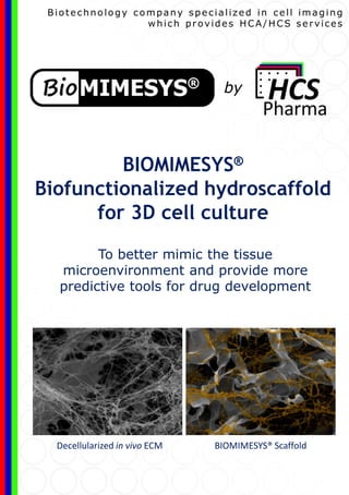
BIOMIMESYS® Biofunctionalized hydroscaffold for 3D cell culture
- 1. To better mimic the tissue microenvironment and provide more predictive tools for drug development BIOMIMESYS® Biofunctionalized hydroscaffold for 3D cell culture Biotechnology company specialized in cell imaging which provides HCA/HCS services BIOMIMESYS® ScaffoldDecellularized in vivo ECM by
- 2. Product Specifications Compositions Elastic modulus BIOMIMESYS® Liver HA + RGDS, Galactosamine, Collagen I, Collagen IV 0.6 kPa BIOMIMESYS® Adipose tissue HA + RGDS, Collagen I, Collagen VI 0.45 kPa BIOMIMESYS® Oncology HA + Collagen I 0.4 kPa Plate specifications: RGDS or Galactosamine grafted Hyaluronic Acid Physiological amount of ECM compound Undenatured collagen Elastic modulus regarding organs Scalable hydroscaffold in vivo like chemical composition and physicochemical properties (porosity , Elastic modulus...) BIOMIMESYS® hydroscaffold Precast matrix in a 96-WELL PLATE The only technology based on HA and ECM compound ready to use
- 3. Cell lines Primary cells iPSCs e.g. HepG2 3 – CHOOSE YOUR READ-OUTS Hoechst ; EDU; mitotracker; colocalization HCT116 Proliferation, apoptosis, cell motility, Cell structure, Stress, Protein analysis, mRNA analysis … MRP2; Actin; colocalization HepG2 1 – CHOOSE YOUR CELLS & MICROENVIRONNEMENT 2 - CHOOSE YOUR INDUCERS & INCUBATION TIME Services ECM: Define - Composition - Elastic modulus - Porosity Perilipin (TG), nucleus Human Preadipocytes (HWP) Small Molecules Biologicals Ingredients Starvations UV Radiations Fixed Cells Live Cells BIOMIMESYS® Liver Hoechst; Phalloidin HepG2 BIOMIMESYS® Adipose Tissue BIOMIMESYS® Oncology
- 4. Contact us for further information www.biomimesys.com hello@biomimesys.com +33 769 999 137 Lille Rennes OUR VALUES Expertise Efficiency Flexibility Transparency Robotic Platform & innovative 3D models for HCA and HCS services Innovative & relevant in vitro models Enriched information on biological content Phenotypic screening Highly visual Automated process Multi-parametric study Big Data / Machine learning