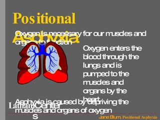
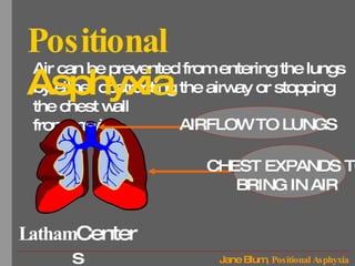
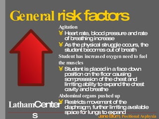
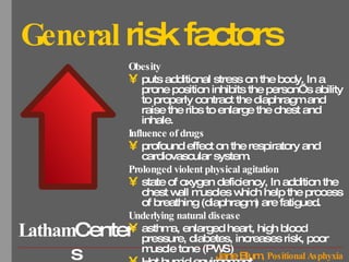
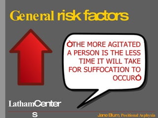
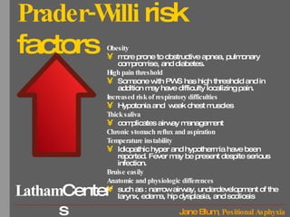
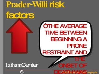
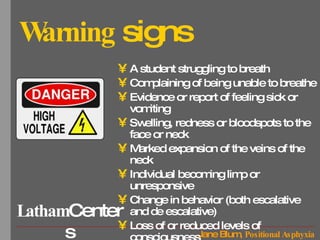
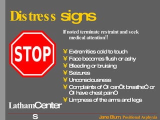
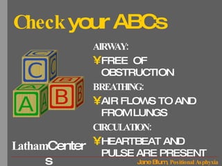
Positional   Asphyxia DEFINITION: “THE POSITION OF ONE’S OWN BODY INTERFERES WITH THE RESTRAINED PERSON’S ABILITY TO BREATHE AND THE PERSON CANNOT GET ENOUGH OXYGEN”
Positional   Asphyxia Oxygen is necessary for our muscles and organs to function Asphyxia is caused by depriving the muscles and organs of oxygen Oxygen enters the blood through the lungs and is pumped to the muscles and organs by the heart
Air can be prevented from entering the lungs by either obstructing the airway or stopping the chest wall from moving Positional   Asphyxia AIRFLOW TO LUNGS CHEST EXPANDS TO BRING IN AIR
General  risk factors Agitation Heart rate, blood pressure and rate of breathing increase As the physical struggle occurs, the student becomes out of breath Student has increased oxygen need to fuel  the muscles Student is placed in a face down position on the floor causing sompresssion of the chest and limiting ability to expand the chest cavity and breathe Abdominal organs pushed up Restricts movement of the diaphragm, further limiting available space for lungs to expand
General  risk factors Obesity puts additional stress on the body. In a prone position inhibits the person’s ability to properly contract the diaphragm and raise the ribs to enlarge the chest and inhale.  Influence of drugs profound effect on the respiratory and cardiovascular system.  Prolonged violent physical agitation state of oxygen deficiency, In addition the chest wall muscles which help the process of breathing (diaphragm) are fatigued. Underlying natural disease asthma, enlarged heart, high blood pressure, diabetes, increases risk, poor muscle tone (PWS) Hot humid environment
General  risk factors “ THE MORE AGITATED A PERSON IS THE LESS TIME IT WILL TAKE FOR SUFFOCATION TO OCCUR ”
Prader-Willi  risk factors Obesity more prone to obstructive apnea, pulmonary compromise, and diabetes.  High pain threshold Someone with PWS has high threshold and in addition may have difficulty localizing pain.  Increased risk of respiratory difficulties Hypotonia and  weak chest muscles Thick saliva complicates airway management Chronic stomach reflux and aspiration Temperature instability Idiopathic hyper and hypothermia have been reported. Fever may be present despite serious infection.  Bruise easily Anatomic and physiologic differences   such as : narrow airway, underdevelopment of the larynx, edema, hip dysplasia, and scoliosis
Prader-Willi  risk factors “ THE AVERAGE TIME BETWEEN BEGINNING A PRONE RESTRAINT AND THE  ONSET OF DEATH WAS ONLY 5.6 MINUTES”
Warning  signs A student struggling to breath Complaining of being unable to breathe Evidence or report of feeling sick or vomiting Swelling, redness or bloodspots to the face or neck Marked expansion of the veins of the neck Individual becoming limp or unresponsive Change in behavior (both escalative and de escalative) Loss of or reduced levels of consciousness Respiratory or cardiac arrest
Common  misconception If an individual can talk then they are able to breathe.  An individual dying from positional asphyxia may well be able to speak or shout prior to collapse MISCONCEPTION TRUTH
Common  misconception ALMOST ALL SUBJECTS WHO HAVE DIED DURING RESTRAINT HAVE ENGAGED IN EXTREME LEVELS OF PHYSICAL RESISTANCE AGAINST THE RESTRAINT FOR A PROLONGED PERIOD OF TIME.
Safety  points Never place weight or  pressure on chest , stomach or back Never put pressure on the neck or put the head in a position that  compresses the neck .  Never  restrain on a soft surface  (mattress) or place a pillow, towel under the head or over the mouth.  If student says “you are hurting me’ or “I can’t breathe” adjust hold and make sure you are not putting pressure or weight on the student. If any  signs of distress  end restraint and seek medical attention.  Never allow student to continue lying on the floor after restraint. Get the student up and  checked by medical staff .  Always keep the  arms close to the floor . Do not raise them up as that might cause pain and decrease ability to breathe.  In the small child, do not lean over or press forward on the back as this could critically  restrict ability to breathe .
Distress  signs If noted terminate restraint and seek medical attention!!  Extremities cold to touch Face becomes flush or ashy Bleeding or bruising Seizures Unconsciousness Complaints of “I can’t breathe” or “I have chest pain” Limpness of the arms and legs
Check  your ABCs AIRWAY:  FREE  OF OBSTRUCTION BREATHING:  AIR FLOWS TO AND FROM LUNGS CIRCULATION:  HEARTBEAT AND PULSE ARE PRESENT
Resources The Gathered View of PWSUSA “Advisory for Care Providers Exploring the Dangers of Positional Asphyxia,” Tina Didino, Mark K. Ziccardi Positional Asphyxia-pso.mhprisonservicd.gov.uk 7/7/2010 Positional Asphyxia-Wikipedia

Positional asphyxia

  • 1.
    Positional Asphyxia DEFINITION: “THE POSITION OF ONE’S OWN BODY INTERFERES WITH THE RESTRAINED PERSON’S ABILITY TO BREATHE AND THE PERSON CANNOT GET ENOUGH OXYGEN”
  • 2.
    Positional Asphyxia Oxygen is necessary for our muscles and organs to function Asphyxia is caused by depriving the muscles and organs of oxygen Oxygen enters the blood through the lungs and is pumped to the muscles and organs by the heart
  • 3.
    Air can beprevented from entering the lungs by either obstructing the airway or stopping the chest wall from moving Positional Asphyxia AIRFLOW TO LUNGS CHEST EXPANDS TO BRING IN AIR
  • 4.
    General riskfactors Agitation Heart rate, blood pressure and rate of breathing increase As the physical struggle occurs, the student becomes out of breath Student has increased oxygen need to fuel the muscles Student is placed in a face down position on the floor causing sompresssion of the chest and limiting ability to expand the chest cavity and breathe Abdominal organs pushed up Restricts movement of the diaphragm, further limiting available space for lungs to expand
  • 5.
    General riskfactors Obesity puts additional stress on the body. In a prone position inhibits the person’s ability to properly contract the diaphragm and raise the ribs to enlarge the chest and inhale. Influence of drugs profound effect on the respiratory and cardiovascular system. Prolonged violent physical agitation state of oxygen deficiency, In addition the chest wall muscles which help the process of breathing (diaphragm) are fatigued. Underlying natural disease asthma, enlarged heart, high blood pressure, diabetes, increases risk, poor muscle tone (PWS) Hot humid environment
  • 6.
    General riskfactors “ THE MORE AGITATED A PERSON IS THE LESS TIME IT WILL TAKE FOR SUFFOCATION TO OCCUR ”
  • 7.
    Prader-Willi riskfactors Obesity more prone to obstructive apnea, pulmonary compromise, and diabetes. High pain threshold Someone with PWS has high threshold and in addition may have difficulty localizing pain. Increased risk of respiratory difficulties Hypotonia and weak chest muscles Thick saliva complicates airway management Chronic stomach reflux and aspiration Temperature instability Idiopathic hyper and hypothermia have been reported. Fever may be present despite serious infection. Bruise easily Anatomic and physiologic differences such as : narrow airway, underdevelopment of the larynx, edema, hip dysplasia, and scoliosis
  • 8.
    Prader-Willi riskfactors “ THE AVERAGE TIME BETWEEN BEGINNING A PRONE RESTRAINT AND THE ONSET OF DEATH WAS ONLY 5.6 MINUTES”
  • 9.
    Warning signsA student struggling to breath Complaining of being unable to breathe Evidence or report of feeling sick or vomiting Swelling, redness or bloodspots to the face or neck Marked expansion of the veins of the neck Individual becoming limp or unresponsive Change in behavior (both escalative and de escalative) Loss of or reduced levels of consciousness Respiratory or cardiac arrest
  • 10.
    Common misconceptionIf an individual can talk then they are able to breathe. An individual dying from positional asphyxia may well be able to speak or shout prior to collapse MISCONCEPTION TRUTH
  • 11.
    Common misconceptionALMOST ALL SUBJECTS WHO HAVE DIED DURING RESTRAINT HAVE ENGAGED IN EXTREME LEVELS OF PHYSICAL RESISTANCE AGAINST THE RESTRAINT FOR A PROLONGED PERIOD OF TIME.
  • 12.
    Safety pointsNever place weight or pressure on chest , stomach or back Never put pressure on the neck or put the head in a position that compresses the neck . Never restrain on a soft surface (mattress) or place a pillow, towel under the head or over the mouth. If student says “you are hurting me’ or “I can’t breathe” adjust hold and make sure you are not putting pressure or weight on the student. If any signs of distress end restraint and seek medical attention. Never allow student to continue lying on the floor after restraint. Get the student up and checked by medical staff . Always keep the arms close to the floor . Do not raise them up as that might cause pain and decrease ability to breathe. In the small child, do not lean over or press forward on the back as this could critically restrict ability to breathe .
  • 13.
    Distress signsIf noted terminate restraint and seek medical attention!! Extremities cold to touch Face becomes flush or ashy Bleeding or bruising Seizures Unconsciousness Complaints of “I can’t breathe” or “I have chest pain” Limpness of the arms and legs
  • 14.
    Check yourABCs AIRWAY: FREE OF OBSTRUCTION BREATHING: AIR FLOWS TO AND FROM LUNGS CIRCULATION: HEARTBEAT AND PULSE ARE PRESENT
  • 15.
    Resources The GatheredView of PWSUSA “Advisory for Care Providers Exploring the Dangers of Positional Asphyxia,” Tina Didino, Mark K. Ziccardi Positional Asphyxia-pso.mhprisonservicd.gov.uk 7/7/2010 Positional Asphyxia-Wikipedia