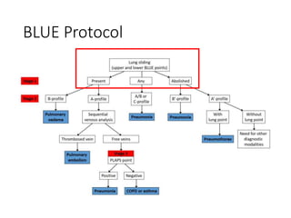
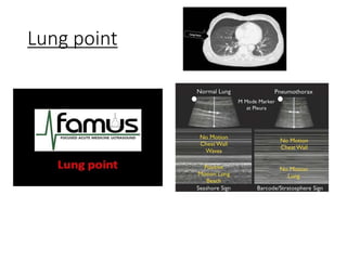
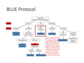
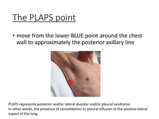
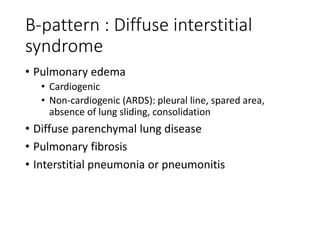
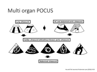
POCUS for dyspnea
Hye Won Kim
• 참고:
• https://www.youtube.com/watch?v=K2ZLjv8I2-U
• http://famus.org.uk/modules/blue-protocol
• Review: Clinically integrated multi-organ POCIS for
undiff respiratory difficulty, chest pain, or shock :
critical
• International evidence based rec for POCUS lung
Choice of probe
• individual choice
• curvilinear (abdominal probe)
• high frequency vascular probe
• greater resolution
• to initially determine the presence of lung sliding
• to see the fairly superficial pleural surface.
The BLUE points
BLUE Protocol
BLUE Protocol
Lung point
BLUE Protocol
non ventilating lung (absent
lung sliding, in this case due
to dense consolidation) with
evidence of increased lung
water/inflammation
evidenced by B lines
=> then the diagnosis is
pneumonia (B’ profile)
BLUE Protocol
if A lines are seen on one side
and B lines on the other, then
the diagnosis should be
pneumonia (the A/B profile).
Again, this makes sense; a
unilateral increase in lung
water/inflammation indicated
by B lines is only likely to be
caused by a local process (eg
pneumonia).The other
diagnosis that can be made at
this point is if anterior
consolidation is seen (C profile)
BLUE Protocol
A simplified version of the BLUE protocol (+sliding)
BLUE Protocol
The PLAPS point
• move from the lower BLUE point around the chest
wall to approximately the posterior axillary line
PLAPS represents posterior and/or lateral alveolar and/or pleural syndrome.
In other words, the presence of consolidation or pleural effusion at the postero-lateral
aspect of the lung.
Bat sign
Probe positions
B-pattern : Diffuse interstitial
syndrome
• Pulmonary edema
• Cardiogenic
• Non-cardiogenic (ARDS): pleural line, spared area,
absence of lung sliding, consolidation
• Diffuse parenchymal lung disease
• Pulmonary fibrosis
• Interstitial pneumonia or pneumonitis
Multi organ POCUS
Ha and Toh Journal of Intensive care (2016) 4:54
POCUS for dyspnea.pptx

POCUS for dyspnea.pptx

  • 1.
  • 2.
    • 참고: • https://www.youtube.com/watch?v=K2ZLjv8I2-U •http://famus.org.uk/modules/blue-protocol • Review: Clinically integrated multi-organ POCIS for undiff respiratory difficulty, chest pain, or shock : critical • International evidence based rec for POCUS lung
  • 3.
    Choice of probe •individual choice • curvilinear (abdominal probe) • high frequency vascular probe • greater resolution • to initially determine the presence of lung sliding • to see the fairly superficial pleural surface.
  • 4.
  • 5.
  • 7.
  • 8.
  • 9.
    BLUE Protocol non ventilatinglung (absent lung sliding, in this case due to dense consolidation) with evidence of increased lung water/inflammation evidenced by B lines => then the diagnosis is pneumonia (B’ profile)
  • 10.
    BLUE Protocol if Alines are seen on one side and B lines on the other, then the diagnosis should be pneumonia (the A/B profile). Again, this makes sense; a unilateral increase in lung water/inflammation indicated by B lines is only likely to be caused by a local process (eg pneumonia).The other diagnosis that can be made at this point is if anterior consolidation is seen (C profile)
  • 11.
  • 12.
    A simplified versionof the BLUE protocol (+sliding)
  • 13.
  • 14.
    The PLAPS point •move from the lower BLUE point around the chest wall to approximately the posterior axillary line PLAPS represents posterior and/or lateral alveolar and/or pleural syndrome. In other words, the presence of consolidation or pleural effusion at the postero-lateral aspect of the lung.
  • 15.
  • 16.
  • 17.
    B-pattern : Diffuseinterstitial syndrome • Pulmonary edema • Cardiogenic • Non-cardiogenic (ARDS): pleural line, spared area, absence of lung sliding, consolidation • Diffuse parenchymal lung disease • Pulmonary fibrosis • Interstitial pneumonia or pneumonitis
  • 19.
    Multi organ POCUS Haand Toh Journal of Intensive care (2016) 4:54

Editor's Notes

  • #20 Ha and Toh Journal of Intensive care (2016) 4:54