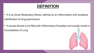
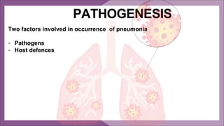
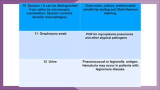
PNEUMONIA
DEFINITION
• It is an Acute Respiratory Illness, defined as an inflammation with exudative
solidification of lung parenchyma.
• It causes Alveoli to be filled with Inflammatory Exudates and usually results in
Consolidation of Lung
• Pathological Definition: Infection of the Alveoli, Distal airway, and
Interstitium of the Lung. Characterized by replacement of normal sponginess
by the Consolidation. Alveoli filled by WBC,RBC and Fibrin
• Clinical Definition: Constellation of Signs and Symptoms (fever, chills,
cough, pleural chest pain, sputum, bronchial breathing, crackles, wheeze)
with at least one opacity on chest X-Ray PA view.
CLASSIFICATION
Pneumonias can be classified in different ways:
1) Classification depending on the anatomic distribution:
A) Lobar Pneumonias
B) Bronchopneumonia
C) Interstitial Pneumonia
2) Etiological Classification:
A) Primary Pneumonia
B) Secondary Pneumonia
C) Suppurative Pneumonia
3) Clinical Setting in which infection occurs (if no pathogen is detected):
A) Community Acquired Pneumonia
B) Nosocomial or Hospital Acquired Pnemonia
C) Pneumonia in Immuno compromised Host
D) Health Care Associated Pneumonia
4) Non Infective Pneumonias:
A) Lipid/Lipoid Pneumonia
B) Radiation Pneumonitis
C) Chemical Pneumonitis
AETIOLOGY
AETIOLOGICAL AGENTS
Bacterial
Non-Bacterial
- Viral
- Fungal
Bacterial causes
They gave been classified under
– Typical (85%): Streptococcus pneumoniae, Haemophilus influenzae,
Staphylococcus aureus, Moraxella catarrhalis
– Atypical (15%): Legionella sp., Mycoplasma pneumoniae, Chlamydia
pneumoniae
Patients age is a differentiating factor. Young adults are more prone to
atypical causes, and very young and older people are predisposed to
typical causes.
Viral Causes
Viral species colonize the nasopharynx of patients with CAP
Whether they are primary cause or contribute to pathogenesis by secondary
bacterial causes
Common viruses associated with CAP include
-Influenza virus,
-Respiratory syncytial virus,
-Parainfluenza and
-Adenoviruses.
Fungal Causes
- In predisposing immunocompromised states like HIV and organ transplant
recipients. Generally by opportunistic group.
Commonly involves
-Aspergillus spp.
-Candida spp.
-Coccidiodes immitis etc
However, often overlooked, some fungal species can cause pneumonia in
immunocompetent individuals for eg Blastomyces, Coccidioides infections
which results in delay in diagnosis and unfavourable prognosis.
Age >65 years
HIV/immunocompromised
Smoking
Recent antibiotic therapy/resistance to antibiotics
Asthma
Cardiovascular Diseases ( CAD, CHF)
Chronic Obstructive Pulmonary Disease (COPD)
Chronic Renal Or Liver Disease
Diabetes Mellitus
Risk Factors
Two factors involved in occurrence of pneumonia
- Pathogens
- Host defences
PATHOGENESIS
The pulmonary defense mechanisms such as
- filtration of pathogens in the upper airways
-cough reflex
-mucociliary clearance
-immune system (humoral and cellular)
They normally prevent development of lower respiratory tract
infections following aspiration of oropharyngeal secretions containing
bacteria or inhalation of infected aerosols .
PATHOGENESIS
Due to defect of one or more of the above mentioned mechanisms, a
foreign pathogen gets access and reaches the alveoli.
Here the resident macrophages serve to protect the lung from foreign
pathogens
The inflammatory reaction triggered by these very macrophages is
what is responsible for histopathological and clinical findings seen in
pneumonia.
Macrophages engulf the foreign
pathogens
Trigger cytokines like TNF-a, IL-8, IL-1
Recruit inflammatory cells like
neutrophils to the site of infection.
Presents these antigens to T cells that
trigger both cellular and humoral
defences, activate complement system
Antibodies against those pathogens.
Causes inflammation of lung
parenchyma
Leaky capillaries
Exudative congestion
PATHOLOGICAL STATES OF PNEUMONIA
1. STAGE OF CONGESTION: INITIAL PHASE
It represents the early acute inflammatory response to bacterial infection that lasts for
1 to 2 days.
In this stage there is just congestion of the vessels without alveolar exudation.
Fine crepitations may be heard
2. RED HEPATISATION : EARLY CONSOLIDATION
This phase lasts for 2 to 4 days. The term hepatisation in pneumonia refers to liver-like
consistency of the affected lobe on cut section.
Intra alveolar exudation especially with RBCs.
Tubular bronchial breathing heard.
3. GREY HEPATISATION:LATE CONSOLIDATION
This phase lasts for 4-10 days
The exudation is of mainly WBCs with minimal RBCs.
Tubular bronchial breathing heard.
4. STAGE OF RESOLUTION
This stage begins by 8th to 9th day if no chemotherapy is administered and is
completed in 1-3 weeks. However antibiotic therapy induces resolution on about
third day. Resolution proceeds in a progressive manner.
The exudate is absorbed or removed by macrophages and proteolytic enzymes.
Coarse crepitations is heard.
Clinical Features
History
• Classical features of CAP are sudden onset of rigors followed by fever, pleuritic
chest pain, cough productive of purulent sputum and haemoptysis.
• These symptoms may be absent in elderly patients who present with confusion.
• Patients with atypical pneumonia may have a dry cough. These patients often
have extrapulmonary features that include myalgias, arthralgias, prominent
headache, mental confusion, abdominal pain and diarrhoea . Haemoptysis is
uncommon in atypical CAP.
Examination
• Approximately 80% are febrile, although this finding is frequently absent in
older patients.
• Respiratory rate is high and this may be the most sensitive sign in the elderly.
• Tachycardia is common. However, relative bradycardia is a typical feature of
Legionnaire's disease.
• Chest examination reveals crepitations in the involved area. About one-third
have bronchial breathing
Investigations in community
acquired pneumonia
INVESTIGATIONS
1 Complete blood count
very high or low
neutrophilc leukocytosis
Hemolytic anemia
Marker of severity
Suggets bacterial pneumonia
Occasionally complicates
mycoplasma.
2 Urea and Electrolytes
Urea more than 7mmol/l
hyponatremia
Marker of severity
Marker of severity, may occur in
patients with legionnairs disease
3 Liver function test.
Raised bilirubin
Hypoalbuminemia
When basal pneumonia inflames
liver or in atypical pneumonia.
Marker of severity
4 Erythrocyte sedimentation rate /
C reactive protein.
Non specifically elevated
5 Blood culture Bacteremia is a marker of severity.
Causative organism may be grown. Blood
cultures are recommended in hospitalized
patients .
6 Serological and antigen detection test. Pneumococcal antigen can be detected in
serum or urine in pneumococcal
pneumonia.
Acute and convalescent titers for
mycoplasma, chlamydia, legionella and
viral infections.
7 Cold agglutinins. Positive in 50% of patients with
mycoplasma.
8 Arterial Blood gases Measure when SaO2 less than 93% or
when severe clinical features to assess
ventilatory failure or acidosis .
9 HIV Testing Since pneumonia is common in
previously undiagnosed HIV infection, a
test should be offered to all patients
with pneumonia.
10 Sputum ( it can be distinguished
from saliva by microscopic
examination. Sputum contains
alveolar macrophages)
Gram stain, culture, antimicrobial
sensitivity testing and Ziehl Neelson
staining.
11 Oropharynx swab
12 Urine Pneumococcal or legionella antigen.
Hematuria may occur in patients with
legionners disease.
PCR for mycoplasma pneumonia
and other atypical pathogens
13 Chest X ray Essential for the confirmation of
diagnosis, follow up and detection of
complications like parapneumonic
effusion and empyema.
14 Aspiration Percutaneous transtracheal aspiration of
secretions
Percutaneous transthoracic needle
aspiration, preferable under CT guidance.
15 Fiberoptic bronchoscopy with BAL
and brushings
Gram stain, AFB stain, culture and
cytology.
16 Biopsy A transbronchial biopsy of lung tissue
for culture and histopathology may be
done in selected cases . Diagnostic
open lung biopsy , which carries a
high risk, is reserved for selected
patients .
17 Pleural fluid Aspirate and culture when present in
more than trivial amounts, preferably
with ultrasound guidance.
TREATMENT
CURB-65 Score
Ø It is a severity score for CAP, comprising of 5 variables, attributing 1 point for each
component.
Ø Estimates the mortality in community-acquired pneumonia to help determine the
outpatient vs inpatient treatment and the need to hospitalize a patient.
Oxygen Therapy
• Oxygen should be administered to all patients with tachypnoea,
hypoxemia, hypotension or acidosis with an aim of maintaining PaO2 >
60 mm Hg or SaO2 > 92%.
• In patients with known COPD, higher concentrations (>35%),
preferably humified oxygen should be administered.
• Continuous positive airway pressure(CPAP) should be considered in
those who remain hypoxic despite high-concentration oxygen therapy.
These patient should be managed in an intensive care environment
where mechanical ventilation can be rapidly employed.
Fluid Balance
Intravenous fluids should be considered in those with –
• Severe illness
• Older patients
• Those with vomiting.
It may be appropriate to discontinue hypertensive agents, temporarily .
And adequate oral intake of fluid should be encouraged.
Treatment of Pleural Pain
Important to relieve pleural pain in order to allow patient to breathe
normally and cough efficiently.
For the majority, simple analgesics like paracetamol, co-codamol or
NSAIDs is sufficient.
Antibiotic Therapy
• Prompt administration of antibiotic improves the outcome.
• The initial choice of antibiotic is guided by clinical context, local knowledge of antibiotic
resistant patterns and epidemiological information, e.g. during a mycoplasma epidemic.
• In most patient with uncomplicated pneumonia, a 5-day course is adequate; although
treatment for longer duration is requiredin patients with Legionella, Staphylococcal or
Klebsiella pneumonia.
Outpatients: Macrolides (eg. Clarithromycin 500mg oral twice daily for 10 days) OR
Doxycycline 100 mg twice daily for 10 days.
Inpatients, non-ICU: A respiratory Fluoroquinolones (Moxifloxacin 400 mg oral,
Gemifloxacin 320 mg oral four times a day) PLUS
A beta-lactam(cefotaxime 1-2 gm i.v , ceftriaxome 1-2gm i.v) PLUS
a Macrolide(dose same as in outpatients)
Inpatients, ICU: A beta-lactam (cefotaxime 1-2 gm i.v, ampicillin-sulbactam 2gm iv) PLUS
Azithromycin or Fluoroquinolones( as listed above for inpatients, Non-ICU)
Complications of pneumonia
• Para-pneumonic pleural effusion- common
• Empyema
• Retention of sputum causing lobar collapse
• DVT and pulmonary embolism
• Pneumothorax, particularly with Staph. Aureus
• Lung abscess
• ARDS, renal failure
• Ectopic abscess formation (Staph. aureus)
• Pyrexia due to drug hypersensitivity
HOSPITAL ACQUIRED PNEUMONIA
• Hospital-acquired or nosocomial pneumonia refers to
pneumonia that occurs at least 48 hours after admission to
hospital and not incubating at the time of admission.
• It is the second most common hospital-acquired infection (HAI)
and the leading cause of HAI associated death.
Organisms Causing HAP
Majority of hospital-acquired infections are caused by gram-
negative bacteria. These are E.coli, Klebsiella and Acineto bacter
species.
Other organisms include Staph. aureus including MRSA forms
and anaerobic organisms.
CLASSIFICATION
• VAP: Ventilator Associated Pneumonia
Pneumonia that develops after 48 hrs of mechanical ventilation and not
incubating at the time intubation.
• HCAP: Health Care Associated Pneumonia.
Health-care-associated pneumonia (HCAP) refers to the development of
pneumonia in a person who has spent at least 2 days in hospital within the last 90
days, or has attended a hemodialysis unit, or received intravenous antibiotics, or
been resident in a nursing home or other long-term care facility.
RISK FACTOR
• Colonization with potential pathogens
Decreased gastric acidity due to use of H-2 blockers and proton pump inhibitors.
Antimicrobial therapy.
Contaminated ventilator circuits or equipment.
• Aspiration of oropharyngeal contents into lower respiratory tract -
Intubation/mechanical ventilation.
• Reduced host defenses :Corticosteroid treatment, diabetes, malignancy.
• Bacteremia.
CLINICAL FEATURES
• THE DIAGNOSIS SHOULD BE CONSIDERED IN ANY HOSPITALISED OR
VENTILATED PATIENT WHO DEVELOPS :
1. Fever, leukocytosis, increase in respiratory secretions, and signs of pulmonary
consolidation on physical examination, along with a new or changing radiographic
infiltrate.
2. Other clinical features may include tachypnea, tachycardia, worsening
oxygenation, and increased minute ventilation if the patient is on ventilator.
INVESTIGATION
• Complete blood count shows increase in leukocyte count.
• Blood cultures.
• Sputum culture and Gram staining.
• Endotracheal aspirate can yield good uncontaminated sample for Gram's stain and
culture.
• Chest X-ray shows a new or changing pulmonary infiltrate.
• CT of the chest if necessary.
• Bronchoscopy may be required sometimes.
MANAGEMENT
• The principles of management are similar to those of CAP. focusing on adequate
oxygenation, appropriate fluid balance and antibiotics.
• The organisms implicated in early-onset HAP (occurring within 4-5 days of
admission) are similar to those involved in CAP.
• In patients who have received no previous antibiotics, co-amoxiclav or cefuroxime
represents a sensible choice. If the patient has received a course of recent
antibiotics, then piperacillin/tazobactam or a third-generation cephalosporin should be
considered.
• Late onset HAP is mostly caused by Gram negative organisms
• Pseudomonas and other gram-negative organisms should be chosen
-Piperacillin/tazobactam
- Cefepime
- Levofloxacin
- Imipenem
- Meropenem .
• If MRSA is highly prevalent in the institution and the patient is at risk for MRSA
infection, add vancomycin or linezolid.
• Aspiration pneumonia can be treated with amoxicillin clavulanic acid 1.2 g 8-hourly
plus metronidazole 500 mg 8-hourly.
• ▪ Physiotherapy for immobile and elderly, and to teach coughing techniques.
PREVENTION
Despite appropriate management, the mortality from HAP is high (30%)
• Healthcare providers must adhere strictly to hand-washing protocol.
• Steps should be taken to minimize the chances of aspiration and to limit the use of
stress ulcer prophylaxis with proton pump inhibitors.
• Oral antiseptic (chlorhexidine 2%) may be used to decontaminate the upper
airway.
• Incentive spirometry is recommended to prevent post-operative pneumonia.
• Use of kinetic bed.
Kinetic bed
Suppurative pneumonia
● Type of pneumonic consolidation characterised by destruction of lung
parenchyma
● Histologically it is characterized by presence of micro abscess
Etiology-
INFECTIOUS
Bacteria-mouth flora anaerobes (fusobacterium, peptostreptococcus),
streptococcus aureus
Fungi- aspergillus species
Parasite-entamoeba histolytica
NON INFECTIOUS
Neoplasm, septic emboli, pulmonary infection
Aspiration pneumonia
Defintion-Aspiration pneumonia develops due to abnormal entry of fluid into the lower
airway
Predisposing factors
● Aspiration of gastric content into the lungs: reduced consciousness
● Mechanical disruption of glottis closure
● Disorders of upper GI tract: esophageal diseases
● Feeding gastrostomy
Types
1. Chemical aspiration pneumonia
2. Bacterial aspiration pneumonia
Chemical aspiration pneumonia
● Here the pneumonia develops due to aspiration of substances which are toxic to
the lower Airways independent of bacterial infection
● Mendelson syndrome
C/f-dyspnea,low grade fever
O/E-cyanosis can be appreciated
Diffuse crepitation can be heard
Chest radiograph
Treatment
● Tracheal suction
● Mechanical ventilation may be
needed
● Antibiotics are used in acute events
Bacterial aspiration pneumonia
RF-patience with depressed gag reflex, nasogastric tube, Endotracheal tube
Etiology-fusobacterium, peptostreptococcus
C/F-depends on host immunity status and the virulence of the organism
Cough, purulent sputum, Dyspnea, fever
Underlying Periodontal disease maybe present in few cases
Diagnosis-sputum-putrid discharge is diagnostic of anaerobic infection
chest x-ray-involvment of dependent pulmonary segments by aspiration
Recumbent position-superior segment of lower lobe/posterior segment of upper lobe
Erect position-basal segment of lower lobe
TREATMENT-antibiotics are the main stay
Penicillin
Clindamycin-DOC for anaerobic infection above diaphragm
Newer drugs-levofloxacin,gemifloxacin
Pneumonia in
Immunocompromised patient
INTRODUCTION
Patients immunocompromised by drugs or a disease like HIV are at
increased risk of pulmonary infection and pneumonia is the most common
cause of death in this group.
The majority of infections are caused by same pathogens that causes
pneumonia in immunocompetent individuals, but in the patients with more
profound immunosuppression less common organisms or those normally
considered to be more virulence or non pathogenic becomes opportunistic
pathogens.
CLINICAL FEATURES
Fever
Cough Breathlessness
But these symptoms are influenced by the degree of immunosuppression
and the presentation may be less specific in the more profoundly
immunosuppressed
The onset of symptoms tends to be swift in those with bacterial infections but
more gradually in patients with oppornutistic organisms such as pnuemocystis
jirovecii and mycobacterial infections .
MANAGEMENT
INVESTIGATION-
The approach is informed by the clinical context and severity of the
Illness The invasive investigations such as:
Bronchoscopy BAL
Transbroncial biopsy Surgical
lung biopsy
The above mentioned investigations are often impractical as many
patients are too ill to undergo this safely however,
Induced sputum offers a relatively safe method of obtaining
microbiological samples
HRCT can be helpful in diagnosing these cases
TREATMENT
• It should be based on established etiological diagnosis, in practice however,
the causative agent is frequently unknown
• Factors that favour bacteriological etiology that includes neutropenia, rapid
onset and deterioation in these circumstances broad spectrum antibiotic
therapy should be commenced immediately eg-third generation cephalosporin
or quinolone plus an anti staphylococcal antibiotic or an anti pseudomonal
penicillin plus an amino glycoside
• Thereafter treatment may be tailrode according to the result of investigation
and clinical response
PNEUMONIA.pdf

PNEUMONIA.pdf

  • 1.
  • 2.
    DEFINITION • It isan Acute Respiratory Illness, defined as an inflammation with exudative solidification of lung parenchyma. • It causes Alveoli to be filled with Inflammatory Exudates and usually results in Consolidation of Lung
  • 3.
    • Pathological Definition:Infection of the Alveoli, Distal airway, and Interstitium of the Lung. Characterized by replacement of normal sponginess by the Consolidation. Alveoli filled by WBC,RBC and Fibrin • Clinical Definition: Constellation of Signs and Symptoms (fever, chills, cough, pleural chest pain, sputum, bronchial breathing, crackles, wheeze) with at least one opacity on chest X-Ray PA view.
  • 4.
    CLASSIFICATION Pneumonias can beclassified in different ways: 1) Classification depending on the anatomic distribution: A) Lobar Pneumonias B) Bronchopneumonia C) Interstitial Pneumonia
  • 5.
    2) Etiological Classification: A)Primary Pneumonia B) Secondary Pneumonia C) Suppurative Pneumonia
  • 6.
    3) Clinical Settingin which infection occurs (if no pathogen is detected): A) Community Acquired Pneumonia B) Nosocomial or Hospital Acquired Pnemonia C) Pneumonia in Immuno compromised Host D) Health Care Associated Pneumonia
  • 7.
    4) Non InfectivePneumonias: A) Lipid/Lipoid Pneumonia B) Radiation Pneumonitis C) Chemical Pneumonitis
  • 8.
  • 9.
  • 10.
    Bacterial causes They gavebeen classified under – Typical (85%): Streptococcus pneumoniae, Haemophilus influenzae, Staphylococcus aureus, Moraxella catarrhalis – Atypical (15%): Legionella sp., Mycoplasma pneumoniae, Chlamydia pneumoniae Patients age is a differentiating factor. Young adults are more prone to atypical causes, and very young and older people are predisposed to typical causes.
  • 11.
    Viral Causes Viral speciescolonize the nasopharynx of patients with CAP Whether they are primary cause or contribute to pathogenesis by secondary bacterial causes Common viruses associated with CAP include -Influenza virus, -Respiratory syncytial virus, -Parainfluenza and -Adenoviruses.
  • 12.
    Fungal Causes - Inpredisposing immunocompromised states like HIV and organ transplant recipients. Generally by opportunistic group. Commonly involves -Aspergillus spp. -Candida spp. -Coccidiodes immitis etc However, often overlooked, some fungal species can cause pneumonia in immunocompetent individuals for eg Blastomyces, Coccidioides infections which results in delay in diagnosis and unfavourable prognosis.
  • 13.
    Age >65 years HIV/immunocompromised Smoking Recentantibiotic therapy/resistance to antibiotics Asthma Cardiovascular Diseases ( CAD, CHF) Chronic Obstructive Pulmonary Disease (COPD) Chronic Renal Or Liver Disease Diabetes Mellitus Risk Factors
  • 14.
    Two factors involvedin occurrence of pneumonia - Pathogens - Host defences PATHOGENESIS
  • 15.
    The pulmonary defensemechanisms such as - filtration of pathogens in the upper airways -cough reflex -mucociliary clearance -immune system (humoral and cellular) They normally prevent development of lower respiratory tract infections following aspiration of oropharyngeal secretions containing bacteria or inhalation of infected aerosols . PATHOGENESIS
  • 16.
    Due to defectof one or more of the above mentioned mechanisms, a foreign pathogen gets access and reaches the alveoli. Here the resident macrophages serve to protect the lung from foreign pathogens The inflammatory reaction triggered by these very macrophages is what is responsible for histopathological and clinical findings seen in pneumonia.
  • 18.
    Macrophages engulf theforeign pathogens Trigger cytokines like TNF-a, IL-8, IL-1 Recruit inflammatory cells like neutrophils to the site of infection. Presents these antigens to T cells that trigger both cellular and humoral defences, activate complement system Antibodies against those pathogens. Causes inflammation of lung parenchyma Leaky capillaries Exudative congestion
  • 19.
    PATHOLOGICAL STATES OFPNEUMONIA 1. STAGE OF CONGESTION: INITIAL PHASE It represents the early acute inflammatory response to bacterial infection that lasts for 1 to 2 days. In this stage there is just congestion of the vessels without alveolar exudation. Fine crepitations may be heard 2. RED HEPATISATION : EARLY CONSOLIDATION This phase lasts for 2 to 4 days. The term hepatisation in pneumonia refers to liver-like consistency of the affected lobe on cut section. Intra alveolar exudation especially with RBCs. Tubular bronchial breathing heard.
  • 20.
    3. GREY HEPATISATION:LATECONSOLIDATION This phase lasts for 4-10 days The exudation is of mainly WBCs with minimal RBCs. Tubular bronchial breathing heard. 4. STAGE OF RESOLUTION This stage begins by 8th to 9th day if no chemotherapy is administered and is completed in 1-3 weeks. However antibiotic therapy induces resolution on about third day. Resolution proceeds in a progressive manner. The exudate is absorbed or removed by macrophages and proteolytic enzymes. Coarse crepitations is heard.
  • 21.
    Clinical Features History • Classicalfeatures of CAP are sudden onset of rigors followed by fever, pleuritic chest pain, cough productive of purulent sputum and haemoptysis. • These symptoms may be absent in elderly patients who present with confusion. • Patients with atypical pneumonia may have a dry cough. These patients often have extrapulmonary features that include myalgias, arthralgias, prominent headache, mental confusion, abdominal pain and diarrhoea . Haemoptysis is uncommon in atypical CAP.
  • 22.
    Examination • Approximately 80%are febrile, although this finding is frequently absent in older patients. • Respiratory rate is high and this may be the most sensitive sign in the elderly. • Tachycardia is common. However, relative bradycardia is a typical feature of Legionnaire's disease. • Chest examination reveals crepitations in the involved area. About one-third have bronchial breathing
  • 23.
  • 24.
    INVESTIGATIONS 1 Complete bloodcount very high or low neutrophilc leukocytosis Hemolytic anemia Marker of severity Suggets bacterial pneumonia Occasionally complicates mycoplasma. 2 Urea and Electrolytes Urea more than 7mmol/l hyponatremia Marker of severity Marker of severity, may occur in patients with legionnairs disease 3 Liver function test. Raised bilirubin Hypoalbuminemia When basal pneumonia inflames liver or in atypical pneumonia. Marker of severity
  • 25.
    4 Erythrocyte sedimentationrate / C reactive protein. Non specifically elevated 5 Blood culture Bacteremia is a marker of severity. Causative organism may be grown. Blood cultures are recommended in hospitalized patients . 6 Serological and antigen detection test. Pneumococcal antigen can be detected in serum or urine in pneumococcal pneumonia. Acute and convalescent titers for mycoplasma, chlamydia, legionella and viral infections.
  • 26.
    7 Cold agglutinins.Positive in 50% of patients with mycoplasma. 8 Arterial Blood gases Measure when SaO2 less than 93% or when severe clinical features to assess ventilatory failure or acidosis . 9 HIV Testing Since pneumonia is common in previously undiagnosed HIV infection, a test should be offered to all patients with pneumonia.
  • 27.
    10 Sputum (it can be distinguished from saliva by microscopic examination. Sputum contains alveolar macrophages) Gram stain, culture, antimicrobial sensitivity testing and Ziehl Neelson staining. 11 Oropharynx swab 12 Urine Pneumococcal or legionella antigen. Hematuria may occur in patients with legionners disease. PCR for mycoplasma pneumonia and other atypical pathogens
  • 28.
    13 Chest Xray Essential for the confirmation of diagnosis, follow up and detection of complications like parapneumonic effusion and empyema. 14 Aspiration Percutaneous transtracheal aspiration of secretions Percutaneous transthoracic needle aspiration, preferable under CT guidance. 15 Fiberoptic bronchoscopy with BAL and brushings Gram stain, AFB stain, culture and cytology.
  • 29.
    16 Biopsy Atransbronchial biopsy of lung tissue for culture and histopathology may be done in selected cases . Diagnostic open lung biopsy , which carries a high risk, is reserved for selected patients . 17 Pleural fluid Aspirate and culture when present in more than trivial amounts, preferably with ultrasound guidance.
  • 31.
  • 32.
    CURB-65 Score Ø Itis a severity score for CAP, comprising of 5 variables, attributing 1 point for each component. Ø Estimates the mortality in community-acquired pneumonia to help determine the outpatient vs inpatient treatment and the need to hospitalize a patient.
  • 33.
    Oxygen Therapy • Oxygenshould be administered to all patients with tachypnoea, hypoxemia, hypotension or acidosis with an aim of maintaining PaO2 > 60 mm Hg or SaO2 > 92%. • In patients with known COPD, higher concentrations (>35%), preferably humified oxygen should be administered. • Continuous positive airway pressure(CPAP) should be considered in those who remain hypoxic despite high-concentration oxygen therapy. These patient should be managed in an intensive care environment where mechanical ventilation can be rapidly employed.
  • 34.
    Fluid Balance Intravenous fluidsshould be considered in those with – • Severe illness • Older patients • Those with vomiting. It may be appropriate to discontinue hypertensive agents, temporarily . And adequate oral intake of fluid should be encouraged. Treatment of Pleural Pain Important to relieve pleural pain in order to allow patient to breathe normally and cough efficiently. For the majority, simple analgesics like paracetamol, co-codamol or NSAIDs is sufficient.
  • 35.
    Antibiotic Therapy • Promptadministration of antibiotic improves the outcome. • The initial choice of antibiotic is guided by clinical context, local knowledge of antibiotic resistant patterns and epidemiological information, e.g. during a mycoplasma epidemic. • In most patient with uncomplicated pneumonia, a 5-day course is adequate; although treatment for longer duration is requiredin patients with Legionella, Staphylococcal or Klebsiella pneumonia. Outpatients: Macrolides (eg. Clarithromycin 500mg oral twice daily for 10 days) OR Doxycycline 100 mg twice daily for 10 days. Inpatients, non-ICU: A respiratory Fluoroquinolones (Moxifloxacin 400 mg oral, Gemifloxacin 320 mg oral four times a day) PLUS A beta-lactam(cefotaxime 1-2 gm i.v , ceftriaxome 1-2gm i.v) PLUS a Macrolide(dose same as in outpatients) Inpatients, ICU: A beta-lactam (cefotaxime 1-2 gm i.v, ampicillin-sulbactam 2gm iv) PLUS Azithromycin or Fluoroquinolones( as listed above for inpatients, Non-ICU)
  • 36.
    Complications of pneumonia •Para-pneumonic pleural effusion- common • Empyema • Retention of sputum causing lobar collapse • DVT and pulmonary embolism • Pneumothorax, particularly with Staph. Aureus • Lung abscess • ARDS, renal failure • Ectopic abscess formation (Staph. aureus) • Pyrexia due to drug hypersensitivity
  • 37.
    HOSPITAL ACQUIRED PNEUMONIA •Hospital-acquired or nosocomial pneumonia refers to pneumonia that occurs at least 48 hours after admission to hospital and not incubating at the time of admission. • It is the second most common hospital-acquired infection (HAI) and the leading cause of HAI associated death. Organisms Causing HAP Majority of hospital-acquired infections are caused by gram- negative bacteria. These are E.coli, Klebsiella and Acineto bacter species. Other organisms include Staph. aureus including MRSA forms and anaerobic organisms.
  • 38.
    CLASSIFICATION • VAP: VentilatorAssociated Pneumonia Pneumonia that develops after 48 hrs of mechanical ventilation and not incubating at the time intubation. • HCAP: Health Care Associated Pneumonia. Health-care-associated pneumonia (HCAP) refers to the development of pneumonia in a person who has spent at least 2 days in hospital within the last 90 days, or has attended a hemodialysis unit, or received intravenous antibiotics, or been resident in a nursing home or other long-term care facility.
  • 39.
    RISK FACTOR • Colonizationwith potential pathogens Decreased gastric acidity due to use of H-2 blockers and proton pump inhibitors. Antimicrobial therapy. Contaminated ventilator circuits or equipment. • Aspiration of oropharyngeal contents into lower respiratory tract - Intubation/mechanical ventilation. • Reduced host defenses :Corticosteroid treatment, diabetes, malignancy. • Bacteremia.
  • 40.
    CLINICAL FEATURES • THEDIAGNOSIS SHOULD BE CONSIDERED IN ANY HOSPITALISED OR VENTILATED PATIENT WHO DEVELOPS : 1. Fever, leukocytosis, increase in respiratory secretions, and signs of pulmonary consolidation on physical examination, along with a new or changing radiographic infiltrate. 2. Other clinical features may include tachypnea, tachycardia, worsening oxygenation, and increased minute ventilation if the patient is on ventilator.
  • 41.
    INVESTIGATION • Complete bloodcount shows increase in leukocyte count. • Blood cultures. • Sputum culture and Gram staining. • Endotracheal aspirate can yield good uncontaminated sample for Gram's stain and culture. • Chest X-ray shows a new or changing pulmonary infiltrate. • CT of the chest if necessary. • Bronchoscopy may be required sometimes.
  • 42.
    MANAGEMENT • The principlesof management are similar to those of CAP. focusing on adequate oxygenation, appropriate fluid balance and antibiotics. • The organisms implicated in early-onset HAP (occurring within 4-5 days of admission) are similar to those involved in CAP. • In patients who have received no previous antibiotics, co-amoxiclav or cefuroxime represents a sensible choice. If the patient has received a course of recent antibiotics, then piperacillin/tazobactam or a third-generation cephalosporin should be considered.
  • 43.
    • Late onsetHAP is mostly caused by Gram negative organisms • Pseudomonas and other gram-negative organisms should be chosen -Piperacillin/tazobactam - Cefepime - Levofloxacin - Imipenem - Meropenem . • If MRSA is highly prevalent in the institution and the patient is at risk for MRSA infection, add vancomycin or linezolid. • Aspiration pneumonia can be treated with amoxicillin clavulanic acid 1.2 g 8-hourly plus metronidazole 500 mg 8-hourly. • ▪ Physiotherapy for immobile and elderly, and to teach coughing techniques.
  • 44.
    PREVENTION Despite appropriate management,the mortality from HAP is high (30%) • Healthcare providers must adhere strictly to hand-washing protocol. • Steps should be taken to minimize the chances of aspiration and to limit the use of stress ulcer prophylaxis with proton pump inhibitors. • Oral antiseptic (chlorhexidine 2%) may be used to decontaminate the upper airway. • Incentive spirometry is recommended to prevent post-operative pneumonia. • Use of kinetic bed. Kinetic bed
  • 45.
    Suppurative pneumonia ● Typeof pneumonic consolidation characterised by destruction of lung parenchyma ● Histologically it is characterized by presence of micro abscess Etiology- INFECTIOUS Bacteria-mouth flora anaerobes (fusobacterium, peptostreptococcus), streptococcus aureus Fungi- aspergillus species Parasite-entamoeba histolytica NON INFECTIOUS Neoplasm, septic emboli, pulmonary infection
  • 46.
    Aspiration pneumonia Defintion-Aspiration pneumoniadevelops due to abnormal entry of fluid into the lower airway Predisposing factors ● Aspiration of gastric content into the lungs: reduced consciousness ● Mechanical disruption of glottis closure ● Disorders of upper GI tract: esophageal diseases ● Feeding gastrostomy Types 1. Chemical aspiration pneumonia 2. Bacterial aspiration pneumonia
  • 47.
    Chemical aspiration pneumonia ●Here the pneumonia develops due to aspiration of substances which are toxic to the lower Airways independent of bacterial infection ● Mendelson syndrome C/f-dyspnea,low grade fever O/E-cyanosis can be appreciated Diffuse crepitation can be heard
  • 48.
    Chest radiograph Treatment ● Trachealsuction ● Mechanical ventilation may be needed ● Antibiotics are used in acute events
  • 49.
    Bacterial aspiration pneumonia RF-patiencewith depressed gag reflex, nasogastric tube, Endotracheal tube Etiology-fusobacterium, peptostreptococcus C/F-depends on host immunity status and the virulence of the organism Cough, purulent sputum, Dyspnea, fever Underlying Periodontal disease maybe present in few cases Diagnosis-sputum-putrid discharge is diagnostic of anaerobic infection
  • 50.
    chest x-ray-involvment ofdependent pulmonary segments by aspiration Recumbent position-superior segment of lower lobe/posterior segment of upper lobe Erect position-basal segment of lower lobe TREATMENT-antibiotics are the main stay Penicillin Clindamycin-DOC for anaerobic infection above diaphragm Newer drugs-levofloxacin,gemifloxacin
  • 51.
  • 52.
    INTRODUCTION Patients immunocompromised bydrugs or a disease like HIV are at increased risk of pulmonary infection and pneumonia is the most common cause of death in this group. The majority of infections are caused by same pathogens that causes pneumonia in immunocompetent individuals, but in the patients with more profound immunosuppression less common organisms or those normally considered to be more virulence or non pathogenic becomes opportunistic pathogens.
  • 53.
    CLINICAL FEATURES Fever Cough Breathlessness Butthese symptoms are influenced by the degree of immunosuppression and the presentation may be less specific in the more profoundly immunosuppressed The onset of symptoms tends to be swift in those with bacterial infections but more gradually in patients with oppornutistic organisms such as pnuemocystis jirovecii and mycobacterial infections .
  • 54.
    MANAGEMENT INVESTIGATION- The approach isinformed by the clinical context and severity of the Illness The invasive investigations such as: Bronchoscopy BAL Transbroncial biopsy Surgical lung biopsy
  • 55.
    The above mentionedinvestigations are often impractical as many patients are too ill to undergo this safely however, Induced sputum offers a relatively safe method of obtaining microbiological samples HRCT can be helpful in diagnosing these cases
  • 56.
    TREATMENT • It shouldbe based on established etiological diagnosis, in practice however, the causative agent is frequently unknown • Factors that favour bacteriological etiology that includes neutropenia, rapid onset and deterioation in these circumstances broad spectrum antibiotic therapy should be commenced immediately eg-third generation cephalosporin or quinolone plus an anti staphylococcal antibiotic or an anti pseudomonal penicillin plus an amino glycoside • Thereafter treatment may be tailrode according to the result of investigation and clinical response