The document presents a method for diagnosing pneumonia from chest X-rays using a convolutional neural network (CNN) based on the VGG19 model, employing techniques such as transfer learning and data augmentation to address class imbalance. The study achieves a recall of 0.96 and precision of 0.87, emphasizing the effectiveness of machine learning in medical image diagnostics as an alternative due to the shortage of qualified radiologists. The methodology includes data pre-processing, modeling, and the use of a cloud-based platform for efficient processing of images.
![Diagnosing Pneumonia from Chest X-Rays Using Neural Networks
Tushar Dalvi Shantanu Deshpande Yash Iyangar Ashish Soni
x18134301 x18125514 x18124739 x18136664
Abstract—Disease diagnosis with radiology is a common prac-
tice in the medical domain but requires doctors to correctly
interpret the results from the images. Over the years due to
the increase in the number of patients and the low availability of
doctors, there was a need for a new method to identify and detect
disease in a patient. In recent years machine learning techniques
have been very effective in image-based disease diagnosis. In
this paper, a method has been proposed to diagnose Pneumonia
using transfer learning with VGG19 model. For pre-processing
online image, augmentation has been used to reduce the class
imbalance in the training data, With this methodology. For pre-
training the VGG19 model, Imagenet weights have been used
and three layers have been stripped off the model and 2 dense
layers have been added with Relu as an activation function in
one layer and softmax in the other layer. We were able to achieve
a recall of 0.96 and a precision of 0.87 with such a setting.
Index Terms—Pneumonia detection, CNN, Image Classification
I. INTRODUCTION
Radiology is one of the many branches of medicine that
basically focuses on making use of medical images for the
detection, diagnosis and characterization of the disease. One of
the main responsibilities of a Radiologist is to view a medical
image and based on the image producing a written report of
the observed findings [1]. Medical experts have made use of
this technique for around several years in order to visualize as
well as explore abnormalities and fractures in the body organs.
In recent years, due to the advancement in the technology
field, we have seen an up-gradation in the healthcare systems.
This has helped the doctors in better diagnosis accuracy and
reducing the fatality rate. The chest is the most important
part of the body as it contains the respiration organs which
are responsible for sustaining important life function of the
body. The count of people being diagnosed with a chest
disease globally is in millions. Some of the diseases that are
diagnosed using chest x-ray images are lung cancer, heart
diseases, Pneumonia, bronchitis, fractures etc.
Pneumonia is one such kind of chest disease wherein there
is an inflammation or infection in the lungs and it is most
commonly caused by a virus or a bacteria. Another cause due
to which a person can acquire Pneumonia is through inhaling
vomit or other foreign substances. If a person is diagnosed
with this disease, the lungs air sacs get filled with mucus,
pus, and few other liquids which do not allow the lungs to
function properly. This causes an obstruction in the path of
oxygen to reach the blood and the cells of the body in an
effective manner. Based on the data provided by the World
Health Organization (WHO), 2.4 million persons die every
year due to Pneumonia. [2]
One of the most tedious tasks for the radiologists is the
classification of X-ray abnormalities to detect and diagnose
chest diseases. As a result of this, several algorithms had
been proposed by researchers to perform this task accurately.
In these couple of decades, computer-aided diagnosis (CAD)
has been developed through various studies to interpret useful
information and assist doctors in getting a meaningful insight
about an X-ray. Several research works were carried out in
past by using artificial intelligence methodologies. Based on
the learnings from artificial intelligence methodologies, we can
also develop an image processing system that will use the X-
ray images and will detect the diseases in the primary stages
with improved accuracy. Some of the neural network method-
ologies that have been effectively used by the researchers in
replacing the traditional pattern recognition methods for the
diagnosis of diseases are probabilistic neural network (PNN),
multi-layer neural network (MLNN), generalized regression
neural network (GRNN), learning vector quantization (LVQ),
and radial basis function. These deep neural networks have
showcased increased accuracy in terms of image classification
and have thereby motivated the researchers to explore more
artificial intelligent techniques.
The convolutional Neural Network is another frequently
used deep learning architecture due to its power of extraction
of various different level features from the images. [3] By go-
ing through the relevant research studies, we hereby propose a
deep convolutional neural network (CNN) in order to improve
the accuracy of the diagnosis of chest disease, particularly
Pneumonia.
II. RESEARCH QUESTION
Can the Recall be improved for Chest X-ray Pneumonia
Detection using CNN based VGG19 model as compared to
the state of the art technique?
III. LITERATURE REVIEW
Owing to the difficulty in manually classifying the X-ray
images for a disease, various studies have evolved around the
use of Computer-aided diagnostics for obtaining meaningful
X-ray information and thereby assist physicians to gain a
quantitative insight of the disease. In this section, we will
review the pieces of literature that supports the current research
work.
In [4], the author examined three methods, Backpropagation
NN, Competitive NN, Convolutional NN. In BPNN, the errors
at output layers are propagated back to the network layer
whereas CpNN is a two-layer NN based on a supervised
algorithm. CNN has the strongest network as it can include
many hidden layers and performs convolutions and subsam-
pling in order to obtain low to a high level of features
from the input data. Also, it has three layers; convolutional](https://image.slidesharecdn.com/pneumoniadetectionreport-191023100300/75/Pneumonia-detection-using-CNN-1-2048.jpg)
![layer, subsampling/pooling layer, full connection layer. Results
showed that CNN took more time and number of learning
iterations however achieved the highest recognition rate and
was also able to get a better generalization power over BPNN
and CpNN.
In the above study, the output in terms of precision, com-
putation time was not as efficient due to no prior image
pre-processing performed. In [5], histogram equalization is
performed for image pre-processing and the images are divided
as well as normalized, further, cross-validation is conducted on
test data. The proposed model achieved an accuracy of 95.3%
on test data.
Further to the outstanding performance of CNN in [4],
another study [6] was conducted that evaluated performance
of three CNN architectures, Sequential CNN, Residual CNN
and Inception CNN. In the residual, loss of information is
prevented by passing information from earlier network layers
downstream thus solving the problem of representational bot-
tlenecks. Six residual blocks have been used and performance
measured based on following metrics, accuracy, precision,
AUC, specificity, recall. Non-linearity is introduced by using
a Rectified Linear Unit (ReLU) layer. The VGC16 model out-
performed all the other models. In [7], like [5], data has been
normalized by dividing all pixels by 255 and thus transformed
them to floating points. CNN model has compared with MLP
wherein each node except the input node is a neuron that
uses non-linear activation function. The technique involved
multiplying inputs by weight and then add them to bias and
dropout technique has been used to avoid overfitting and
reduce the training time. CNN outperformed MLP based on
evaluation metrics like cross-validation and confusion matrix.
A different approach can be seen in [8] where the network
layer utilizes the final layer as the convolutional implementa-
tion of a fully connected layer that allows a 40-fold speedup.
For dealing with imbalanced label distributions, a two-phase
training procedure has been proposed. Performance of three
different optimizers has been compared in [9], Adam opti-
mizer, momentum optimizer and stochastic gradient descent.
Filters are applied to the input image and the matrices thus
formed are known as feature maps. Filters of 5x5 size used
to extract features. In order to prevent overfitting, half of
the neurons are disabled using the dropout technique. Adams
optimizer outperformed the other two optimizers. Utilization
of ResNet-50 for embedding the features has been done in
[10] and CNN model thus built is able to get overall AUC of
0.816.
A different and novel approach is visible in [11] where
the author used Transfer Learning to train the model. The
advantage of TL is that a model can be trained even with
limited data. A dataset of 5232 images has been used and
the model achieved accuracy of 92.8% with sensitivity and
specificity of 93.2% and 90.1% respectively. In [12], the pre-
processing steps involved increasing the images and rotating
them by 40 degrees. By following a similar model building
process as [11], the author obtained the accuracy of 95.31%
and is better than [11].
Two different CNN techniques were used in [13] and [14],
the GoogleNet and AlexNet. During the pre-processing stage,
multiple rotations at 90, 180 and 270 degrees have been
performed and able to get AUC of 0.99. A rather different
approach than CNN can be seen in [15] where the author
used a technique called Multiple-instance learning (MIL). The
research overcomes some of the drawbacks like no proper
estimation of positive instances by reducing the number of
iterations. The slow configuration is avoided by avoiding the
reclassification and the AUC achieved is 0.86%. In [16],
the author tackled the issue of class imbalance using image
augmentation methods like rotating and flipping the images.
In the last dense layer, the sigmoid function is used to avoid
the problem of overfitting. Like [9], Adam optimizer has been
used to reduce losses and batch size is set to 64 whereas epoch
set to 100. The recall obtained is 96.7% and is better than state
of art techniques. A 121 layer convolutional neural network
has been trained on a large dataset of 100,000 X-Ray images
with 14 diseases in [17]. Weights initialized using pre-trained
weights from ImageNet and the model is trained using Adam.
An extension of the algorithm is undertaken to detect multiple
diseases and it is found that this model outperforms the state
of the art.
A deep neural network algorithm, Mask-RCNN is used
in [18]. This method uses lung opacity as a feature for
binary classification and Pneumonia detection. The final output
obtained is an ensemble of two models. The base network
is trained with COCO weights based on ResNet50 and
ResNet101 models to extract features from the images. An
improved result due to an ensemble approach is 0.21. In [19],
for extracting representative and discriminative features from
the Chest X-ray images for the effective classification into
various body parts, CNN’s were explored and the capability
of CNN in terms of capturing the image structure from feature
maps is portrayed and hand-engineered features were easily
outperformed by CNNs.
The usefulness of transfer learning [11] is further stated
in [20] where the authors used a pre-trained ChexNet model
with 121-dense layers and applied it in 4 blocks. The output
of pretrained model is transferred to the new model and Adam
optimizer is used. The model was trained on multiple blocks
and multiple layers and best validation accuracy obtained was
90.38% for the model with 6 layers. The use of demographic
features like age, weight and height is used in addition to
image features in [21] and the balanced dataset is used for
training. Five different algorithms have been used viz. In-
ceptionResNetV2, DenseNet121, InceptionV3, ResNet50 and
VGG19. The batch size used was 16 and Stochastic Gradient
Descent was chosen as optimizer function. VGG19 model
outperformed other models with AUC 0.9714 for training and
0.9213 for testing data.
Like [20] and [11], transfer learning is used in [22], how-
ever, in this study due to the high resolution of CXR images,
an extra convolution layer is added. Proposed system showed
enhanced screening performance. A multi-CNN model is used
in [23] which consists of three CNN components. The output](https://image.slidesharecdn.com/pneumoniadetectionreport-191023100300/75/Pneumonia-detection-using-CNN-2-2048.jpg)
![of each CNN component is a probability which classifies
the image as normal or abnormal. The outcome of all three
components is calculated by using the association fusion rule.
The fusion rule consists of eight cases which lead to the final
classification of the image. The model has been trained using
the association fusion rule for classification of the images.
Their model has achieved an accuracy of 96%.
IV. METHODOLOGY
From above Literature Review, we can say that in the medi-
cal domain it is hard for a layman to identify a disease by just
looking at the images of the body part. To identify disease from
x-rays, MRI Scans well-trained doctors or properly trained
computer-based system are required because a layman doesn’t
possess the knowledge of the disease like the doctor. Even
though we are building a model to detect Pneumonia we would
want our model to detect different diseases with an adequate
amount of training examples and model weights. So we will be
using the CRISP-DM methodology [24]. It stands for Cross
Industry Standard Process for Data Mining. It has six steps
out of which we will be using Business Understanding, Data
Understanding, Data Preparation, Modeling and Evaluation.
We won’t be deploying our model for real-world use as it is
a model developed for this project and further methodologies
need to be tested.
1) Setup: In this study all the data is image formatted, so
we need to process the images to identify the patterns and
based on that patterns need to classify the different classes.
To perform all these tasks, we need a proper hardware system
which can handle image processing task without system fail-
ure. Also, the requirement of proper GPU is must to train the
model within minimum time. Python Programming Language
has been used for this research, but due to lack of hardware
processing power, normal laptops or systems might be not able
to handle the model building process smoothly. To overcome
this issue, for this study we are using cloud service-based
platform named Google Collaboratory provided by Google. It
is a free Jupyter notebook environment that requires no prior
setup and runs entirely on cloud [25]. In this environment 12
GB of ram and 50 GB of storage space is provided initially
as freemium service, also Google provides Free Tesla K80
Graphical processor of about 12 GB for fast computing. To
perform this study data is uploaded on the Google drive
for maximum time availability. And all the task which are
mentioned in the next section is performed on Google Colab
notebook.
V. DATA UNDERSTANDING AND PRE-PROCESSING
While training the neural network, we face a common
problem that not enough data is available to train the model
to maximize the capability of the neural network [26]. In
the medical domain data sets having more class imbalance
as compared to a dataset of other domains. The data which
we have gathered for our study is collected from Kaggle and is
already divided into training, validation and testing sets. So we
don’t have to create the partition for the data. As mentioned
above the data set is highly imbalanced, training data contains
a total of 5216 images out of which 1341 are images of normal
lungs and 3875 are images of Pneumonia infected lungs, which
means for every image of normal lungs we have 3 images of
Pneumonia infected lungs.
Fig. 1. Normal Vs Pneumonia Cases.
In such cases, the evaluation cannot rely only on metrics
like Accuracy. There are many techniques available to reduce
the recurring problem of class imbalance in the data which
includes random undersampling, random oversampling and
Synthetic Minority Over-sampling Technique(SMOTE). But
there is a flaw in using these methods as there is a high
probability of information loss as we have to detect Pneumonia
and ignoring any Pneumonia case from the dataset will lead
to loss of information for training purpose. Hence in data
pre-processing Data augmentation is a technique used to deal
with the class imbalance in the data without the need to
collect new data. In this method the images of the class with
fewer numbers in the data are duplicated by flipping, rotating,
padding the available images of that class. There are two types
of augmentation techniques one is offline and another is online,
offline create a new dataset in the mentioned directory and it
is time-consuming. But in Online Augmentation with the help
of Keras, we can create a batch of images and that image gets
duplicated and used as input, also online augmentation doesnt
need that much space and it consumes less computational
power. For code reproducibility we have set the initial seed to
100 with the function random.seet(100). In augmentation, we
have performed rescaling, rotation, width shift, height shift,
and horizontal flip using the ImageDataGenerator function
from the Keras package [27]. In Rescaling, image is divided by
255 and converted into floating value. Also, images are rotated
by 20 degrees. Width and height is shifted by 0.2 fractions
of the total width, and with the horizontal flip parameter
set to true, the images are flipped horizontally. The images
in the dataset had varying size in height and width, which
was not proper to provide as an input to the model, hence
all images were downsized to 128 x 128 pixel by using
the flow from directory function and also the shuffle flag
parameter was set to true, so while training phase model](https://image.slidesharecdn.com/pneumoniadetectionreport-191023100300/75/Pneumonia-detection-using-CNN-3-2048.jpg)
![should pick the random image form the data. Providing all
images at the same time will take a lot of computational power
to analyze images hence we set the batch size to 16 images per
batch and set the class mode as categorical. With the help of
the above steps, we were able to reduce the class imbalance in
the dataset. For testing and validation data set only rescaling
was performed during augmentation and also batch size was
set to 16 images.
VI. MODELING
As this study aims to diagnose Pneumonia from Chest
X-Ray images, a supervised learning technique is suitable
for further study. Previous studies like [16] and [6] used
Convolutional Neural Network and got promising results.
A study conducted by [21] is used as our base paper to
continue our research. As mention earlier in section IV-1
we are using Python programming language to complete this
study, the main reason to use python is, it has large amount
of libraries like NumPy, Matplotlib, and TensorFlow used to
carry out various task, and with cloud environment like Google
Collaboratory, and Google drive helps to keep personal system
available for another task. To build a model it requires suitable
computational power, with reference to [21] study, we are
using VGG19 model for further process. Here VGG stands
for the Visual Geometry group from the Oxford University
who developed the VGG16 model for an image recognition
competition and won it. The number ’16’ and ’19’ stands for
the number of layers in the CNN model. This CNN model
was developed and trained on multiple images for image
classification. It consists of 3x3 layers of convolution stacked
onto each other with the activation function as Rectifier Linear
Unit (ReLU), to reduce the volumetric size max-pooling layers
are used and are followed by fully connected layers again
with ReLU as their activation function. It can classify images
from almost one thousand different categories and can take
an input image with dimensions 224x224 pixels. The model’s
application in this project is explained in the next section.
1) Model Building: Building a model or selecting a model
is one of the crucial tasks in data mining techniques. Choosing
the right model not only enhance the result but also reduce the
time required to train the model. In this study, we use transfer
learning to build out CNN model, in transfer learning a pre-
built model store the knowledge which is gained while solving
one problem and use and apply this knowledge on another
related program [28]. In our study, we use MobileNetV2
as a base model for transfer learning. MobileNets improve
the performance of mobile models on multiple tasks. We
have created a custom model with the help of VGG19.
As mentioned above it is a renowned Convolutional Neural
Network Architecture for object recognition task developed
and trained by Oxford’s renowned Visual Geometry Group
[29]. In this study VGG19() function from the Keras package
imports VGG19 model. Here we use a novel approach by
excluding the top 3 layers of the model, add two dense layers
and we provide the input images with the dimensions of 128
x 128 x 3. Also, pretraining is performed on the Imagenet
weights by allocating Imagenet attribute to the weights. Lastly,
we set False flag for trainable attribute so while training the
model no new weight will be updated. After importing VGG19
model we call the GlobalAveragePooling2D() function which
performs global average pooling and calculates the average
map of features from the previous CNN layer to reduce the
effect of overfitting. Two dense layers are added to the model
with activation function as ReLU and softmax respectively.
The reason behind using ReLU activation function is, it is
non-linear and doesn’t produce back propagation errors like
the Sigmoid function. It also speeds the model building process
for a large Neural Networks. Softmax activation function helps
to get output in 0 to 1 range, therefore the output from softmax
falls under the probability distribution function and helps in
classifying multiple classes as it outputs the probability for
each class between 0 and 1. Also, while using a dense layer
512 units have been provided for the output space [30]. The
Dropout rate is set to 0.7 to reduce the effect of overfitting.
2) Model Training: In the previous section, we build a
custom model by adding 2 dense layers in the imported
VGG19 model. But the model is configured for better per-
formance and to minimize the loss function with the use
of the compile function. While configuring compile function
we need to use optimize our model to reduce training loss.
In this study, Adam optimizer has been used because it
computes the individual learning rates for different parameter.
Secondly, for the loss function, categorical crossentropy is
used. categorical crossentropy is a loss function that is used
for categorization of single label that means the single example
from our dataset should only belong to a single class. Hence
our model will not classify the both Normal and Pneumonia
class for a single image and for the metrics we have used
accuracy for evaluation of the model on the training data.
The model training phase is the most time-consuming phase
in the entire data mining process. This phase takes time to
perform depending on the size of the dataset and the number
of iteration. To configure this process fit generator() function
from Keras is used to fit the entire training data into the
memory, by setting the Epochs to 20 and verbose to 1.
The main task of the fit generator function is to discard the
previous data which was already used for training purpose
and create a new one for the next process. This task repeats
the same process with the number of epochs that have been
provided. Model training takes a lot of computing cost and
its a heavy process when it comes to performing image-based
data classification. Hence we run this process on Google collab
with 12 GB of Ram and with Tesla K80 graphical processor
with 12 GB graphical memory [31]. Even with this hardware
configuration, it took around 9 hrs to complete 20 Epochs.
While performing this training we store the log of each epoch
to evaluate the performance of the model which is explained
in the next section.
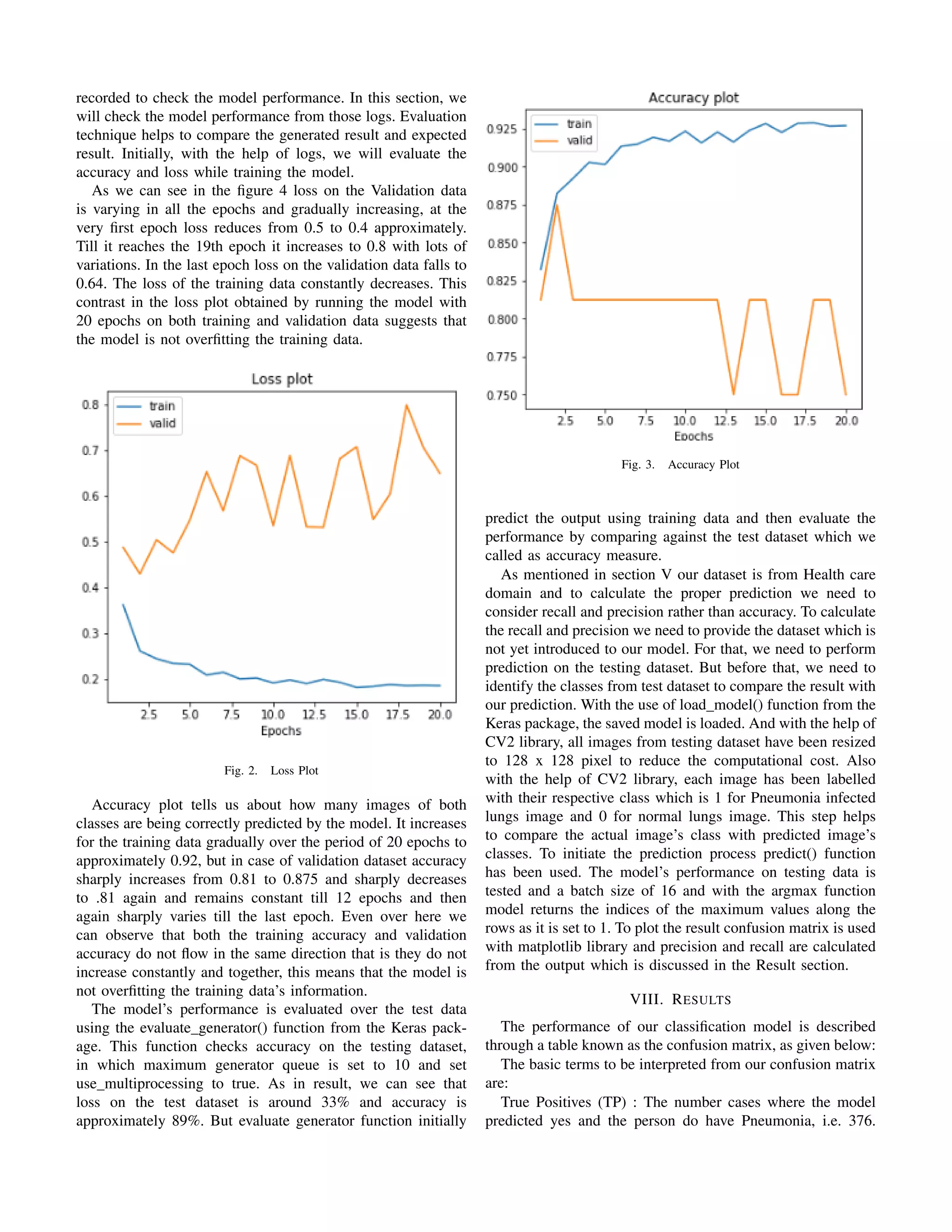
VII. EVALUATION
In the previous section, we created our model and trained it
on the training dataset, during the training period, logs were](https://image.slidesharecdn.com/pneumoniadetectionreport-191023100300/75/Pneumonia-detection-using-CNN-4-2048.jpg)
![Fig. 4. Confusion Matrix
True Negatives (TN) : the number of cases where the model
predicted no and the person does not have Pneumonia. i.e.
177. False positives (FP) : Model predicted yes, but actually
they do not have Pneumonia. i.e. 57. False Negatives (FN) :
Model predicted no, but actually they do have Pneumonia. i.e.
34.
How often our model is correct when it predicts yes, is
given by precision and it is calculated as:
Precision = TP / (TP + FP) i.e. 0.87
Also, for our study, we will be focusing on Recall as a
metric and expect it to be high. Recall would be a good
measure of fit for our case as our goal is to correctly classify
all the Pneumonia cases to save the patient’s life. How many
images the model has classified correctly out of all the positive
classes is given by Recall and it should be as high as possible.
Recall “
TP
pTP ` FNq
As for our case, achieved Recall is 0.96. A high recall is
expected but whenever there is a high recall, the precision
reduces. This means an increase in the number of false
positive. This means the model is wrongly classifying normal
lung images as Pneumonia infected lung images. Thus there
is a trade-off between recall and precision which needs to be
maintained and depends on the domain and industry standards.
IX. CONCLUSION AND FUTURE WORK
In this study, a methodology has been proposed to diagnose
Pneumonia from chest x-ray images. The dataset selected
had a high number of Pneumonia images and comparative
less number of normal images. The imbalance ratio was
3 is to 1, so the image augmentation procedure was used
to reduce the class imbalance. Due to time constraints and
limited availability of resources other pre-processing methods
like pixel brightness transformation and image segmentation
could not be performed we suggest these techniques to be
implemented in the future. The VGG19 model was used and
three layers of the model were removed and two dense layers
were added. Building a CNN model from scratch requires high
computational capacity and as there was a shortage of time,
we would suggest building a CNN model from scratch and
fine-tuning the parameters for the future works. We were able
to achieve a decent output with 0.96 recall, with a different
approach, the result can be improved.
REFERENCES
1 Kohli, M., Prevedello, L. M., Filice, R. W., and Geis, J. R., “Implementing
machine learning in radiology practice and research,” American Journal
of Roentgenology, vol. 208, no. 4, pp. 754–760, 2017.
2 Er, O., Yumusak, N., and Temurtas, F., “Chest diseases diagnosis
using artificial neural networks,” Expert Systems with Applications,
vol. 37, no. 12, pp. 7648–7655, 2010. [Online]. Available: http:
//dx.doi.org/10.1016/j.eswa.2010.04.078
3 Bengio, Y., Lamblin, P., Popovici, D., and Larochelle, H., “Greedy
layer-wise training of deep networks,” in Advances in Neural Information
Processing Systems 19, Sch¨olkopf, B., Platt, J. C., and Hoffman, T., Eds.
MIT Press, 2007, pp. 153–160. [Online]. Available: http://papers.nips.cc/
paper/3048-greedy-layer-wise-training-of-deep-networks.pdf
4 Tom`e, D., Monti, F., Baroffio, L., Bondi, L., Tagliasacchi, M.,
and Tubaro, S., “Deep convolutional neural networks for pedestrian
detection,” vol. 2018, 2015. [Online]. Available: http://arxiv.org/abs/1510.
03608tz%u0Ahttp://dx.doi.org/10.1016/j.image.2016.05.007
5 Saraiva, A., Ferreira, N., Lopes de Sousa, L., Costa, N., Sousa, J.,
Santos, D., Valente, A., and Soares, S., “Classication of Images of
Childhood Pneumonia using Convolutional Neural Networks,” pp. 112–
119, 2019.
6 Rajaraman, S., Candemir, S., Kim, I., Thoma, G., and Antani, S., “Visual-
ization and Interpretation of Convolutional Neural Network Predictions in
Detecting Pneumonia in Pediatric Chest Radiographs,” Applied Sciences,
vol. 8, no. 10, p. 1715, 2018.
7 Hand, D. J., “Measuring classifier performance: A coherent alternative to
the area under the ROC curve,” Machine Learning, vol. 77, no. 1, pp.
103–123, 2009.
8 Havaei, M., Davy, A., Warde-Farley, D., Biard, A., Courville, A.,
Bengio, Y., Pal, C., Jodoin, P. M., and Larochelle, H., “Brain tumor
segmentation with Deep Neural Networks,” Medical Image Analysis,
vol. 35, pp. 18–31, 2017. [Online]. Available: http://dx.doi.org/10.1016/j.
media.2016.05.004
9 Hooda, R., Sofat, S., Kaur, S., Mittal, A., and Meriaudeau, F., “Deep-
learning: A potential method for tuberculosis detection using chest ra-
diography,” Proceedings of the 2017 IEEE International Conference on
Signal and Image Processing Applications, ICSIPA 2017, pp. 497–502,
2017.
10 Guan, Q. and Huang, Y., “Multi-label chest X-ray image classification via
category-wise residual attention learning,” Pattern Recognition Letters,
no. xxxx, 2018. [Online]. Available: https://doi.org/10.1016/j.patrec.2018.
10.027
11 Kermany, D. S., Goldbaum, M., Cai, W., Valentim, C. C., Liang, H.,
Baxter, S. L., McKeown, A., Yang, G., Wu, X., Yan, F., Dong, J.,
Prasadha, M. K., Pei, J., Ting, M., Zhu, J., Li, C., Hewett, S., Dong, J.,
Ziyar, I., Shi, A., Zhang, R., Zheng, L., Hou, R., Shi, W., Fu, X., Duan, Y.,
Huu, V. A., Wen, C., Zhang, E. D., Zhang, C. L., Li, O., Wang, X.,
Singer, M. A., Sun, X., Xu, J., Tafreshi, A., Lewis, M. A., Xia, H., and
Zhang, K., “Identifying Medical Diagnoses and Treatable Diseases by
Image-Based Deep Learning,” Cell, vol. 172, no. 5, pp. 1122–1131.e9,
2018. [Online]. Available: https://doi.org/10.1016/j.cell.2018.02.010
12 Stephen, O., Sain, M., Maduh, U. J., and Jeong, D. U., “An Efficient Deep
Learning Approach to Pneumonia Classification in Healthcare,” Journal
of Healthcare Engineering, vol. 2019, 2019.
13 Lakhani, P. and Sundaram, B., “Radiol.2017162326,” vol. 000, no. 0,
2017.](https://image.slidesharecdn.com/pneumoniadetectionreport-191023100300/75/Pneumonia-detection-using-CNN-6-2048.jpg)
![14 Gu, X., Pan, L., Liang, H., and Yang, R., “Classification of Bacterial and
Viral Childhood Pneumonia Using Deep Learning in Chest Radiography,”
pp. 88–93, 2018.
15 Melendez, J., Van Ginneken, B., Maduskar, P., Philipsen, R. H., Re-
ither, K., Breuninger, M., Adetifa, I. M., Maane, R., Ayles, H., and
S´anchez, C. I., “A novel multiple-instance learning-based approach to
computer-aided detection of tuberculosis on chest X-rays,” IEEE Trans-
actions on Medical Imaging, vol. 34, no. 1, pp. 179–192, 2015.
16 Liang, G. and Zheng, L., “A transfer learning method with deep
residual network for pediatric pneumonia diagnosis,” Computer Methods
and Programs in Biomedicine, no. xxxx, 2019. [Online]. Available:
https://doi.org/10.1016/j.cmpb.2019.06.023
17 Rajpurkar, P., Irvin, J., Zhu, K., Yang, B., Mehta, H., Duan, T.,
Ding, D., Bagul, A., Langlotz, C., Shpanskaya, K., Lungren, M. P.,
and Ng, A. Y., “CheXNet: Radiologist-Level Pneumonia Detection on
Chest X-Rays with Deep Learning,” pp. 3–9, 2017. [Online]. Available:
http://arxiv.org/abs/1711.05225
18 Jaiswal, A. K., Tiwari, P., Kumar, S., Gupta, D., Khanna, A., and
Rodrigues, J. J., “Identifying pneumonia in chest X-rays: A deep learning
approach,” Measurement: Journal of the International Measurement
Confederation, vol. 145, pp. 511–518, 2019. [Online]. Available:
https://doi.org/10.1016/j.measurement.2019.05.076
19 Srinivas, M., Roy, D., and Mohan, C. K., “Discriminative feature ex-
traction from X-ray images using deep convolutional neural networks,”
ICASSP, IEEE International Conference on Acoustics, Speech and Signal
Processing - Proceedings, vol. 2016-May, pp. 917–921, 2016.
20 Pardamean, B., Cenggoro, T. W., Rahutomo, R., Budiarto, A.,
and Karuppiah, E. K., “Transfer learning from chest x-ray pre-
trained convolutional neural network for learning mammogram
data,” Procedia Computer Science, vol. 135, no. Breast cancer:
basic and clinical research 9 2015, pp. 400–407, 2018.
[Online]. Available: https://app.dimensions.ai/details/publication/pub.
1106388589andhttps://doi.org/10.1016/j.procs.2018.08.190
21 Heo, S. J., Kim, Y., Yun, S., Lim, S. S., Kim, J., Nam, C. M., Park, E. C.,
Jung, I., and Yoon, J. H., “Deep learning algorithms with demographic
information help to detect tuberculosis in chest radiographs in annual
workers’ health examination data,” International Journal of Environmental
Research and Public Health, vol. 16, no. 2, 2019.
22 Hwang, S., Kim, H.-E., Jeong, J., and Kim, H.-J., “A novel approach for
tuberculosis screening based on deep convolutional neural networks,” 03
2016, p. 97852W.
23 Kieu, P. N., Tran, H. S., Le, T. H., Le, T., and Nguyen, T. T. T.,
“Applying multi-cnns model for detecting abnormal problem on chest
x-ray images,” 2018 10th International Conference on Knowledge and
Systems Engineering (KSE), pp. 300–305, 2018.
24 Wirth, R. and Hipp, J., “CRISP-DM : Towards a Standard Process Model
for Data Mining,” Proceedings of the Fourth International Conference on
the Practical Application of Knowledge Discovery and Data Mining, no.
24959, pp. 29–39, 1995.
25 Carneiro, T., Da Nobrega, R. V. M., Nepomuceno, T., Bian, G. B., De
Albuquerque, V. H. C., and Filho, P. P. R., “Performance Analysis of
Google Colaboratory as a Tool for Accelerating Deep Learning Applica-
tions,” IEEE Access, vol. 6, pp. 61 677–61 685, 2018.
26 Lemley, J., Bazrafkan, S., and Corcoran, P., “Smart Augmentation Learn-
ing an Optimal Data Augmentation Strategy,” IEEE Access, vol. 5, pp.
5858–5869, 2017.
27 Fujita, K., Kobayashi, M., and Nagao, T., “Data Augmentation using Evo-
lutionary Image Processing,” 2018 International Conference on Digital
Image Computing: Techniques and Applications, DICTA 2018, pp. 1–6,
2019.
28 Ling Shao, Fan Zhu, and Xuelong Li, “Transfer Learning for Visual
Categorization: A Survey,” IEEE Transactions on Neural Networks and
Learning Systems, vol. 26, no. 5, pp. 1019–1034, 2014.
29 Shaha, M. and Pawar, M., “Transfer Learning for Image Classification,”
Proceedings of the 2nd International Conference on Electronics, Commu-
nication and Aerospace Technology, ICECA 2018, no. Iceca, pp. 656–660,
2018.
30 Lau, M. M. and Lim, K. H., “Review of adaptive activation function
in deep neural network,” 2018 IEEE EMBS Conference on Biomedical
Engineering and Sciences, IECBES 2018 - Proceedings, pp. 686–690,
2019.
31 Garg, A., Gupta, D., Saxena, S., and Sahadev, P. P., “Validation of Random
Dataset Using an Efficient CNN Model Trained on MNIST Handwritten
Dataset,” 2019 6th International Conference on Signal Processing and
Integrated Networks, SPIN 2019, pp. 602–606, 2019.](https://image.slidesharecdn.com/pneumoniadetectionreport-191023100300/75/Pneumonia-detection-using-CNN-7-2048.jpg)


![Coded Agents – with UiPath SDK + LangGraph [Virtual Hands-on Workshop]](https://cdn.slidesharecdn.com/ss_thumbnails/codedagentsdeck-251215155422-5497c599-thumbnail.jpg?width=640&height=640&fit=bounds)


![Vibe Coding vs. Spec-Driven Development [Free Meetup]](https://cdn.slidesharecdn.com/ss_thumbnails/vibecodingvsspecdrivendevelopment-251209105622-43f455e7-thumbnail.jpg?width=640&height=640&fit=bounds)
