Chest Disease Detectionusing CNN Algorithm
Name of the student:
A.Rajesh Reddy (21H51A66E3)
B.Lokesh (21H51A66E4)
D.Simhadri (21H51A66E6)
Under esteemed guidance of
Ms.Sana Afreen
Assistant Professor(CSE(AI&ML)
CMR COLLEGE OF ENGINEERING & TECHNOLOGY
Kandlakoya, Medchal, Hyderabad - 501401
Department of CSE(AI&ML)
Batch No:15
Batch: 2021-2025 Major Project
HOD:
Dr. P. Sruthi
(Associate Professor)
2.
Outline
• Abstract
• Introduction
•Research Objective
• Problem Definition
• Scope of the Project
• Literature Review
• Flowchart
• Result
• Conclusion
• References
3.
ABSTRACT
Chest diseasessuch as pneumonia, fibrosis, and inflammation pose significant health
risks, requiring accurate and timely diagnosis for effective treatment.
This project introduces a Convolutional Neural Network (CNN)-based system
designed to detect and classify chest conditions into four categories: normal,
pneumonia, fibrosis, and inflammation, utilizing chest X-ray images.
With the advancement of Deep Learning, Convolutional Neural Networks (CNNs)
have emerged as a powerful tool in medical image analysis, providing a reliable and
automated approach for detecting chest diseases.
The proposed system leverages a robust CNN architecture featuring convolutional
layers for feature extraction, pooling layers for dimensionality reduction, and fully
connected layers for classification.
4.
Introduction
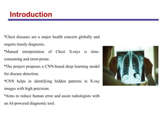
•Chest diseases area major health concern globally and
require timely diagnosis.
•Manual interpretation of Chest X-rays is time-
consuming and error-prone.
•The project proposes a CNN-based deep learning model
for disease detection.
•CNN helps in identifying hidden patterns in X-ray
images with high precision.
•Aims to reduce human error and assist radiologists with
an AI-powered diagnostic tool.
5.
Problem Definition
• Chestdiseases, including pneumonia, fibrosis, and inflammation, are major global
health concerns due to their high morbidity and mortality rates. Accurate and timely
diagnosis is critical for effective treatment .
• Developing an automated system using Convolutional Neural Networks (CNNs) can
improve detection accuracy, reducing the dependency on skilled radiologists,
especially in underserved areas.
• Tiredness and mental distractions can cause doctors to miss small details or make
mistakes when diagnosing chest X-rays.
• A CNN-based detection system can provide diagnostic support in remote and
resource-limited settings, improving accessibility to quality healthcare.
• CNN algorithms enhance the efficiency and consistency of chest X-ray analysis,
reducing misdiagnosis risks due to human fatigue or oversight.
6.
Research Objective
•Improve DiagnosticAccuracy: Enhance the precision and reliability of chest
disease diagnosis through the application of CNN-based AI algorithms.
•Early Detection: Enable the early identification of various chest diseases such as
COPD, pneumonia, asthma, tuberculosis, and lung cancer to facilitate timely
treatment.
•Support Radiologists: Provide a supportive tool for radiologists by automating the
analysis of chest X-rays, thus reducing workload and minimizing the risk of human
error.
•Efficiency: Speed up the diagnostic process, allowing for quicker decision-making
and treatment initiation, which is critical in managing and treating chest diseases.
7.
Literature Review
1.Computer-Aided Diagnosis(CAD) Systems
•Early CAD systems used image processing techniques such as edge detection,
texture analysis, and feature extraction.
•Example: Ginneken et al. (2001) developed a CAD system for tuberculosis
detection using thresholding and rule-based classification.
•Limitations:
• Handcrafted feature extraction is complex and less effective.
• Not robust for large-scale applications.
8.
Literature Review
2.AI &Machine Learning-Based Approaches
Machine Learning (ML) Techniques
•Support Vector Machines (SVM), Decision Trees, k-NN, and Random Forest have
been used for X-ray classification.
•Wang et al. (2017) used SVM with handcrafted features to classify lung abnormalities.
•Limitations:
• Feature engineering is complex and requires domain expertise.
• Lower accuracy compared to deep learning.
9.
Scope of theProject
• Disease Detection: Focuses on identifying chest conditions like pneumonia, fibrosis,
inflammation, and normal cases from chest X-ray images.
• Smartphone-Based Diagnosis: Mobile app for AI-based chest disease detection from
scanned X-ray images.
• Improved Accuracy: Utilizes CNN models with advanced techniques like data
augmentation and transfer learning to achieve high diagnostic accuracy.
• Multi-Disease Classification: Extend the model to detect TB, COVID-19, Lung
Cancer, and Asthma.
• Early Diagnosis: Helps detect diseases at an early stage, reducing severe health risks.
10.
Proposed Solution
•A chestX-ray dataset with four categories was collected: Normal, Pneumonia,
Fibrosis, and Inflammation.
•Images were preprocessed and resized to make them suitable for training.
•A Convolutional Neural Network (CNN) was developed using Python and Keras for
disease classification.
•Random Forest and Decision Tree algorithms were also implemented for performance
comparison.
•All models were evaluated using metrics like accuracy, precision, recall, and F1-score.
•A simple Tkinter-based GUI was designed to allow users to test images and view
predictions.
Result
• The systemincludes a GUI with functionalities like uploadingdatasets,preprocessing,
running CNN, Random Forest, and Decision Tree algorithms, and performing predictions.
Comparison graphs allow users to evaluate the performance
of different algorithms.
• Using the CNN algorithm, we achieved accuracy around
93% detecting chest diseases from X-ray images, while
Random Forest achived 85% and Decision Tree
Achieved 82%.
• This highlights CNNs' superiority in extracting complex
features for medical image classification, reinforcing the role of deep learning in disease
diagnosis.
13.
Conclusion
In conclusion, currentsystems for chest disease detection utilizing Deep Learning and
other algorithms have demonstrated significant advancements in automating diagnostic
processes, particularly in identifying diseases such as pneumonia, tuberculosis, and other
thoracic conditions. Studies have shown that CNN models, especially those enhanced
through transfer learning, achieve high accuracy levels, often matching or exceeding
radiologist-level diagnostic capabilities.
However, the performance of these models is largely dependent on the quality, diversity,
and size of the datasets used. Large-scale datasets like ChestX-ray14 have been
instrumental in advancing the field, yet issues such as variability in image quality,
limited access to annotated datasets, and a need for multi-label classification continue to
pose challenges.
14.
References
• Ambati A,Dubey SR. Ac-covidnet: Attention guided contrastive cnn for
recognition of covid-19 in chest x-ray images. In: International Conference on
Computer Vision and Image Processing, pp. 71–82 (2022)
• Nasser AA, Akhloufi M. Chest diseases classification using cxr and deep
ensemble learning. In: 19th International Conference on Content-based
Multimedia Indexing, pp. 116–120 (2022).
• Miyazaki, A. et al. Computer-aided diagnosis of chest X-ray for covid-19
diagnosis in external validation study by radiologists with and without deep
learning system. Sci. Rep. 13, 17533 (2023).
• Kermany, D. Labeled optical coherence tomography (oct) and chest X-ray
images for classification. https://data.mendeley.com/datasets/rscbjbr9sj/2 (2018).