Download to read offline

![Introduction
It is given that the new generation of drugs that will be used to manage MDR-, XDR-, and
TDR-TB must result in cidality under in-vivo conditions [1]. However, the factors that govern
in-vitro to the in-vivo translation of cidality are far from obvious. Mtb encounters complex
physiological situations due to inflammatory immune pressures in the human host, starting
with phagocytosis by the macrophages and ending in the same niche- the macrophages. The
phagosome-lysosome fusion causes an acidic environment [2], and a strong chemistry of
nitroxidative free radicals [3,4] produced by the macrophages in the granuloma; accompanied
by gradual deficiency of nutrients: Carbon [5], Nitrogen [6], Oxygen [7], etc. However, some
Mtb populations may be replicating logarithmically [8]. Thus, Mtb faces multiple milieus, in
the host that makes its survival more complex and challenging [9]. Finally, a narrow window of
“decision” between the infecting/ persisting pathogen and the adaptive/innate host immune
response (immuno-competent/immuno-compromised), determines the judgement: disease or
no disease [9]. Rest is the paradoxical hide-n-seek between the two, with their ambush (inva-
sion and phagocytoses) and artillery (triggering or blocking various anti-inflammatory
responses).
Conventionally, the drug discovery starts with the in-vitro screening of inhibitors. It is
important to have the right assay condition/s for selecting potent molecules that must translate
into in-vivo animal efficacy, the final proof of concept (POC). However, the standard in-vitro
screens often do not adequately represent in-vivo physiologies. Therefore, one has to find an
appropriate in-vitro model to predict efficacy, because every inhibitor cannot be validated
through animal models. Usually, the failure of drugs to reach the clinic is attributed to two pri-
mary reasons: right efficacy and the right safety. It is economically and strategically prudent to
fail at an early stage of inhibitor-screening rather than at a later stage of drug development.
In the present study, we tried to mimic the entire in-vivo relevant physiological milieu
under in-vitro conditions. Our objective was to find out a robust link for in-vitro to in-vivo
translation.
We used antisense-RNA (AS-RNA) silencing to inhibit the selected potential cidal targets
from TB genome [10,11], including rpoB- the target of the tuberculocidal drug rifampicin;
under all the simulated in-vitro conditions, as well as the in-vivo in the immunocompetent
mice BALB/c. It was followed by a correlation of cidality from in-vitro screens with in-vivo cid-
ality data. AS-RNA has a great potential in validating the therapeutically cidal vs. static targets
for drug intervention in human diseases and selecting cidal anti-mycobacterials [8,11–14].
Here, we report the application of in-vivo AS-repression to demarcate the ideal assay condi-
tions and tuberculocidal targets.
Our comprehensive AS-RNA silencing studies on the translation of target cidality from in-
vitro to in-vivo revealed that AroK is the in-vivo validated target that culminated from the cid-
ality SCORE. It emerged as an “in-vitro total” and “in-vivo” cidal target, whose inhibition is
expected to be lethal to Mtb clinically. This study also raises the possibility of developing
AS-RNA based therapeutics for treating TB patients in the long run. The low pH assay appears
to be a critical in-vitro physiological condition that predicts the bactericidal potential of targets
and correlates positively with in-vivo efficacy.
Materials and Methods
Bacterial strains, media, and antisense recombinants
Bacterial strains of Escherichia coli (MOS Blue cells {F’endA1 hsdR17 (rK2 mK+), supE44 thi-1
recA1 gyrA96 relA1 lac [F’ lacIqZDM15 proAB + Tn10 (TetR)]}, Amersham), Mycobacterium
Antisense Unveils Mycobacterial Cidality
PLOS ONE | DOI:10.1371/journal.pone.0154513 May 4, 2016 2 / 23
employed by Bugworks Research. This commercial
affiliation has no competing interests relating to
employment, consultancy, etc. by the affiliated
institutes or commercial companies. There are no
patents, products in development or marketed
products to declare. This does not alter the authors’
adherence to all the PLOS ONE policies on sharing
data and materials, as detailed online in the guide for
authors.](https://image.slidesharecdn.com/457f3d5b-b6ef-429f-84d9-a99676deb689-160506012528/85/PLoSONE_AS_Mtb_2016-PDF-2-320.jpg)
![smegmatis mc2
155, and Mycobacterium tuberculosis H37Rv ATCC 27294 were used for this
study. We rationally selected five target genes for this study from the list of Sassetti’s classifica-
tion on essential targets [11] i.e. rpoB, rpoC, aroK, ilvB, and ppk. The recombinants of these tar-
get genes for AS-RNA were generated by cloning full-length genes (sequences were taken from
KEGG and Tuberculist databases) of these in the reverse/ antisense orientation into the vector
pAZI9018b by replacing the lacZ gene [8].Three different independent transformations were
performed for the target AS constructs. The AS recombinants of Mtb were selected from 7H10
agar plates supplemented with 50μg/ml Hygromycin (Hyg50) and were grown in 7H9 broth
containing Hyg50. The O.D.600 nm was adjusted to 0.1, and the cells were induced at 10 μM
IPTG. Transformants showed a slower growth rate, so wherever required; the suspensions
were concentrated to match the required O.D. (O.D.600nm to 0.1, approximately 107
cells/ml).
Target selection for in-vivo validation
Though there are about 600 in-vitro essential genes in Mtb [11], we made a particular choice
by selecting a few key target genes from different pathways: 1). Transcription: RpoB (Rv0667)
and rpoC(Rv0668), encoding β and β’-subunits of RNA polymerase. RpoB is a well known clini-
cally validated target, hence was also used as a bactericidal target control for this study. Its part-
ner rpoC which is involved in the same process of transcription was selected to compare if it
has an equal partnership and cidality potential. 2). Fundamental amino acid (aa) biosynthesis
pathways: We chose two targets, aroK, and ilvB, one each from two amino acid biosynthesis
pathways; aromatic (shikimate) and branched chain aa pathways. Both the targets aroK and
ilvB, being in-vitro essential and have no human homologs, present a great opportunity
towards the development of non-toxic drugs [15–18]. AroK (Rv2539c), from aromatic (shiki-
mate) aa pathway, encodes the 5th
enzyme shikimate kinase, phosphorylating shikimic acid to
shikimate-3-phosphate during chorismate biosynthesis. IlvB (ilvB1, Rv3003c)from branched-
chain aa pathway, encodes Acetohydroxyacid synthase, (AHAS), the 1st
and most important
enzyme [19] out of the several genes (ilvB1, ilvB2, ilvG, and ilvX). Among all the genes in shiki-
mate pathway, aroK is the only gene which is exclusive and in-vitro essential in Mtb. The
essential amino acids can conditionally modulate bacterial growth [8,15,16], hence may act as
regulatory tools to delineate their essentiality. Mtb exists in multiple milieu in-vivo [9], so, the
targets of amino acid biosynthesis pathway were chosen as suitable tools for concept validation
under various physiologies encountered in-vivo. In our earlier report [8], we have demon-
strated in-vitro cidality of ilvB under replicating growth condition [8,12]. It is regulatable with
the addition of physiological concentrations of ILV and P (Isoleucine, Leucine, Valine and Pan-
tothenate) showing auxotrophy [20,21]. 3) The central energy metabolism: Ppk (Rv2984)or
polyphosphate kinase plays an important regulatory role in the transition of the bacteria to the
persistence phase under the growth-limiting conditions (phosphate depletion, amino acid star-
vation, or osmotic stress). It tries to accumulate polyP intracellularly, modulating several bacte-
rial processes (protein synthesis, nucleotide balance, lipid metabolism, energy utility, and
susceptibility to antibiotics)[12]. Ppk was chosen as an unvalidated target for this study because
this target gene has not been characterised as per Sassetti’s list [11]. It is neither reported as
essential (in-vitro or in-vivo) nor a non-essential target [11]. Earlier, we had established a par-
tial validation of ppk as essential and cidal target under in-vitro replication condition only [12].
Hence, we chose ppk for this study as an un-validated target gene for further characterisation
of its cidality under in-vivo condition.
Other than the clinically validated target from transcription subunit rpoB, rest of the targets
ppk, aroK, rpoC, and ilvB are unvalidated targets since their in-vivo essentiality in Mtb is not
reported yet. Therefore, in order to delineate in-vivo bactericidal potential of these targets in
Antisense Unveils Mycobacterial Cidality
PLOS ONE | DOI:10.1371/journal.pone.0154513 May 4, 2016 3 / 23](https://image.slidesharecdn.com/457f3d5b-b6ef-429f-84d9-a99676deb689-160506012528/85/PLoSONE_AS_Mtb_2016-PDF-3-320.jpg)
![the present study, we used IPTG (Isopropyl β-D-1-thiogalactopyranoside) inducible AS-RNA
silencing [8] as a gene-specific inhibitor for the chosen targets. It was used under multiple
physiologies of granuloma simulated in-vitro (replicating conditions, and, under the stressful
condition of nitrosative free radicals, acidic, hypoxic, or under starvation of C and N2) as well
as under in-vivo conditions (experimental TB of mice).
In-vitro cidality of Mtb targets under replicating conditions using survival
kinetics after AS-RNA inhibition
The antisense effect was estimated in triplicate from 3 independent transformants for AS
recombinants of rpoB, rpoC, and aroK in Mtb. The conditional AS-gene-silencing was induced
by 10 or 100μM [8] IPTG under an in-vitro replicating growth condition. The survivors (from
AS-silencing) were enumerated by plating on different generation times (Day 0, 1, 7, 14, 21, up
to 63 days with a gap of 1 week), and the data was analysed using Prism software (Graph Pad
Software, Inc., San Diego, Calif.). Appropriate controls were used. Two earlier established
cidal targets (ppk, ilvB) for AS-repression under only the in-vitro ‘replicating growth condi-
tions’[8,12]; were used as positive controls in this entire replication condition AS-silencing
study. The clinically validated target, rpoB, was another cidal positive control throughout for
all the validations. The negative controls for antisense repression were the WT = wild type
Mtb; and the V = empty vector in Mtb (also the control for the recombinants). The genes aroK,
ilvB, and rpoC were selected as un-validated target genes along with another uncharacterised
target gene ppk.
In-vitro cidality of Mtb targets under different physiologies
A total of seven different Mtb strains, i.e. five AS-recombinants (rpoB, rpoC, aroK, ilvB, ppk)
and two control strains (WT and vector in Mtb) were taken for this study. We had to profile
the survival kinetics of all the selected seven Mtb strains/ AS-recombinants, in triplicate, under
six different assay conditions, (Platings/day = 7strainsX 3plicateX 6assaysX 5dil.sX 9time
points) by plating 5 dilutions for cfu, up-to 35 days, under the constraints of bio-safety level-3
containment facility. A throughput method was ideal to conduct and compare all the experi-
ments with replicates in parallel. Hence, a validated throughput SPOT-MBC assay [22] was
used for this study because of its efficient way of testing a large number of samples/replicates
from various in-vitro models, to be explored in parallel under BSL3 containment. Moreover,
this relatively straightforward throughput method has been validated for different phenotypes
of Mtb (AS-recombinants, vector controls in Mtb as well as WT, sensitive or resistant Mtb) as
well; and had yielded cfu counts similar to conventional plating method [22]. The standard
conventional plating method would have given an identical output; hence, it was prudent to
use this efficient SPOT-MBC assay system. The ppk target was validated earlier [22] with
SPOT-MBC and hence was taken as a positive control for this validation. The rest of the
selected uncharacterised targets (rpoB, rpoC, aroK, ilvB) were validated in this study by conven-
tional vs. SPOT-MBC methods in parallel under in-vitro replicating growth conditions, before
using it for various physiological screens.
We further investigated the vulnerability of selected genes by checking cidality under all the
in-vivo simulated multiple physiological conditions (the equivalent of macrophages and the
lung granuloma) by performing survival kinetics in-vitro. All the strains were tested under six
different physiological in-vitro assay conditions for investigations in parallel, in triplicate: repli-
cating, hypoxia [7], nutrient starvation [5], low pH [2], nitric oxide stress [3], and nitrogen
starvation [6]. The survivors were monitored by enumerating up to 35 days in a kinetic manner
from all the screens in parallel using SPOT-MBC assay [22]. Since our previously reported ppk
Antisense Unveils Mycobacterial Cidality
PLOS ONE | DOI:10.1371/journal.pone.0154513 May 4, 2016 4 / 23](https://image.slidesharecdn.com/457f3d5b-b6ef-429f-84d9-a99676deb689-160506012528/85/PLoSONE_AS_Mtb_2016-PDF-4-320.jpg)
![AS-repression (SPOT vs. conventional assay) experiments were performed only under prolifer-
ating assay conditions [12,22], and no other alternate physiological environments; we, there-
fore, investigated ppk AS-repression under all other the physiological conditions as well.
In-vivo cidality of Mtb targets after AS-RNA repression
All the seven Mtb strains, including the controls and AS recombinants, were investigated for
their growth or survival kinetics under in-vivo conditions in BALB/c mice, for confirmation of
in-vitro cidality. This important in-vivo evaluation provided us with the proof of concept on
cidality of targets under experimental TB in mice, the actual disease condition.
Ethics statement. The in-vivo study was carried out in strict accordance with the recom-
mendations of the Institutional Animal Ethics Committee (IAEC), registered with the Com-
mittee for the Purpose of Control and Supervision (CPCSEA), Government of India
(registration no. CPCSEA1999/5). All the protocols for animal experimentation and animal
usage were reviewed and approved in advance by the IAEC. Carbon dioxide (CO2) was used
for euthanasia.
Animals. Male BALB/c mice were purchased from RCC Hyderabad, India. Mice (6–
8weeks) with an average body weight of 20–25 grams were randomly assigned to groups of
three per cage and were allowed two weeks of acclimatisation before experimentation. The ani-
mals were housed under standard conditions in the facility with a 12 hr. day-night cycle. The
infected mice were maintained in individually ventilated cages (Allentown Technologies, USA)
in bio-safety level 3 (BSL-3) facilities. All the procedures including dosing of the infected mice
were performed under strict BSL-3 bio-containment guidelines. Feed (the sterile commercial
diet) and water were given ad libitum. We provided IPTG to mice in their drinking water at a
5mM concentration, as per the earlier established methods[23,24], for in-vivo target induction/
repression. The animals were euthanized with CO2at the respective time points, and the lungs
were sterically removed, homogenised and were plated for cfu enumeration of survivor Mtb
bacilli.
Course of Infection (COI) studies in mouse-tuberculosis infection model. BALB/c mice
were infected in the bio-safety level 3 (BSL3) facility, via inhalation in an aerosol infection
chamber [25] with the suitable modification that delivered ~104
bacilli/mouse lung, a higher
dose of infection [26]. This method was standardised and validated in our laboratory [25,26].
The groups of mice (n = 3) were infected with different Mtb strains (WT and AS-recombinants
of Mtb). High dose bacterial infection to lungs was delivered by increasing the strength of the
bacterial inoculum (109
cfu/ml) used for inhalation [26] that delivered 104
cfu/lung, to allow
evaluation of cfu reduction by capturing enough window following AS-repression. Instillation
of 104
cfu to lung/mouse was enumerated by harvesting and plating the lungs at day 3 post
infection. The course of infection (COI) of all the strains was monitored on 3, 7, 14, 28, 42, and
56 days post infection by cfu enumeration.
Rifampicin was used at 30mg/kg bw as a reference drug control (for the WT Mtb) formu-
lated in CMC (Carboxymethyl Cellulose) suspension [25]. Drug treatment with rifampicin
started after 3 days following the establishment of acute infection. The drug was administered
by oral gavage. AS-repression was induced with IPTG, given as 5mM in drinking water ad libi-
tum beginning on day 3, based on the previously validated and established method [23,24].
The water bottles containing IPTG were replaced every 48 hrs. At every time point, the mice
were euthanized with CO2, and the lungs were harvested and homogenized [25] in PBS con-
taining 0.1% bovine gelatine and 0.1% triton-X100 using tissue grinders (W012576; Wheaton).
Each suspension was serially diluted in 10-fold steps and plated on Middlebrook 7H11 agar
supplemented with 10% albumin-dextrose-catalase. Plates were incubated at 37°C with 5%
Antisense Unveils Mycobacterial Cidality
PLOS ONE | DOI:10.1371/journal.pone.0154513 May 4, 2016 5 / 23](https://image.slidesharecdn.com/457f3d5b-b6ef-429f-84d9-a99676deb689-160506012528/85/PLoSONE_AS_Mtb_2016-PDF-5-320.jpg)
![CO2 for 3 weeks, and colony forming unit (CFU) counts were enumerated on 3, 7, 14, 28, 42,
and 56 days.
Transcriptome analysis of in-vivo antisense repression of target genes. Lung homoge-
nates of AS-recombinants of Mtb along with the WT and vector control cultures were collected
from different time points (day 3, 7, 14, 28, 42, 56), and pelleted. One ml of TrizolH was added
to the cell pellets (from 2 ml homogenate/s) to stabilize and arrest the mRNA. These samples
were flash frozen in dry ice and stored at -70°C until further processing. Wild-type (WT) M.
tuberculosis culture control as well as the rifampicin-treated homogenates were also included.
While processing, pellets were thawed on ice, cells were disrupted by bead beating using 0.1
mm diameter zirconium beads (Biospec), followed by a 5 min centrifugation at 14,000 g. Total
RNA was isolated, and qRTPCR was performed using SYBR Green chemistry (Brilliant II
SYBR1 Green QPCR Master Mix) in the Mx3005P Stratagene system as reported [8,12] using
respective forward and reverse primers (Table A in S1 File). The transcript levels were mea-
sured against SigA (Rv2703) as a validated house-keeping control gene, from our previous
study of AS-repression [12], as well as taking clues from other reports including a detailed anal-
ysis by Manganelli et al. [27,28]. The fold difference was calculated against vector control using
Delta-delta Ct method [29].
Statistical Analysis
The data from in-vitro as well as in-vivo experimentation was analysed using Prism software
(Graph Pad Software, Inc., San Diego, Calif.) for all the calculations for data. In-vitro data
included survival plots as well as the correlation of cfu/ml from the SPOT-MBC vs. conven-
tional plating method. The in-vivo data included the COI of various Mtb strains tested after
AS-repression in-vivo. Two-way analysis of variance (ANOVA) with Bonferroni post hoc test
correction was used to compare the net bacterial loads at each time point using Prism software
(Graph Pad Software, Inc., San Diego, Calif.), to check the significance of AS repression for in-
vivo cidality. The correlation between the outcomes from various in-vitro screens vs. in-vivo
studies was analysed to narrow down to the best in-vitro screen, the predictor of in-vivo
cidality.
Results
Antisense-silencing under in-vitro replicating condition
We selected the potential cidal target genes [11] rpoB, rpoC, aroK, ppk, ilvB of Mtb, and started
with evaluating AS-gene-silencing [8] under in-vitro replicating growth condition. Comple-
mentation of the antisense (AS) and the sense (S) sequences de-stabilized the mRNA. Depend-
ing on the target vulnerability, translational blocking resulted in varying degrees of target
specific AS-repression driven cidality of Mtb-recombinants as enumerated by log10 cfu/ml (Fig
1). The controls behaved as expected (vector as negative; and rpoB, ppk, and ilvB as the in-
vitro-cidal positive controls). The known cidal ones were specifically cidal (rpoB). The order of
cidality (!2log10 cfu reduction) under replicating condition as observed on day-63 was: ilvB
(5.4)> ppk(4.8)> rpoC(3.5)> rpoB(2.5)> aroK(2.2). On day-63, ilvB showed the maximum
cidality (5.4 log10 cfu reduction) while in replicating condition, without hitting the maximum
extent (Emax) yet.
Throughput cfu enumeration assay for AS- silencing in-vitro
Validation data of all the AS-recombinants by the throughput “SPOT-MBC assay” vs. conven-
tional [22] plating for cfu enumeration (S1 Fig) demonstrated an overlapping data with a
Antisense Unveils Mycobacterial Cidality
PLOS ONE | DOI:10.1371/journal.pone.0154513 May 4, 2016 6 / 23](https://image.slidesharecdn.com/457f3d5b-b6ef-429f-84d9-a99676deb689-160506012528/85/PLoSONE_AS_Mtb_2016-PDF-6-320.jpg)
![strong positive correlation (Pearson’s): rpoB (R2
= 0.5857), rpoC (R2
= 0.7770), and aroK
(R2
= 0.9863) vs. vector controls. However, in the case of ilvB, we found a variation in the Emax
(SPOT = 3.3 log10 and conventional = 5.2 log10 reduction), probably, the smaller surface area of
the agar media in SPOT-assay, allowed the colonies to grow in proximity. It enabled socializing
through the exchange of the required amino-acids between the healthy and the sick or nearly
dead bacilli, helping them to rejuvenate. Overall, the SPOT-MBC assay was aptly and signifi-
cantly suitable (EEE) [22] for kinetic profiling of Mtb AS-recombinants and controls under
multiple tests. Henceforth, spot assay was used for cfu enumeration for all the in-vitro screens.
AS-silencing under physiological stress conditions in-vitro
AS-driven survival kinetics, under various alternate physiological in-vitro conditions (Fig 2),
was a yardstick to understand the overall importance (cidality) of respective targets. As
Fig 1. AS-silencing of Mtb targets under replicating in-vitro growth conditions. The survival kinetics of target AS-recombinants of
Mtb enumerated up to 63-days = almost ~70 generations; are shown here as log10 cfu/ml vs. the vector control. Under replicating growth
condition, ilvB demonstrated the maximum AS-repression among different magnitude of cidality in comparison to ppk. The order of
cidality (log10 cfu/ml) was ilvB (5.4)> ppk(4.8)> rpoC(3.5)> rpoB(2.5)> aroK(2.2).
doi:10.1371/journal.pone.0154513.g001
Antisense Unveils Mycobacterial Cidality
PLOS ONE | DOI:10.1371/journal.pone.0154513 May 4, 2016 7 / 23](https://image.slidesharecdn.com/457f3d5b-b6ef-429f-84d9-a99676deb689-160506012528/85/PLoSONE_AS_Mtb_2016-PDF-7-320.jpg)
![expected, the WT and V controls exhibited condition-specific, normal growth under all the
physiologies. Since ppk was reported cidal under proliferating assay conditions [13,22]; in this
study, under different physiological conditions, ppk exhibited the maximum total AS-repres-
sion among the tested. Whereas, under the replication condition alone, ilvB had demonstrated
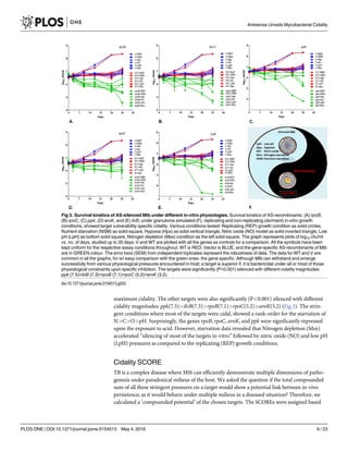
Fig 2. TB or No TB: A delicate balance between host-pathogen interactions. There are various hostile or stringent conditions
encountered by Mtb in-vivo in the host. Mtb enters the host via inhalation. The first encounter in-vivo is with the immune cells: the
macrophages which phagocytose and attack Mtb with their ammunition like low pH, hydrolases, free-radicals, etc. This first encounter is
unanimously responded by all the hosts irrespective of their immune status. Beyond this point, the countering of the pathogen is host
population specific. The outcome as TB or no-TB is a delicate balance and is outcome result of the battles between the pathogen and
the immuno-competence of the host to outsmart. Further, it progresses into a larger immune structure: the granuloma. During the
process, the other stresses in the granuloma, especially in the caseating/ necrotizing lesions are the gradually decreasing levels of O2,
N, C, etc. representing hypoxia, the arrest of various biosynthesis processes and poor nutrition nearly close to starvation. If the host
wins, the pathogen is contained in the solid granuloma, which may gradually heal with time. But in case the pathogen Mtb overpowers
the immune pressure, the granuloma progresses as a caseating and necrotising granuloma. In this case Mtb bacilli multiply to finally
break open from granuloma, and disseminate to other organs of the body or come out into the bronchi and get coughed out to infect
other hosts. We attempted to simulate some of these stress conditions in the form of various in-vitro screens to identify the ideal in-vitro
screen/ condition.
doi:10.1371/journal.pone.0154513.g002
Antisense Unveils Mycobacterial Cidality
PLOS ONE | DOI:10.1371/journal.pone.0154513 May 4, 2016 8 / 23](https://image.slidesharecdn.com/457f3d5b-b6ef-429f-84d9-a99676deb689-160506012528/85/PLoSONE_AS_Mtb_2016-PDF-8-320.jpg)
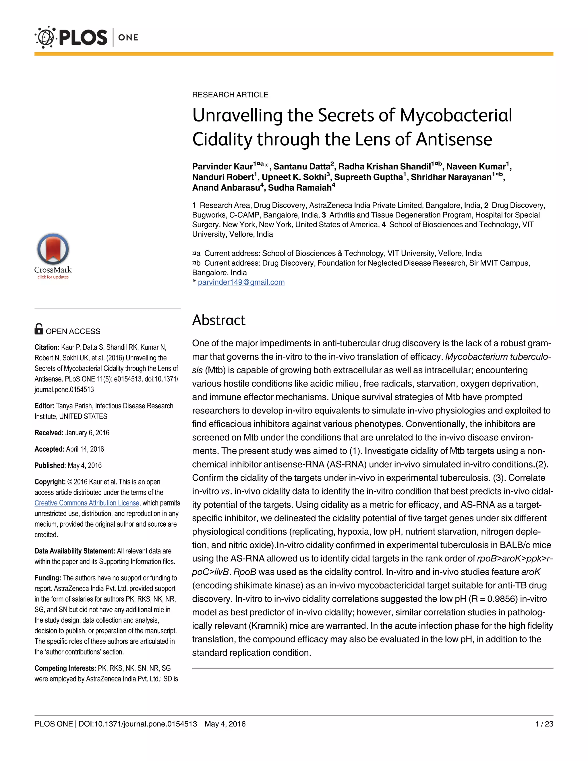
![(Fig 4). This outcome of the in-vitro alternate physiological conditions established specifically
few targets (rpoB, aroK, ilvB) cidal under all the conditions (Fig 4). Our next objective step was
to validate cidality of these targets under the in-vivo experimental model of tuberculosis.
In-vivo AS-silencing of in-vitro cidal targets in mice
Validation of the SCORE based in-vitro cidality was confirmed in-vivo by infecting BALB/c
mice with the respective Mtb strains (AS-recombinants and WT and vector controls) via an
aerosol route [25,26]. In-vivo studies were approved by IAEC (CPCSEA), as mentioned in the
ethics statement. The in-vivo AS-silencing was achieved by providing IPTG (5mM) to mice in
drinking water [23,24], a non-invasive technique of IPTG delivery in drinking water; thereby
minimising the handling of infected mice in the bio-containment set-up.
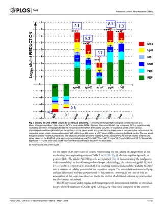
In-vivo cidality of the targets on 28th
day
The first visible and quantitative in-vivo trends of cidality appeared in the lungs after four
weeks post-infection. The control Mtb strains grew as expected, with fully developed visible
granulomas in the lungs (Fig 5), whereas the AS-recombinants showed reduced growth on the
28th
day, due to target specific AS-repression and cidality. RpoB showed the maximum cidality
in-vivo (3.9 log10 reduction), despite only 2.5 log10cfu reduction in the replicating in-vitro
model. The culture plates were incubated up to 45 days, and the colony characteristics were
observed. None of the AS-recombinants, except ilvB, showed any signs of re-growth or delayed
appearance of additional colonies on the media plates. The colony counts remained constant
upon further incubation; suggesting cidality due to AS-repression in ppk, rpoB, rpoC, and
aroK. On the contrary, in the case of ilvB, the appearance of additional colonies on extended
incubation point towards attenuation rather than cidality. IlvB showed a negligible cidality
(0.36 log10cfu reduction) under the in-vivo condition on day 28 and exhibited granuloma
formation similar to the control strains. Targets like ppk, rpoC and aroK were repressed by
their respective AS-counterparts and showed significant (two-way ANOVA,ÃÃ
P = 0.0086 to
Ã
P = 0.0402), but varying levels of log10 cfu reduction: rpoB(2.5)> aroK(1.9)> ppk(1.2)> rpoC
(1.5). The in-vivo model was validated by including a positive treatment control; using the
rifampicin treatment (30mg/kg) of WT Mtb that demonstrated ~3.8 log10cfu reduction. This
drug treatment control was a non-AS-RNA based inhibition control.
In-vivo cidality of targets on 56th
day
The bacterial reduction in the lungs clearly delineated cidal targets from the static ones. The rank
order of log10cfu reduction magnitude was: rpoB(3.9)> aroK(2.4)> ppk(1.6)> rpoC(1.59); signif-
icantly different (ÃÃÃ
P = <0.0001 to 0.0004) when compared to the WT. The vector control Mtb
demonstrated no difference from the WT strain (ns, P = 0.6283) (Fig 5). In contrast, ilvB
(ÃÃ
P = 0.0012, two-way ANOVA) behaved as a static-to no-effect target under the in-vivo condi-
tions. It demonstrated a marginal cfu reduction (0.36 log10 cfu reduction) vs. the control Mtb.
The treatment (IPTG and Rifampicin) was initiated 3-day post-infection (104
cfu/Lung), to sub-
stantiate and compare the killing kinetics of AS induction vs. the drug treatment. It was “more
reflective of treating individuals who are recently infected” and not the chronic TB infection.
Pharmacokinetics (PK) of AS-RNA. We evaluated the levels of AS-RNA generated by the
Mtb-recombinants (during the span of infection in mice), indirectly, by measuring the net fold
target repression, representing bio-availability of AS-RNA. It was estimated by RTPCR
(Table C in S1 File, Fig 6) of transcriptome from Mtb strains recovered from the infected lung
homogenates. Each target was repressed, though with different magnitudes (13- to 103-fold),
proving that Mtb AS-RNA had no PK issues. Although the order of cidality was
rpoB>aroK>ppk>rpoC>ilvB; the transcript down-regulation rank order was:
ppk>rpoC>aroK>ilvB>rpoB (Fig 6). The Rifampicin treated WT Mtb transcript showed a
Antisense Unveils Mycobacterial Cidality
PLOS ONE | DOI:10.1371/journal.pone.0154513 May 4, 2016 11 / 23](https://image.slidesharecdn.com/457f3d5b-b6ef-429f-84d9-a99676deb689-160506012528/85/PLoSONE_AS_Mtb_2016-PDF-11-320.jpg)
![mere ~3-fold down-regulation of the target gene (rpoB, maximum 2.6 fold) in-vivo, deficient
enough to show cidality of 3.8 log10 cfu reduction. Maximum cidality demonstrated by rpoB
had the minimum fold transcript down-regulation, hence vulnerable. The maximum transcript
was down-regulated in ppk that showed the least cidality. The safety and bioavailability of
IPTG are reported earlier [23].
Correlation of in-vitro score vs. the in-vivo cidality
The in-vitro replicating/non-replicating data had a varied cidality/gene repression response
under in-vivo conditions with different magnitudes. Most of the ‘in-vitro cidal’ targets (4 out of
Fig 5. Cidality of Mtb targets by AS-silencing in lung infection in mice. (A). Overall survival kinetics of Mtb AS-recombinants and
the WT and vector controls in the lungs of mice (n = 3) on the days-3, 7, 14, 28, 42, 56. The control strains showed expected course of
infection in the lungs; there was no difference in both these strains (WT and vector, ns, P = 0.3105, two-way ANOVA). The treatment
control (Rifampicin treatment of WT Mtb) showed expected cidality pattern of ~3.8 log10 cfu reduction, represented by the turquoise and
brown chequered bars on day-3 and day-28. Target ilvB (however, significantly different from control, *P = 0.0402, two-way ANOVA)
was non-cidal in-vivo. The graph is a plot of log10 cfu/lung of mice vs. number of days. The cidality emerged in the rank order of
rpoB>aroK>ppk>rpoC. Data was statistically significant (**P = 0.0086 to *P = 0.0402, two-way ANOVA) from 14 day onwards. (B).
Lung pictures on the day 28 (4th
week), visually demonstrate in-vivo cidality by a clearing of infection in the lungs with the healing of
granuloma due to the killing of the respective AS-recombinant of Mtb in the order of rpoB>aroK>rpoC>ppk; correlating with the cfu data
outcome (panel C). The maximum healing was visible in the rifampicin treated lungs, correlating with the cidality shown in the cfu
histogram. The control strains (WT and V) demonstrated the fully formed visible granulomas in the lungs. (C). Final histograms of AS-
based cidality on Day-56 (8th
week) graphs. A statistically significant robust data with error bars (SEM) from triplicates (n = 3), shows the
cidality pattern in the order of rpoB(3.9)> aroK(2.4)> ppk(1.6)> rpoC(1.59)>ilvB(0.36).
doi:10.1371/journal.pone.0154513.g005
Antisense Unveils Mycobacterial Cidality
PLOS ONE | DOI:10.1371/journal.pone.0154513 May 4, 2016 12 / 23](https://image.slidesharecdn.com/457f3d5b-b6ef-429f-84d9-a99676deb689-160506012528/85/PLoSONE_AS_Mtb_2016-PDF-12-320.jpg)
![cidality. It prompted us to identify the targets that are bactericidal under all in-vivo physiolo-
gies, like rpoB, the target of rifampicin.
In this study, we investigated various in-vivo simulated physiologies of the granuloma-like
environment, under in-vitro conditions. Using AS-RNA-silencing as a cidality tool (genetic
inhibitor), we examined five rationally selected targets cidal under all the in-vitro screens and
correlated the outcome from in-vitro versus the in-vivo data. Our data demonstrated rpoB and
aroK as the best mycobactericidal targets under all the physiologically relevant conditions.
RpoB was used as a clinically proven cidal target control.
AroK(encoding shikimate kinase) emerged as an in-vivo validated Mtb-cidal and vulnerable
target under all the physiologically relevant conditions, suggesting its potential for TB-drug dis-
covery. Shikimate pathway has been previously reported as in-vivo essential only for aroA and
aroC in the ESKAPE pathogen, Acinetobacter baumannii [30]. Few pathogens like E. coli, Sal-
monella typhimurium, and Yersinia pestis have one extra shikimate kinase gene isoform (aroL),
which is catalytically superior, better regulated and plays a pivotal role [30–32]. On the con-
trary, no clarity on the physiological function of aroK suggests its non-essentiality in these
pathogens [33]. On the contrary, in M. tuberculosis shikimate kinase is unique and exclusive
enzyme essential for its survival; as is the finding reported in another intracellular pathogen
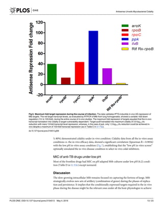
Fig 7. Correlation of Cidality under physiology of in-vitro vs. in-vivo. The low pH condition appears to be the ideal screening system
(R = 0.9856) showing linearity during a correlation of different in-vitro screens of alternate stress responses versus the in-vivo outcome
for selection of cidal inhibitors. REP = Replicating growth condition, Hpx = Hypoxia, Msx = Nitrogen depletion using L-methionine
sulphoxide, NO = Nitric oxide, NS = Nutrient starvation of Carbon, and LpH = low pH condition. A statistically positive correlation
(Spearman R = 0.9856) was observed between the in-vitro Low pH condition and the in-vivo outcome. The counts of in-vitro cidality
SCORE are available in Table B in S1 File.
doi:10.1371/journal.pone.0154513.g007
Antisense Unveils Mycobacterial Cidality
PLOS ONE | DOI:10.1371/journal.pone.0154513 May 4, 2016 14 / 23](https://image.slidesharecdn.com/457f3d5b-b6ef-429f-84d9-a99676deb689-160506012528/85/PLoSONE_AS_Mtb_2016-PDF-14-320.jpg)
![Helicobacter pylori [15,16,34]. It presents an excellent opportunity for exploring aroK target for
TB drug discovery. The in-vivo essentiality of aroK in Mtb has not been reported yet. However,
in the case of Acinetobacter baumannii, a very recent report has confirmed aroK as an in-vivo
essential enzyme [30,32].
In the present study, Mtb AroK performed consistently well across all the simulated physiol-
ogies in-vitro (cidality SCORE = 3.2) as well asunder in-vivo condition (2.4 log10cfu reduc-
tion;39.7-fold transcript repression in-vivo, Figs 1 and 3–5) despite a comparatively lower in-
vitro cidality SCORE, thus, delineating its cidality. Blocking aroK target kills Mtb under in-
vivo, clearly indicating nearly zero availability of amino acids (tryptophan, tyrosine, and phe-
nylalanine) in the in-vivo milieu. However, whether it will translate into a therapeutically valid
target for clinical usage is worth further investigations.
Our studies confirmed rpoB as the most vulnerable target with a mere 13-fold transcript
repression in-vivo (Table C in S1 File) translating into an excellent in-vivo cidality (3.9 log10
cfu reduction). Emerging from a good in-vitro cidality SCORE (7.1, Fig 4) with cidality under
low pH, it established that inhibiting rpoB with AS-therapeutics (unique MOA of blocking
mRNA specifically) is equipotent to rifampicin treatment, validating our experimental
approach. However, it’s another RNAP partner, the rpoC, demonstrated a lower “cidality
SCORE” (and a negligible cidality in low pH) compared to rpoB (5.2vs. 7.1, Fig 4), though mar-
ginally better cidality under replicating condition than rpoB (Fig 1). Despite a high (43-fold)
in-vivo transcript repression for rpoC, the cidality SCORE could, in fact, delineate rpoC as just
a marginally in-vivo bactericidal target (1.6 log10 cfu reduction) as compared to rpoB (Table C
in S1 File, Fig 5).
Targets, ilvB, and ppk were “highly in-vitro bactericidal” (Figs 1 and 4), but comparatively
less cidal targets under in-vivo condition (Fig 5); despite their best repressed in-vivo transcript
levels (ilvB = 31.1-fold, ppk = 103.6-fold; Table C in S1 File, Fig 6). One of the best in-vitro-
cidal targets, ilvB, demonstrated negligible in-vivo cidality (0.36 log10 cfu reduction, Fig 5).
This outcome is attributable to its auxotrophic nature [8,35] and suggests compensation of the
effect by the availability of trace amino acids (isoleucine, leucine, valine, and pantothenate)
under in-vivo milieu. It is hard to delineate attenuation and cidality, but probably ilvB flaunted
target attenuation under the in-vitro condition as well, because of the revival of additional colo-
nies upon extended incubation. The presence of trace amino acids (ILVP) nullified its cidality
potential [8] leading to attenuation. Other targets studied (ppk, rpoB, rpoC, aroK), did not dem-
onstrate this phenomenon, suggesting their bactericidal nature.
Next best in-vitro cidal target ppk (Figs 1 and 4), was marginally in-vivo-cidal (1.6 log10 cfu
reduction, Fig 5) despite its maximum in-vivo transcript repression (103.6-fold; Table C in S1
File, Fig 6). Since ppk is proven to be a stationary phase specific target [12], a long-term infec-
tion model in mice may be required to demonstrate its cidality. The in-vivo model used for the
present study was a hybrid model encompassing from acute to chronic infection states. We
intentionally infected mice with high dose (104
cfu/lung) to facilitate a measurable window for
cfu enumeration, whether the bacterial numbers increase (up to 108
cfu/lung) or reduce. To
begin with, this mouse infection model is an acute model harbouring replicating bacilli. But
with time (beyond four weeks) bacterial growth of WT control and the recombinant strains
slows down, it acquires more of a nonreplicating state in-vivo while the AS effect is still on.
Hence, the later part of the course of infection represents the AS-effect on a chronic disease.
Since AS-repression based cidality was observed beyond 4 weeks as well (Fig 5), it suggested a
possibility of killing Mtb even during the chronic phase. However, AS-based cidality studies in
a chronic mouse infection model are warranted. Upon a continuous expression of AS-RNA
under IPTG induction, the respective targets were being silenced during the course of infection
Antisense Unveils Mycobacterial Cidality
PLOS ONE | DOI:10.1371/journal.pone.0154513 May 4, 2016 15 / 23](https://image.slidesharecdn.com/457f3d5b-b6ef-429f-84d9-a99676deb689-160506012528/85/PLoSONE_AS_Mtb_2016-PDF-15-320.jpg)
![in mice from acute to chronic; hence, demonstrating a stage-specific cidality effect, if the target
was essential under the respective conditions.
Interestingly during 4–8 weeks period (chronic stage of infection) in particular, a further
growth of ppk recombinant was significantly inhibited (ns, P = 0.1080, one-way ANOVA, Dun-
nett’s multiple comparisons) by the antisense. Whether the bacterial count will reduce further
beyond 8-weeks, needs to be determined. However, we need to delayer it separately, with care-
ful investigations in a long term course-of-infection model beyond 8-weeks.
The cidality SCORE correlated well with the in-vivo data outcome in 4 out of 5 targets
(80%) confirming in-vivo-cidality. AroK target, which showed a moderate in-vitro cidality
under replicating condition, would have been missed out if had not been checked under alter-
nate physiological conditions as well. It outlines the importance of cidality-SCORE. A consis-
tent performance of aroK under all the physiological conditions tested demonstrated a cidality
SCORE of 3.2, and revealed its cidality potential; and hence, selected for in-vivo validation. It
emerged as a bactericidal target under in-vivo condition also, probably because it was cidal
under all the physiologies studied. RpoB was far more superior to rpoC. The cidality-SCORE
approach was a worthwhile investigation, implying that a target is preferred if it is cidal under
all the physiologies encountered in the host. It was apparently confirmed by rpoB and aroK,
but not by rpoC. The cidality SCORE could intricately delineate the in-vivo bactericidal poten-
tial of rpoB and rpoC targets. The only target which did not show in-vitro to in-vivo correlation
was ilvB, because of other issues like auxotrophy and attenuation observed in this study. Over-
all, the in-vitro cidality SCORE could predict the in-vivo cidality of the targets, irrespective of
their magnitudes. Since frontloading of all alternate physiological screens in parallel may not
be feasible; we questioned which screen out of the six conditions tested is the best in-vivo cidal-
ity predictor?
Antimycobacterials are tested under the replicating growth conditions in 7H9 media
[8,36,37], and confer limited translation of in-vitro cidality into the in-vivo efficacy, thus result-
ing in high attrition rate in drug discovery. This physiology of ‘in-vitro replication’ growth con-
dition does not represent the ‘actual microenvironment encountered under host immune
pressures’ to a large extent, thus overlooking the selection of potential in-vivo cidal com-
pounds. As a consequence, inappropriate compounds get selected. During the paradoxical
intra-macrophagic phase, the acidification renders Mtb almost inactive, even before it under-
stands the in-vivo milieu to adapt in [38]. The activation-specific pH of macrophage (~6.2),
further drops upon phagosome-lysosomal (P-L) fusion to ~4.5 within 15 to 60 minutes under
various immune pressures [39]. Despite this acidified external milieu of macrophage, the inter-
nal pH of Mtb (~7) remains neutral [40]. In order to refrain from the P-L fusion (Fig 2), and to
retain itself within the macrophage to progress further, Mtb invariably fools the system by sev-
eral mechanisms of interference by inducing virulence proteins and efficiently expressing mac-
rophage-specific survival genes [39,41]. The stage-specific transcriptome levels in macrophage
[42] fluctuate with the physiological transitions of replication and death rate (Table C in S1
File) in the host. The macrophage attack on Mtb is the 1st battle to be won by the host (Fig 2)
representing the most significant universal host-pathogen interaction. It highlights the poten-
tial link of low pH to the intraphagosomal survival of Mtb [42–45], urging researchers to simu-
late this ‘in-host’ stage under a single in-vitro screen to select bactericidal agents [43,44]. We
conducted comprehensive studies on in-vivo simulated microenvironments under in-vitro
conditions and their correlation versus in-vivo cidality data. This correlation from a limited set
of five genes investigated in the acute infection in BALB/c mouse point to low pH as probably
the most unfavourable microenvironment (R = 0.9856) encountered by Mtb, among the vari-
ous immune pressures (low pH, hypoxia, nutrient starvation, free radicals, etc.). However, sim-
ilar studies using a larger set of genes under all the in-vitro physiologies as well as more
Antisense Unveils Mycobacterial Cidality
PLOS ONE | DOI:10.1371/journal.pone.0154513 May 4, 2016 16 / 23](https://image.slidesharecdn.com/457f3d5b-b6ef-429f-84d9-a99676deb689-160506012528/85/PLoSONE_AS_Mtb_2016-PDF-16-320.jpg)
![relevant mouse models with human-like pathology may unleash these cidality correlations
better.
Target aroK, like rpoB, the best in-vivo-cidal target, demonstrated a predominant cidality in
the low pH screen (Table B in S1 File). It was interesting to discover that a seldom used, low
pH screen, correlating best with in-vivo confirmation of cidal targets (rpoB and aroK); can sta-
tistically (R = 0.9856) predict in-vivo cidality (Fig 7). Does it mean that the replicating condi-
tion (7H9 media) and the rest of the in-vitro conditions are obsolete? May be/ maybe not.
These correlations need to be verified in other mice strains like Kramnik that endows human
like lung lesions and chronic disease.
Under the starvation of essential nutrients (Carbon, Nitrogen) in the granuloma, the non-
replicating Mtb undergoes a global metabolic shift for energy conservation, shutting down
some of its pathways, re-routing Carbon flow from central fatty acid metabolism to lipid and
glutamate biosynthesis. Nitrogen is required in both replicating and non-replicating Mtb for
biosynthesis of amino acids, nucleotides, organic cofactors to control critical molecular events
(asparagine hydrolysis, ammonia release, pH buffering, growth) in the acidic environment,
with α-ketoglutarate/glutamate as the key ‘nodal-point of pathways’[46–50]. In this study,
most of the targets were cidal under in-vitro Nitrogen depletion condition (R = 0.4928), that
enhances acid stress in granuloma; and restores the activity of lysosomal hydrolases to kill Mtb
[46–50]. Thus, a “chicken or egg” conundrum, what triggers first? Are this Nitrogen depletion
and low pH conditions, part of a vicious cycle; or, operate in tandem metabolically, along with
other hostile conditions? Whatsoever, it requires a careful investigation and needs to be teased
apart meticulously. However, difficult to crack, but it will be exciting and path-breaking.
The low pH, is probably subsidiary to another useful host defence mechanism, nitric oxide
(NO, R = 0.5508), attacking Mtb with free radicals from innate immune cells; as the iNOS-/-
(inducible NO synthase-deficient) mice, are reported to succumb to Mtb infection [4]. How-
ever, the cidality data from hypoxia or replicating growth conditions failed to correlate with in-
vivo cidality outcome from BALB/c mouse model studies (Fig 7). Alternatively, it justifies the
use of an appropriate in-vivo model that exhibits hypoxic lesions (not formed in BALB/c
mouse) for better correlations.
Mtb infection in the BALB/c mouse leads to solid-granulomas harbouring primarily intra-
cellular bacteria, but these lesions are not hypoxic and do not best represent lung pathology of
human-TB [51]. Whereas, in the case of the human host, where mixes of both intracellular, as
well as extracellular Mtb populations, exist, other conditions like hypoxia, and nutrient starva-
tion may also play important roles. Though, the BALB/c mouse model has its limitations but is
still better than investigating the in-vitro conditions alone.
In the recent years, development of several improvised gene knock-out mouse models has
lead to better understanding of TB pathophysiology. These models display a human-like dis-
ease pathology in the lungs with hypoxia or other microenvironments of granuloma; harbour-
ing mixed populations of Mtb: 1) Kramnik mice strains (C3HeB/FeJ [52]; 2). NOD-SCID/γc
null of NSG model [53]; 3). iNOS-/- for NO synthase [4], etc. Each of these models has their
pros and cons [54].
BALB/c mice have a strong precedence of use for testing drug efficacy including the investi-
gational new drugs in the current pipeline, like TMC 207, PA824, SQ109, ADZ-5847, and Ben-
zothiazinones, [55–60] and for target essentiality [61,62]. Hence, we used BALB/c mice strain
in these studies. Although, BALB/c mice models are well validated for tuberculosis and are the
standard route for translation in drug discovery, but, for cidality studies under different physi-
ologies, it may not best represent the micro-environments and pathophysiology of human-TB.
The solid granulomas harbouring primarily intracellular bacteria do not particularly develop
hypoxia in BALB/c mice.
Antisense Unveils Mycobacterial Cidality
PLOS ONE | DOI:10.1371/journal.pone.0154513 May 4, 2016 17 / 23](https://image.slidesharecdn.com/457f3d5b-b6ef-429f-84d9-a99676deb689-160506012528/85/PLoSONE_AS_Mtb_2016-PDF-17-320.jpg)
![The low pH in-vitro screen emerging as the best-correlated predictor of in-vivo cidality,
from BALB/c mouse model, may be a partial conclusion because of the two reasons: 1). Despite
an effort to establish a hybrid of acute-chronic infection model in BALB/c, the high dose infec-
tion established represented mostly an acute-like infection with replicating bacteria analogous
to early human disease. Hence, in the acute phase of infection for the high fidelity translation,
the compound efficacy may also be evaluated in the low pH, in addition to the standard replica-
tion condition.2). The BALB/c mice used in the present study primarily form solid granuloma
(only a single pathophysiological lesion type) harbouring largely the intracellular Mtb, unlike
those observed (intra/extra-cellular Mtb with hypoxic microenvironment) in the cavitary
lesions of human TB patients. Therefore, it is pertinent to perform such correlation studies
using AS-recombinants of a larger set of target genes in humanised chronic infection models
(Kramnik-C3HeB/FeJ) that may unleash these cidality correlations better. Kramnik model rep-
resents both extra/intra-cellular pathogen populations, along with most of the cavitary lesion
types, as well as multiple stringent μ-environments of human-like granuloma that influence
the pathogen to modulate its survival-specific genes for adaptation [51,54].
Our treatment therapy is effective on the acute phase of infection; it may not reflect what
happens in the chronic phase. The low pH may not be important in general, as we earlier envis-
aged, but may be only relevant to the acute phase. The importance of the low pH or the appro-
priate screen in the chronic phase may be separately investigated.
Treatment of TB is complex; eradication of the dormant Mtb takes longer to treat due to
hide-n-seek being played by this pathogen [63]. It cannot be attributed only to poor bio-avail-
ability of drugs [64,65], but may primarily be dictated by either the physiologic heterogeneity
of bacteria in the tissues [66] or penetration of drugs in caseating foci/granuloma[66]. Rifampi-
cin and PZA, the best sterilizing drugs responsible for shortening treatment duration in
humans, penetrate better in to granulomas vs. moxifloxacin that largely concentrates in the
periphery [66,67]. Interestingly all of these best tuberculocidals exhibit good MIC in acidic pH
(Table D in S1 File), except isoniazid, which is inactive under acidic environment, hence, dem-
onstrates a reduced activity on intracellular Mtb. The alarming statistics [1] on drug resistance
in TB demand novel PZA-like sterilizing drugs that work best in low/acidic pH, kill non-repli-
cating populations of Mtb and shorten the treatment duration [38,68]. In a separate study, we
have demonstrated the importance of low pH screens by selecting PZA-hybrid molecules in-
vitro [69]. Recently, PZA was also reported to enhance the cidality of various combinations of
existing drugs [70]and is an integral component of emerging novel combinations in the clinic
like PAMZ (PA-824, Moxi, PZA), in STAND (Shortening Treatment by Advancing New
Drugs, www.clinicaltrial.com) clinical trial. Our findings unequivocally suggest that anti-TB
activity in the low pH environment may be predictive of in-vivo tuberculocidality, especially
for the acute phase of infection.
The low pH condition in-vivo appears to be a cumulative sum of various triggers and several
secondary mechanisms. These triggers need to be explored in intricate details, and their appro-
priateness to identify tuberculocidal therapeutics may be strengthened further using large com-
pound libraries and potential targets. The targets of stationary phase like ppk, need to be
explored in the long-term infection models of experimental TB.
Our studies on in-vivo AS-repression of cidal targets lead to the killing of Mtb in-vitro and
in-vivo, hence, demonstrating the bactericidal effect. It suggests that in the long run, AS-thera-
peutics can be explored in patients suffering from drug-sensitive and drug-resistant TB, for
which currently there are only a few effective drugs available. It is a futuristic goal, but AS-ther-
apeutics is an emerging radical approach to treating various diseases like anti-viral infections
[71] (Fomivirsen or Vitravene) anti-cholesterol [72] (mipomersen), or even anti-cancer [73]
(AP 12009) without any safety issues. However, as antibacterial, there is a need to develop
Antisense Unveils Mycobacterial Cidality
PLOS ONE | DOI:10.1371/journal.pone.0154513 May 4, 2016 18 / 23](https://image.slidesharecdn.com/457f3d5b-b6ef-429f-84d9-a99676deb689-160506012528/85/PLoSONE_AS_Mtb_2016-PDF-18-320.jpg)
![improved AS-delivery systems for its successful applications. A lot of investigations are under-
way to overcome significant obstacles towards the development of efficient delivery systems
like CPP (cell penetrating peptides), nanoparticles, or nanotubes, etc. [74,75] in the long pro-
cess. Some companies are developing the AS delivery systems for bacterial therapeutic AS, ISIS
Pharmaceuticals [ISIS Pharmaceuticals Inc.; now called Ionis pharmaceuticals http://www.
ionispharma.com] is the leader in the field of AS-therapeutics.
An astute observation through the lens of antisense into a kaleidoscope of physiology-spe-
cific gene expression under in-vivo pressures in mouse; or, simulated in-vitro screens; unrav-
elled a few secrets while a lot remain unrevealed.
Our studies with in-vivo AS-RNA-silencing of mycobacterial targets unravelled following
features on target cidality: 1). AroK emerged as the in-vivo cidal target under diverse physiolo-
gies. These findings suggest aroK a potential target for developing mycobactericidal agents. 2).
Use of rpoB target validated the entire concept on in-vitro to in-vivo translation. Like rpoB, aroK
demonstrated that a druggable target needs to be cidal under all the physiological states in the
host. 3). Cidality-SCORE is a rational approach to rank order cidality potential of the targets.
Cidality-SCORE positively correlated in-vitro to the in-vivo translation of 4 out of 5 targets
(rpoB, aroK, rpoC, and ppK) expressed under multiple physiologies. However, ilvB failed to trans-
late to in-vivo cidality due to its auxotrophic nature and hence attenuation. 4). Low pH appeared
to be an in-vivo predictor of cidality in acute infection. Hence, in the acute phase of infection for
the high fidelity translation, the compound efficacy may also be evaluated in the low pH, in addi-
tion to the standard replication condition. Further, in-vitro to in-vivo translation correlations
need to be studied in the chronic humanised models to narrow-down to a single in-vitro screen
that alone can predict in-vivo (acute-chronic) cidality potential of the targets/ inhibitors.
We used a small subset of only five, potentially cidal target genes. Studies with a larger set of
mixes of validated and unvalidated genes and their investigation in physiologically more rele-
vant humanised mouse disease models like Kramnik and others are required to unravel other
unrevealed shades.
Supporting Information
S1 Fig. Correlation of SPOT vs. cfu data of survival kinetics.
(TIF)
S1 File. “Table A in S1 File”, “Table B in S1 File”, “Table C in S1 File”, and “Table D in S1
File”.
(DOCX)
Acknowledgments
The authors acknowledge the contributions of Rajanna Subhash, Thimaiah Rajkumar, Zaheer
Basha for their technical support throughout the study; Sandeep Kannirasan for the artwork;
Sambandamurthy Vasan, Anirban Ghosh, Chandan Narayan, Bibhuti Mishra and Anisha
Ambady for various scientific contributions from time to time. The authors acknowledge the
anonymous reviewers for their critical review and enriching comments that helped in improv-
ing this manuscript.
Author Contributions
Conceived and designed the experiments: PK SD RKS SN AA SR. Performed the experiments:
PK UKS SG RKS NK NR. Analyzed the data: PK SD RKS SN AA SR. Contributed reagents/
materials/analysis tools: PK UKS SG NK NR. Wrote the paper: PK SD RKS AA SR SN.
Antisense Unveils Mycobacterial Cidality
PLOS ONE | DOI:10.1371/journal.pone.0154513 May 4, 2016 19 / 23](https://image.slidesharecdn.com/457f3d5b-b6ef-429f-84d9-a99676deb689-160506012528/85/PLoSONE_AS_Mtb_2016-PDF-19-320.jpg)
![22. Kaur P, Ghosh A, Krishnamurthy RV, Bhattacharjee DG, Achar V, Datta S, et al. A High-Throughput
Cidality Screen for Mycobacterium tuberculosis. PLoS One. 2015; 10:e0117577. doi: 10.1371/journal.
pone.0117577 PMID: 25693161
23. Cronin CA, Gluba W, Scrable H. The lac operator-repressor system is functional in the mouse. Genes
Dev. 2001; 15:1506–1517. doi: 10.1101/gad.892001 PMID: 11410531
24. Stevenson M, Carlisle R, Davies B, Preece C, Hammett M, Liu WL, et al. Development of a Positive-
readout Mouse Model of siRNA Pharmacodynamics. Mol Ther Nucleic Acids. 2013; 19:e133. doi: 10.
1038/mtna.2013.63
25. Jayaram R, Gaonkar S, Kaur P, Suresh BL, Mahesh BN, Jayashree R, et al. Pharmacokinetics-phar-
macodynamics of rifampin in an aerosol infection model of tuberculosis. Antimicrob Agents Chemother.
2003; 47:2118–2124. doi: 10.1128/AAC.47.7.2118–2124.2003 PMID: 12821456
26. Solapure S, Dinesh N, Shandil R, Ramachandran V, Sharma S, Bhattacharjee D, et al. In Vitro and In
Vivo Efficacy of β-Lactams against Replicating and Slowly Growing/Nonreplicating Mycobacterium
tuberculosis. Antimicrob Agents Chemother. 2013; 57: 2506–2510. doi: 10.1128/AAC.00023-13
PMID: 23507276
27. Choudhary E, Thakur P, Pareek M, Agarwal N. Gene silencing by CRISPR interference in mycobacte-
ria. Nat Commun. 2015; 6: 6267. doi: 10.1038/ncomms7267 PMID: 25711368
28. Manganelli R, Dubnau E, Tyagi S, Kramer FR, Smith I. Differential expression of 10 sigma factor genes
in Mycobacterium tuberculosis. Mol Microbiol. 1999; 31: 715–24. PMID: 10027986
29. Livak KJ, Schmittgen TD. Analysis of relative gene expression data using Real-Time Quantitative PCR
and the 2-[Delta][Delta]CT method. Methods. 2001; 25:402–408. PMID: 11846609
30. Sutton KA, Breen J, MacDonald U, Beanan JM, Olson R, Russo TA, et al. Structure of shikimate kinase,
an in vivo essential metabolic enzyme in the nosocomial pathogen Acinetobacter baumannii, in com-
plex with shikimate. Acta Crystallogr D Biol Crystallogr. 2015; 71:1736–1744. doi: 10.1107/
S139900471501189X PMID: 26249354
31. Umland TC, Schultz LW, MacDonald U, Beanan JM, Olson R, Russo TA. In vivo-validated essential
genes identified in Acinetobacter baumannii by using human ascites overlap poorly with essential
genes detected on laboratory media. MBio. 2012; 3(4). pii: e00113–112. doi: 10.1128/mBio.00113-12
PMID: 22911967
32. Sutton KA, Breen J, Russo TA, Schultz LW, Umland TC. Crystal structure of 5-enolpyruvylshikimate-3-
phosphate (EPSP) synthase from the ESKAPE pathogen Acinetobacter baumannii. Acta Crystallogr F
Struct Biol Commun. 2016; 72: 179–187. doi: 10.1107/S2053230X16001114 PMID: 26919521
33. Whipp MJ, Pittard AJ. A reassessment of the relationship between aroK- and aroL-encoded shikimate
kinase enzymes of Escherichia coli. J Bacteriol. 1995; 177:1627–1629. PMID: 7883721
34. Han C, Zhang J, Chen L, Chen K, Shen X, Jiang H. Discovery of Helicobacter pylori shikimate kinase
inhibitors: Bioassay and molecular modelling. Bioorg Med Chem. 2007; 15: 656–662. PMID: 17098431
35. Awasthy D, Gaonkar S, Shandil RK, Yadav R, Bharath S, Marcel N, et al. Inactivation of the ilvB1 gene
in Mycobacterium tuberculosis leads to branched-chain amino acid auxotrophy and attenuation of viru-
lence in mice. Microbiol. 2009; 155: 2978–2987. doi: 10.1099/mic.0.029884–0
36. Chung GA, Aktar Z, Jackson S, Duncan K. High-Throughput Screen for Detecting Antimycobacterial
Agents. Antimicrob Agents Chemother. 1995; 39:2235–2238. PMID: 8619574
37. Forbes L, Ebsworth-Mojica K, DiDone L, Li S-G, Freundlich JS, Connell N, et al. A High Throughput
Screening Assay for Anti-Mycobacterial small molecules based on adenylate kinase release as a reporter
of cell lysis. PLoS ONE. 2015; 10:e0129234. doi: 10.1371/journal.pone.0129234 PMID: 26098625
38. Zhang Y, Shi W, Zhang W, Mitchison DA. Mechanisms of pyrazinamide action and resistance, Micro-
biol Spectrum. 2014; 2: MGM2-0023-2013(2014). doi: 10.1128/microbiolspec.MGM2-0023-2013
39. Vandal OH, Nathan CF, Ehrt S. Acid Resistance in Mycobacterium tuberculosis. J Bacteriol. 2009a;
191:4714–4721.
40. Zhang Y, Scorpio A, Nikaido H, Sun Z. Role of Acid pH and Deficient Efflux of Pyrazinoic Acid in Unique
Susceptibility of Mycobacterium tuberculosisto Pyrazinamide. J Bacteriol. 1999; 181: 2044–2049.
PMID: 10094680
41. MacMicking JD, Taylor GA, McKinney JD. Immune control of tuberculosis by IFN-gamma-inducible
LRG-47. Science. 2003; 302:654–659. PMID: 14576437
42. Rohde KH, Veiga DFT, Caldwell S, Bala´zsi G, Russell DG. Linking the Transcriptional Profiles and the
Physiological States of Mycobacterium tuberculosis during an Extended Intracellular Infection. PLoS
Pathog. 2012; 8:e1002769. doi: 10.1371/journal.ppat.1002769 PMID: 22737072
43. Vandal OH, Pierini LM, Schnappinger D, Nathan CF, Ehrt S. A membrane protein preserves intrabac-
terial pH in intraphagosomal Mycobacterium tuberculosis. Nat Med. 2008; 14:849–854. doi: 10.1038/
nm.1795 PMID: 18641659
Antisense Unveils Mycobacterial Cidality
PLOS ONE | DOI:10.1371/journal.pone.0154513 May 4, 2016 21 / 23](https://image.slidesharecdn.com/457f3d5b-b6ef-429f-84d9-a99676deb689-160506012528/85/PLoSONE_AS_Mtb_2016-PDF-21-320.jpg)

This study aimed to investigate the bactericidal potential of Mycobacterium tuberculosis targets under various in vivo simulated in vitro conditions and in vivo in mice. Using antisense RNA to inhibit five target genes, the study evaluated target cidality under six physiological conditions in vitro and in vivo. It identified aroK, which encodes shikimate kinase, as an in vivo bactericidal target based on correlations between in vitro and in vivo cidality data. The study suggests that the low pH in vitro model best predicts in vivo cidality and identifies targets with potential for anti-tuberculosis drug development.


