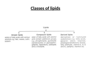
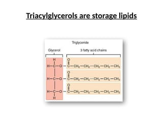
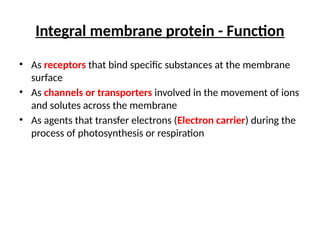
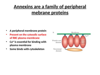
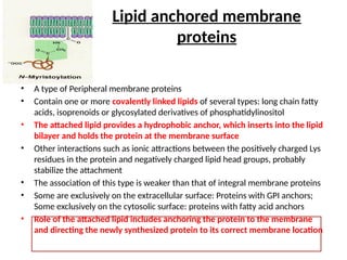
Classes of lipids
Triacylglycerols are storage lipids
Phospholipid
Glycerophospholipids
Sphingomyelins
Glycolipid
Cerebroside is a glycosphingolipid
Ganglioside is another glycosphingolipid
Sterol
• Cholesterol in animals
• Stigmasterol in plants
• Egrosterol in fungi
CELL MEMBRANE
Cytoplasmic
membrane
• Thin structure (8nm) that completely surrounds the cell
• Critical barrier separating the cytoplasm from its environment
• Impermeable to most polar or charged solutes, but permeable to nonpolar
compounds
• Fluid mosaic model structure
• Individual lipid and protein molecules free to move laterally in the plane of the
membrane
• Leakage or lysis causes cell death
• Highly selective barrier, enabling a cell to concentrate specific metabolites and
excrete waste materials
• Appear trilaminar when viewed in cross section with the electron microscope
•The principal components of the plasma membrane are lipids (predominantly
phospholipids and cholesterol; sphingolipids), proteins and carbohydrate groups
that are attached to some of the lipids and proteins
•Phospholipids contain both highly hydrophobic and relatively hydrophilic
moieties
•Different types of phospholipids due to variation in the nature of (i) fatty acids
and (ii) phosphate containing group attached to the glycerol backbone
•Cholesterol, another lipid composed of four fused carbon rings, is found
alongside phospholipids in the core of the membrane
•Carbohydrate groups are present only on the outer surface of the plasma
membrane and are attached to proteins, forming glycoproteins or lipids,
forming glycolipids. The proportions of proteins, lipids, and carbohydrates in the
plasma membrane vary between different types of cells. For a typical human cell,
however, proteins account for about 50 percent of the composition by mass, lipids
(of all types) account for about 40 percent, and the remaining 10 percent comes
from carbohydrates
Fluid Mosaic Model
• Proposed by Singer and Nicholson in 1972
• Phospholipids and sterols form a lipid bilayer in which the non-polar
regions of the lipid molecules face each other at the core of the bilayer
and their polar head groups face outward
• In this bilayer sheet, proteins are embedded at irregular intervals, held by
hydrophobic interactions between the membrane lipid and hydrophobic
domains of the proteins.
• The individual lipid and protein units in a membrane form a fluid mosaic
• Some proteins protrude from one side of the membrane only, others have
domains exposed on both sides.
• The membrane mosaic is fluid because most of the interactions among
its components is non-covalent, leaving individual lipid and protein
molecules free to move laterally in the plane of the membrane
The relative proportion of proteins and lipids
vary
• The relative proportion of proteins and lipids vary with the type of
membranes.
• Proportion of lipids and proteins in the membrane is based on the
function of that membrane
• Membrane with more proportion of lipids than proteins: myelin sheath
in neuron. Here lipids function as insulator and helps in transmission of
electrical impulses.
• Membrane with more proteins than lipids: membranes in bacterial cell,
mitochondria, chloroplasts. In bacterial cell membrane the proteins
function as enzymes producing energy, synthesizing lipids, export of
proteins, cell division etc.
Distribution of lipids (Asymmetric)
• Each kingdom, each species, each tissue or each cell type and the organelles
of each type have a characteristic set of membrane lipids.
• The basis of the experimental proof: Lipid digesting enzymes can not penetrate
the plasma membrane and so they digest lipids that are present in the outer
leaflet of the bilayer only
• Lipids are asymmetrically distributed between the two monolayers of the
bilayer
• Glycolipids mostly in the outer layer of membrane
• The carbohydrates of the glycolipids of the RBC plasma membrane determine
ABO blood-group
As in erythrocyte membrane
Role of lipids in cell structure and function
1) Because of hydrophobic tail lipid bilayer is never exposed to aqueous
solution, consequently membranes have no free edge; they are always
continuous, unbroken structures. As a result, membranes form
extensive, interconnected networks within the cell
2) Because of flexibility of lipid bilayer, membranes are deformable and
their overall shape can change during locomotion, cell division etc
3) Lipid bilayer is thought to facilitate the regulated fusion or budding of
membranes
Liposome – a novel drug
delivery system
• Greek words, “lipos” meaning fat and “soma” meaning body.
• When lipids are placed in contact with water, the unfavorable interactions of the
hydrophobic segments of the molecule with the solvent result in the self
assembly of lipids, often in the form of liposomes.
• Liposomes consist of an aqueous core surrounded by a lipid bilayer, much like a
membrane, separating the inner aqueous core from the bulk outside.
• The unique feature of liposomes is their ability to compartmentalize and
solubilize both hydrophilic and hydrophobic materials by nature
• This unique feature, coupled with biocompatibility and biodegradability make
liposomes very attractive as drug delivery vehicles
• Specific proteins in walls to bind selectively to the surface of particular target cell
• Outer coating of PEG to inhibit phagocytosis
Distribution of proteins is asymmetric
• The protein composition of membranes from different sources varies
even more widely then their lipid composition, reflecting functional
specialization.
• FOR EXAMPLE: >90% of the membrane proteins in the light absorbing
region of rod cells is glycoprotein RHODOPSIN
• Orientation of proteins in the bilayer is asymmetric, giving the
membrane sidedness and reflecting functional asymmetry
• Glycoproteins are present in outer layer of cell membrane but rarely in
intracellular membranes like membranes of mitochondria or chloroplast
Membrane lipids are in constant motion
• Predominantly, 4 types of motion
 Swinging movement
 Rotation
 Lateral diffusion
 Transverse diffusion or flip-flop diffusion (catalyzed by flippase) RARE
• Motion of lipids and sterol imparts fluidity of membrane
Lipid Aggregates
Depending on precise conditions and shape of the lipids, three types of lipid
aggregates can form when amphipathic lipids are mixed with water
(i) micelles (when cross-sectional area of the head group is greater than that of
the acyl side chains). Free fatty acids
(ii) bilayer (when cross-sectional area of the head and acyl side chains are
similar). Glycerophospholipid
(iii) liposome (formed spontaneously from bilayer because the hydrophobic
regions at its edges are transiently in contact with water)
First living cell probably resembled liposomes
Membrane Proteins
Membrane proteins
• Proteins account for approximately 70% of the mass of the membrane in
prokaryotes and approximately 50% in eukaryotes
• Lipid rich membrane is myelin sheath in neuron
• Three types of membrane proteins
• Integral membrane proteins
• Peripheral membrane proteins
• lipid anchored membrane proteins
Integral /intrinsic protein
• Like membrane lipids are amphipatihic
• Their hydrophobic regions are buried in the lipid while the hydrophilic
portions project from the membrane surface
• Associated with the fatty acid layer in lipid core forming hydrophobic bonds
where the predominant amino acids are hydrophobic in nature
• Some are transmembrane traversing the membrane throughout
• Extended surfaces at both ends contain hydrophilic amino acids resulting
ionic interaction with polar heads and other proteins
• Strongly bound to the membrane and can not be easily extracted from
membranes
• Can be released by treating membrane with agents that interfere with
hydrophobic interactions like detergents (SDS), organic solvents or
denaturants
• Insoluble in aqueous solution when even freed of lipids
• 70-80% of total membrane proteins
• Can diffuse laterally in the membrane to new locations but can not flip-flop
Integral membrane protein - Function
• As receptors that bind specific substances at the membrane
surface
• As channels or transporters involved in the movement of ions
and solutes across the membrane
• As agents that transfer electrons (Electron carrier) during the
process of photosynthesis or respiration
Glycophorin is an integral membrane protein
• Present in human RBC
• N-terminal domain is outside of the
membrane, containing polar amino acids and
highly glycosylated
• 15 O-linked tetrasaccharides
• 1 N-linked oligosaccharide
• Transmembrane domain has hydrophobic
amino acids
• C-terminal domain is cytosolic and contains
polar amino acids
• Rich in sialic acids, a negatively charged sugar,
that carry blood group antigenic
determinants and serve as ligands for
influenza virus
• The carbohydrate units of glycophorins give
RBC a very hydrophilic, anionic coat, which
enables them to circulate without adhering to
other cells and endothelial walls
Peripheral/extrinsic protein
• Associate with the membrane through electrostatic interactions and
hydrogen bonding with the hydrophilic domain of integral proteins and with
the polar head groups of membrane lipids
• Loosely connected to the membrane and can be easily removed
• Can be released by relatively mild treatments that interfere with
electrostatic interactions or break hydrogen bonds like changes in pH, Ca2+
chelating agent, urea, carbonate etc.
• Easily soluble in aqueous solution and make up about 20-30% of total
membrane proteins
• May be present in the outer surface (environment or periplasm) and inner
side of the membrane (cytoplasm. Do not span the membrane.
Peripheral protein - Function
• Provide mechanical support for the membrane
• Function as an anchor for integral membrane protein
• Serve as regulators of membrane-bound enzymes
• Serve as specialized coats
• Factors that transmit transmembrane signals
Annexins are a family of peripheral
mebrane proteins
• A peripheral membrane protein
• Present on the cytosolic surface
of RBC plasma membrane
• Ca2+
is essential for binding with
plasma membrane
• Some binds with cytoskeleton
Integral and peripheral membrane proteins
are different
Lipid anchored membrane
proteins
• A type of Peripheral membrane proteins
• Contain one or more covalently linked lipids of several types: long chain fatty
acids, isoprenoids or glycosylated derivatives of phosphatidylinositol
• The attached lipid provides a hydrophobic anchor, which inserts into the lipid
bilayer and holds the protein at the membrane surface
• Other interactions such as ionic attractions between the positively charged Lys
residues in the protein and negatively charged lipid head groups, probably
stabilize the attachment
• The association of this type is weaker than that of integral membrane proteins
• Some are exclusively on the extracellular surface: Proteins with GPI anchors;
Some exclusively on the cytosolic surface: proteins with fatty acid anchors
• Role of the attached lipid includes anchoring the protein to the membrane
and directing the newly synthesized protein to its correct membrane location
GPI linked proteins are lipid anchored
membrane proteins
Experiment to determine localization of
membrane proteins
IEA: isoethionylacetimidate,
impermeable to hydrophobic core
EA: ethylacetimidate, permeable
Membrane
strengthening agent
• Sterols in the eukaryotic membrane and hopanoids in the prokaryotic
membrane
• Sterols are four ringed, rigid, planar molecule.
• Sterol content of membrane determine transition temperature (temperature
above which paracrystalline solid changes to fluid state)
• FUNCTION- Regulate the extremes of solidity and fluidity of the membrane
(i) Below transition temperature, sterol insertion prevents highly ordered
packing of fatty acyl chains and thus increase membrane fluidity
(ii) Above thermal transition point, the rigid ring system of sterol reduces the
freedom of neighbouring fatty acyl chains to move by rotation and thus
reduces fluidity in the core of the bilayer
Lipid Rafts
• Lipid rafts are self-assembled microdomains of cholesterol and
sphingolipids seen to form when artificial lipid bilayers are prepared
• These areas are more gelated and highly ordered than surrounding
regions consisting primarily of phosphoglycerides
• When added to these artificial blayers, certain proteins tend to become
concentrated in the lipid rafts (example GPI-anchored protein)
• Presence of lipid rafts in living cells could not be demonstrated
• Lipid rafts concentrate cell surface receptors for signal transduction
END

cell membrane (structure) and function.pptx

  • 1.
  • 3.
  • 4.
  • 5.
  • 6.
  • 8.
  • 9.
    Cerebroside is aglycosphingolipid
  • 10.
    Ganglioside is anotherglycosphingolipid
  • 11.
    Sterol • Cholesterol inanimals • Stigmasterol in plants • Egrosterol in fungi
  • 12.
  • 13.
    Cytoplasmic membrane • Thin structure(8nm) that completely surrounds the cell • Critical barrier separating the cytoplasm from its environment • Impermeable to most polar or charged solutes, but permeable to nonpolar compounds • Fluid mosaic model structure • Individual lipid and protein molecules free to move laterally in the plane of the membrane • Leakage or lysis causes cell death • Highly selective barrier, enabling a cell to concentrate specific metabolites and excrete waste materials • Appear trilaminar when viewed in cross section with the electron microscope
  • 15.
    •The principal componentsof the plasma membrane are lipids (predominantly phospholipids and cholesterol; sphingolipids), proteins and carbohydrate groups that are attached to some of the lipids and proteins •Phospholipids contain both highly hydrophobic and relatively hydrophilic moieties •Different types of phospholipids due to variation in the nature of (i) fatty acids and (ii) phosphate containing group attached to the glycerol backbone •Cholesterol, another lipid composed of four fused carbon rings, is found alongside phospholipids in the core of the membrane •Carbohydrate groups are present only on the outer surface of the plasma membrane and are attached to proteins, forming glycoproteins or lipids, forming glycolipids. The proportions of proteins, lipids, and carbohydrates in the plasma membrane vary between different types of cells. For a typical human cell, however, proteins account for about 50 percent of the composition by mass, lipids (of all types) account for about 40 percent, and the remaining 10 percent comes from carbohydrates
  • 17.
    Fluid Mosaic Model •Proposed by Singer and Nicholson in 1972 • Phospholipids and sterols form a lipid bilayer in which the non-polar regions of the lipid molecules face each other at the core of the bilayer and their polar head groups face outward • In this bilayer sheet, proteins are embedded at irregular intervals, held by hydrophobic interactions between the membrane lipid and hydrophobic domains of the proteins. • The individual lipid and protein units in a membrane form a fluid mosaic • Some proteins protrude from one side of the membrane only, others have domains exposed on both sides. • The membrane mosaic is fluid because most of the interactions among its components is non-covalent, leaving individual lipid and protein molecules free to move laterally in the plane of the membrane
  • 20.
    The relative proportionof proteins and lipids vary • The relative proportion of proteins and lipids vary with the type of membranes. • Proportion of lipids and proteins in the membrane is based on the function of that membrane • Membrane with more proportion of lipids than proteins: myelin sheath in neuron. Here lipids function as insulator and helps in transmission of electrical impulses. • Membrane with more proteins than lipids: membranes in bacterial cell, mitochondria, chloroplasts. In bacterial cell membrane the proteins function as enzymes producing energy, synthesizing lipids, export of proteins, cell division etc.
  • 21.
    Distribution of lipids(Asymmetric) • Each kingdom, each species, each tissue or each cell type and the organelles of each type have a characteristic set of membrane lipids. • The basis of the experimental proof: Lipid digesting enzymes can not penetrate the plasma membrane and so they digest lipids that are present in the outer leaflet of the bilayer only • Lipids are asymmetrically distributed between the two monolayers of the bilayer • Glycolipids mostly in the outer layer of membrane • The carbohydrates of the glycolipids of the RBC plasma membrane determine ABO blood-group As in erythrocyte membrane
  • 22.
    Role of lipidsin cell structure and function 1) Because of hydrophobic tail lipid bilayer is never exposed to aqueous solution, consequently membranes have no free edge; they are always continuous, unbroken structures. As a result, membranes form extensive, interconnected networks within the cell 2) Because of flexibility of lipid bilayer, membranes are deformable and their overall shape can change during locomotion, cell division etc 3) Lipid bilayer is thought to facilitate the regulated fusion or budding of membranes
  • 23.
    Liposome – anovel drug delivery system • Greek words, “lipos” meaning fat and “soma” meaning body. • When lipids are placed in contact with water, the unfavorable interactions of the hydrophobic segments of the molecule with the solvent result in the self assembly of lipids, often in the form of liposomes. • Liposomes consist of an aqueous core surrounded by a lipid bilayer, much like a membrane, separating the inner aqueous core from the bulk outside. • The unique feature of liposomes is their ability to compartmentalize and solubilize both hydrophilic and hydrophobic materials by nature • This unique feature, coupled with biocompatibility and biodegradability make liposomes very attractive as drug delivery vehicles • Specific proteins in walls to bind selectively to the surface of particular target cell • Outer coating of PEG to inhibit phagocytosis
  • 24.
    Distribution of proteinsis asymmetric • The protein composition of membranes from different sources varies even more widely then their lipid composition, reflecting functional specialization. • FOR EXAMPLE: >90% of the membrane proteins in the light absorbing region of rod cells is glycoprotein RHODOPSIN • Orientation of proteins in the bilayer is asymmetric, giving the membrane sidedness and reflecting functional asymmetry • Glycoproteins are present in outer layer of cell membrane but rarely in intracellular membranes like membranes of mitochondria or chloroplast
  • 25.
    Membrane lipids arein constant motion • Predominantly, 4 types of motion  Swinging movement  Rotation  Lateral diffusion  Transverse diffusion or flip-flop diffusion (catalyzed by flippase) RARE • Motion of lipids and sterol imparts fluidity of membrane
  • 26.
    Lipid Aggregates Depending onprecise conditions and shape of the lipids, three types of lipid aggregates can form when amphipathic lipids are mixed with water (i) micelles (when cross-sectional area of the head group is greater than that of the acyl side chains). Free fatty acids (ii) bilayer (when cross-sectional area of the head and acyl side chains are similar). Glycerophospholipid (iii) liposome (formed spontaneously from bilayer because the hydrophobic regions at its edges are transiently in contact with water) First living cell probably resembled liposomes
  • 27.
  • 28.
    Membrane proteins • Proteinsaccount for approximately 70% of the mass of the membrane in prokaryotes and approximately 50% in eukaryotes • Lipid rich membrane is myelin sheath in neuron • Three types of membrane proteins • Integral membrane proteins • Peripheral membrane proteins • lipid anchored membrane proteins
  • 30.
    Integral /intrinsic protein •Like membrane lipids are amphipatihic • Their hydrophobic regions are buried in the lipid while the hydrophilic portions project from the membrane surface • Associated with the fatty acid layer in lipid core forming hydrophobic bonds where the predominant amino acids are hydrophobic in nature • Some are transmembrane traversing the membrane throughout • Extended surfaces at both ends contain hydrophilic amino acids resulting ionic interaction with polar heads and other proteins • Strongly bound to the membrane and can not be easily extracted from membranes • Can be released by treating membrane with agents that interfere with hydrophobic interactions like detergents (SDS), organic solvents or denaturants • Insoluble in aqueous solution when even freed of lipids • 70-80% of total membrane proteins • Can diffuse laterally in the membrane to new locations but can not flip-flop
  • 31.
    Integral membrane protein- Function • As receptors that bind specific substances at the membrane surface • As channels or transporters involved in the movement of ions and solutes across the membrane • As agents that transfer electrons (Electron carrier) during the process of photosynthesis or respiration
  • 32.
    Glycophorin is anintegral membrane protein • Present in human RBC • N-terminal domain is outside of the membrane, containing polar amino acids and highly glycosylated • 15 O-linked tetrasaccharides • 1 N-linked oligosaccharide • Transmembrane domain has hydrophobic amino acids • C-terminal domain is cytosolic and contains polar amino acids • Rich in sialic acids, a negatively charged sugar, that carry blood group antigenic determinants and serve as ligands for influenza virus • The carbohydrate units of glycophorins give RBC a very hydrophilic, anionic coat, which enables them to circulate without adhering to other cells and endothelial walls
  • 35.
    Peripheral/extrinsic protein • Associatewith the membrane through electrostatic interactions and hydrogen bonding with the hydrophilic domain of integral proteins and with the polar head groups of membrane lipids • Loosely connected to the membrane and can be easily removed • Can be released by relatively mild treatments that interfere with electrostatic interactions or break hydrogen bonds like changes in pH, Ca2+ chelating agent, urea, carbonate etc. • Easily soluble in aqueous solution and make up about 20-30% of total membrane proteins • May be present in the outer surface (environment or periplasm) and inner side of the membrane (cytoplasm. Do not span the membrane.
  • 36.
    Peripheral protein -Function • Provide mechanical support for the membrane • Function as an anchor for integral membrane protein • Serve as regulators of membrane-bound enzymes • Serve as specialized coats • Factors that transmit transmembrane signals
  • 37.
    Annexins are afamily of peripheral mebrane proteins • A peripheral membrane protein • Present on the cytosolic surface of RBC plasma membrane • Ca2+ is essential for binding with plasma membrane • Some binds with cytoskeleton
  • 38.
    Integral and peripheralmembrane proteins are different
  • 39.
    Lipid anchored membrane proteins •A type of Peripheral membrane proteins • Contain one or more covalently linked lipids of several types: long chain fatty acids, isoprenoids or glycosylated derivatives of phosphatidylinositol • The attached lipid provides a hydrophobic anchor, which inserts into the lipid bilayer and holds the protein at the membrane surface • Other interactions such as ionic attractions between the positively charged Lys residues in the protein and negatively charged lipid head groups, probably stabilize the attachment • The association of this type is weaker than that of integral membrane proteins • Some are exclusively on the extracellular surface: Proteins with GPI anchors; Some exclusively on the cytosolic surface: proteins with fatty acid anchors • Role of the attached lipid includes anchoring the protein to the membrane and directing the newly synthesized protein to its correct membrane location
  • 40.
    GPI linked proteinsare lipid anchored membrane proteins
  • 41.
    Experiment to determinelocalization of membrane proteins IEA: isoethionylacetimidate, impermeable to hydrophobic core EA: ethylacetimidate, permeable
  • 43.
    Membrane strengthening agent • Sterolsin the eukaryotic membrane and hopanoids in the prokaryotic membrane • Sterols are four ringed, rigid, planar molecule. • Sterol content of membrane determine transition temperature (temperature above which paracrystalline solid changes to fluid state) • FUNCTION- Regulate the extremes of solidity and fluidity of the membrane (i) Below transition temperature, sterol insertion prevents highly ordered packing of fatty acyl chains and thus increase membrane fluidity (ii) Above thermal transition point, the rigid ring system of sterol reduces the freedom of neighbouring fatty acyl chains to move by rotation and thus reduces fluidity in the core of the bilayer
  • 45.
    Lipid Rafts • Lipidrafts are self-assembled microdomains of cholesterol and sphingolipids seen to form when artificial lipid bilayers are prepared • These areas are more gelated and highly ordered than surrounding regions consisting primarily of phosphoglycerides • When added to these artificial blayers, certain proteins tend to become concentrated in the lipid rafts (example GPI-anchored protein) • Presence of lipid rafts in living cells could not be demonstrated • Lipid rafts concentrate cell surface receptors for signal transduction
  • 46.