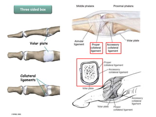
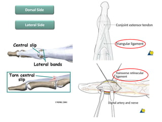
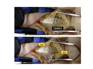
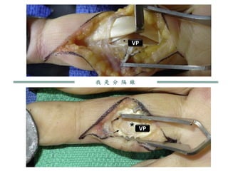
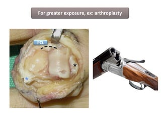
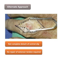
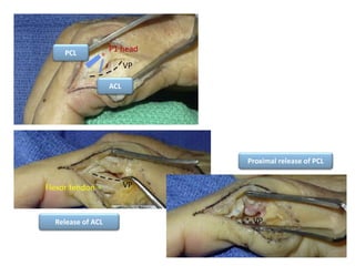
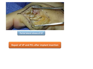
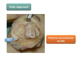
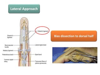
Volume 41, published 2016
Received for publication August 11, 2015
Accepted in revised form November 15, 2015
Impact Factor: 1.64
Andre Eu-Jin Cheah, MD, MBA
Assistant Professor
Department of Orthopaedic Surgery
Yong Loo Lin School of Medicine
National University of Singapore
Consultant
Department of Hand & Reconstructive
Microsurgery
University Orthopaedics, Hand & Reconstructive
Microsurgery Cluster
National University Health System, Singapore
Jeffrey Yao, MD
Associate Professor
Robert A. Chase Hand and Upper Limb Center,
Department of Orthopaedic Surgery,
Stanford University Medical Center,
Redwood City, CA
Affiliation
International Wrist Investigators Workshop
American Academy of Orthopaedic Surgeons
American Society for Surgery of the Hand
American Association for Hand Surgery
Arthroscopy Association of North America
Outline
• Surgical Anatomy
• Introduction
• Surgical Approach
– Volar approach
– Dorsal approach
– Lateral approach
• Pearls and Pitfalls
• Complications
• Recommendations
• Surgical Anatomy
• Introduction
• Surgical Approach
– Volar approach
– Dorsal approach
– Lateral approach
• Pearls and Pitfalls
• Complications
• Recommendations
Three sided box
Ventral Side
Dorsal Side
Lateral Side
• Surgical Anatomy
• Introduction
• Surgical Approach
– Volar approach
– Dorsal approach
– Lateral approach
• Pearls and Pitfalls
• Complications
• Recommendations
PIPJ:
Arthropathy
Fracture
Dislocation
Malunion
Traditional:
Dorsal approach
Extensor
damage
Post OP
swelling
PIPJ:
Arthropathy
Fracture
Dislocation
Malunion
Traditional:
Dorsal approach
Extensor
damage
Post OP
swelling
Volar approach
Lateral approach
• Surgical Anatomy
• Introduction
• Surgical Approach
– Volar approach
– Dorsal approach
– Lateral approach
• Pearls and Pitfalls
• Complications
• Recommendations
Volar Approach
Implant arthroplasty
ORIF
Corrective osteomy
Hemi-hamate arthroplasty
Volar plate arthroplasty
Bruner
Bruner-midlaterral
hybrid
Zigzag
C2A3C1
A2
A4
我 是 分 隔 線
VP
VP
ACL
VP
ACL
VP
For greater exposure, ex: arthroplasty
PCL
Alternate Approach by Simmen
Dorsal Approach
Implant arthroplasty
ORIF
Corrective osteomy
Midline longitudinal
Curvilinear
Lazy S
Preserve dorsal vein as
much as possible
Central slip insertion
Lateral band
Approach by Swanson
Central slip insertion
P1 head
Central slip insertion
P1 head
Central slip insertion
P1 head
Collateral ligament
Reattach central slip
Alternate Approach by Chamay
Restore tension of
extensor apparatus
Collateral ligament
Alternate Approach
Not complete detach of central slip
No repair of extensor tendon required
Lateral Approach
Implant arthroplasty
ORIF
Middle of P1
Middle of P2
PCL
ACL
VP
P1 head
Release of ACL
Proximal release of PCL
VPFlexor tendon
VP
Partial lateral release of VP
Repair of VP and PCL after implant insertion
VP
• Surgical Anatomy
• Introduction
• Surgical Approach
– Volar approach
– Dorsal approach
– Lateral approach
• Pearls and Pitfalls
• Complications
• Recommendations
Preserve neurovascular
bundle
Volar Approach
PCL
Single FDS slip release
Proximal spliting of FDS
Dorsal Approach
Conservation of the central slip
Chamay appraoch Restore tension
Bias dissection to dorsal half
Lateral Approach
Shotgunned?
Recession but not complete release
Without affecting finger function or lateral stability
• Surgical Anatomy
• Introduction
• Surgical Approach
– Volar approach
– Dorsal approach
– Lateral approach
• Pearls and Pitfalls
• Complications
• Recommendations
Extensor lag of 5’-20 despite approach
AROM poor in arthroplasty compared with short term
fracture fixation
Decrease of DIP AROM disturbance to lateral band
• Surgical Anatomy
• Introduction
• Surgical Approach
– Volar approach
– Dorsal approach
– Lateral approach
• Pearls and Pitfalls
• Complications
• Recommendations
Central slip reconstruction
FDS tenodesis
Access to base of P2
Access to head of P1
Dorsal
Volar
Volar
Dorsal
Guided by Surgeon’s experience !!!
PIP joint approach

PIP joint approach

Editor's Notes

  • #4 Impact factor 0.47
  • #10 lateral bands are stabilized centrally by the triangular ligament. And laterally by TRL TRL: originates from the flexor sheath at the level of the PIP joint and inserts into the lateral bands
  • #15 2, 3 more surgical exposure
  • #18 Left:Proximal interphalangeal joint VP (asterisk) is accessed after retracting the flexor tendons to one side. Right:Alternative route to the VP (asterisk) between the 2 slips of the FDS
  • #21 the proximal attachment of the PCLs may be released proximally or distally to allow the joint to be shotgunned open
  • #22 The flexor sheath and ACLs are incised between the A2 and A4 pulley on either side of the VP
  • #23 2, 3 more surgical exposure
  • #25 Longitudinal split of the entire central slip with elevation of the 2 halves laterally with each lateral band, to expose the articular surface and the collateral ligaments Black and red asterisks show the central slip insertion and P1 head, respectively. Dashed lines indicate the split extensor tendon. proximal release of one collateral ligament (blue asterisk). Black asterisk shows the central slip insertion; dashed line shows the intact collateral ligament.
  • #26 Black and red asterisks show the central slip insertion and P1 head, respectively. Dashed lines indicate the split extensor tendon. proximal release of one collateral ligament (blue asterisk). Black asterisk shows the central slip insertion; dashed line shows the intact collateral ligament.
  • #27 Black and red asterisks show the central slip insertion and P1 head, respectively. Dashed lines indicate the split extensor tendon. proximal release of one collateral ligament (blue asterisk). Black asterisk shows the central slip insertion; dashed line shows the intact collateral ligament.
  • #28 2 drill holes are made at the base of P2 at the insertion of the central slip, a nonabsorbable 3-0 or 4-0 suture is passed through these holes, and a subsequent mattress repair of the central slip is done
  • #29 Distally based triangular extensor tendon flap designed (dashed line). Asterisk shows the central slip attachment. The triangular tendon flap is then raised to expose the distal P1, collateral ligaments (blue asterisks), and articular surface while preserving the central slip attachment (black asterisk).
  • #30 Interval between the central slip and lateral bands can be used as access to the PIP joint. The black asterisk shows the central slip attachment, the red asterisk indicates the P1 head, and the dashed line shows the tendon split.
  • #31 A midlateral skin incision is made from the middle of the P1 to the middle of the P2
  • #39 A window is made in the lateral surface of the flexor sheath to identify and protect the flexor tendons before the VP (black asterisk) is incised
  • #40  recession but not complete release
  • #42 Extensor lag of 5’-20’in every approach AROM poor in arthroplasty compared with short term fracture fixationrecurring soft tissue change in arthrosis Decrease of DIP AROM disturbance to lateral band