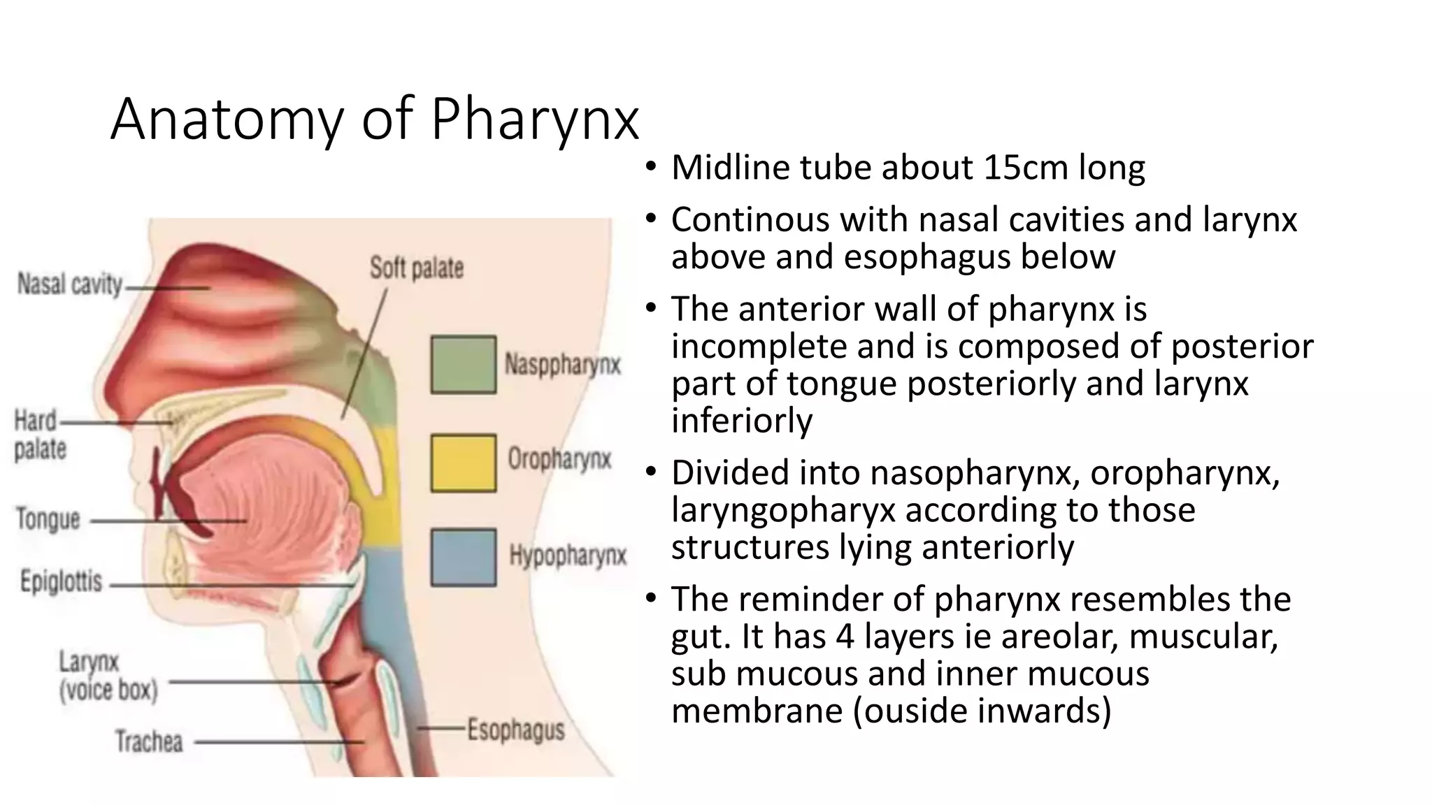
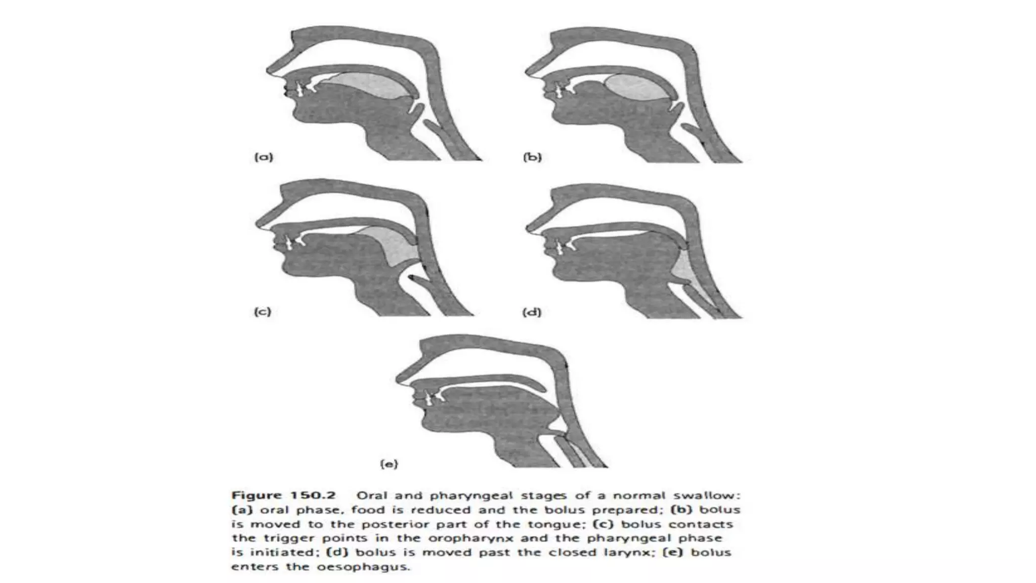
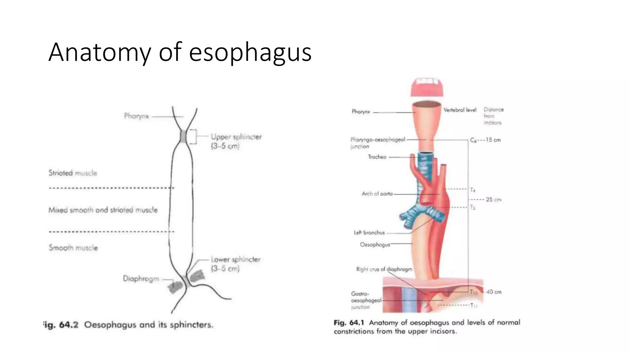
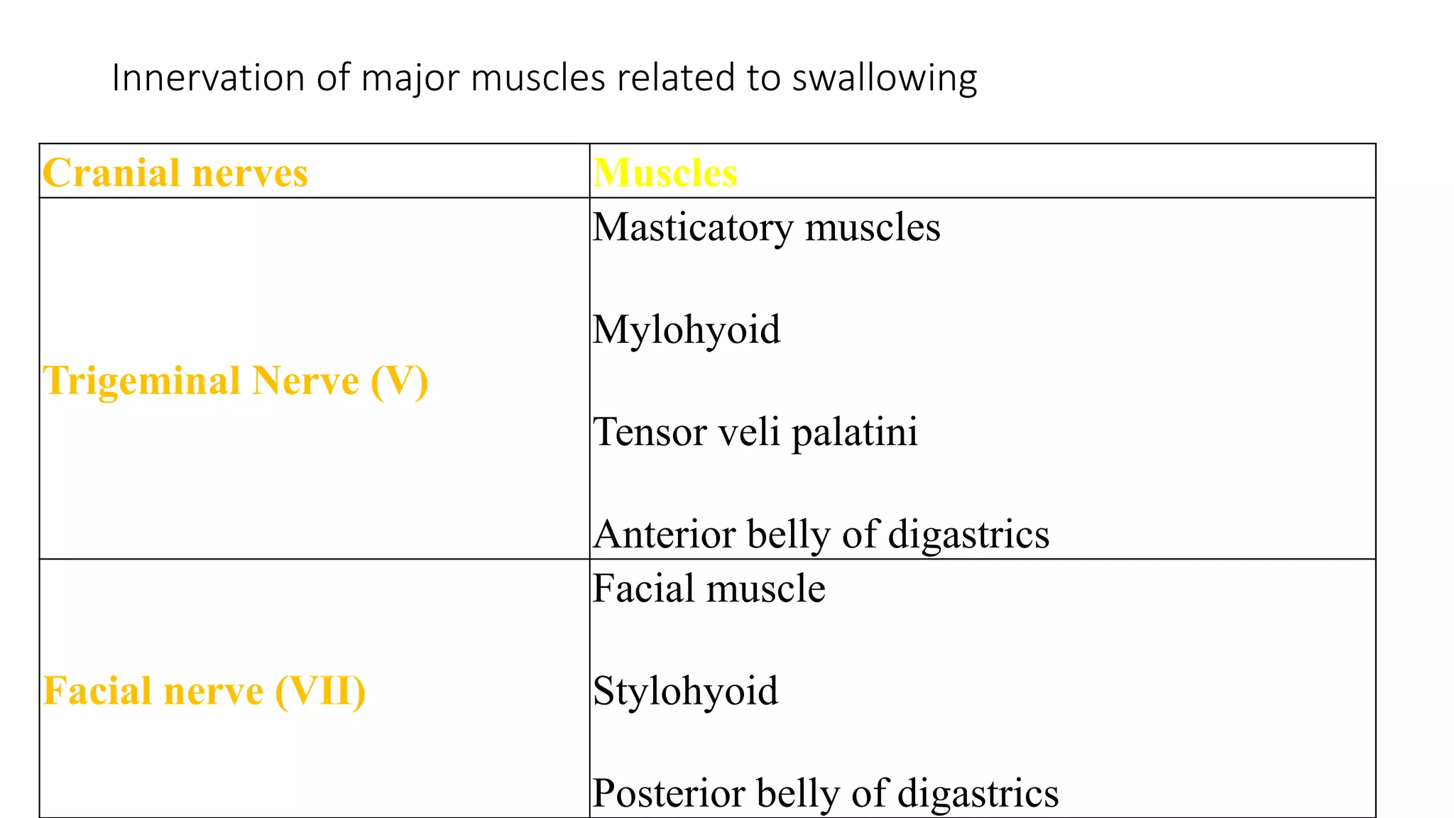
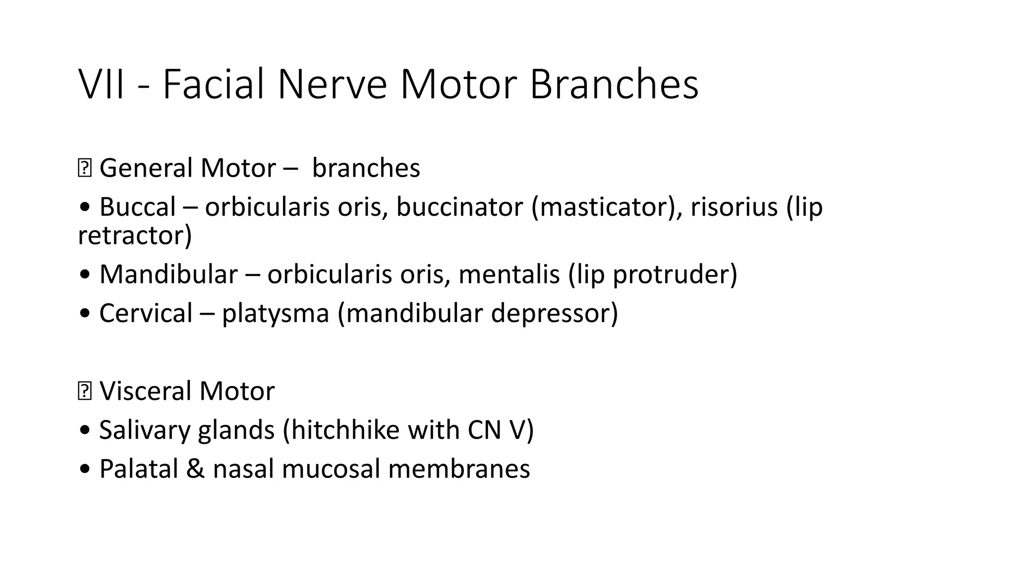
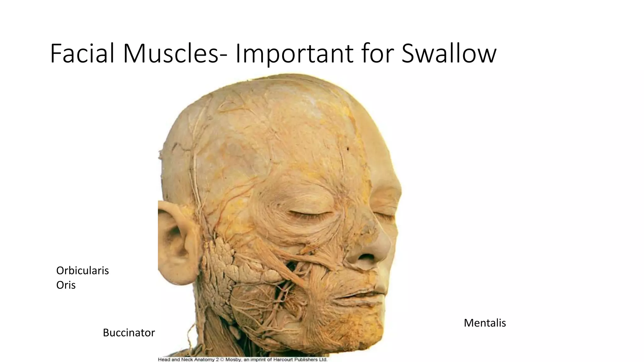
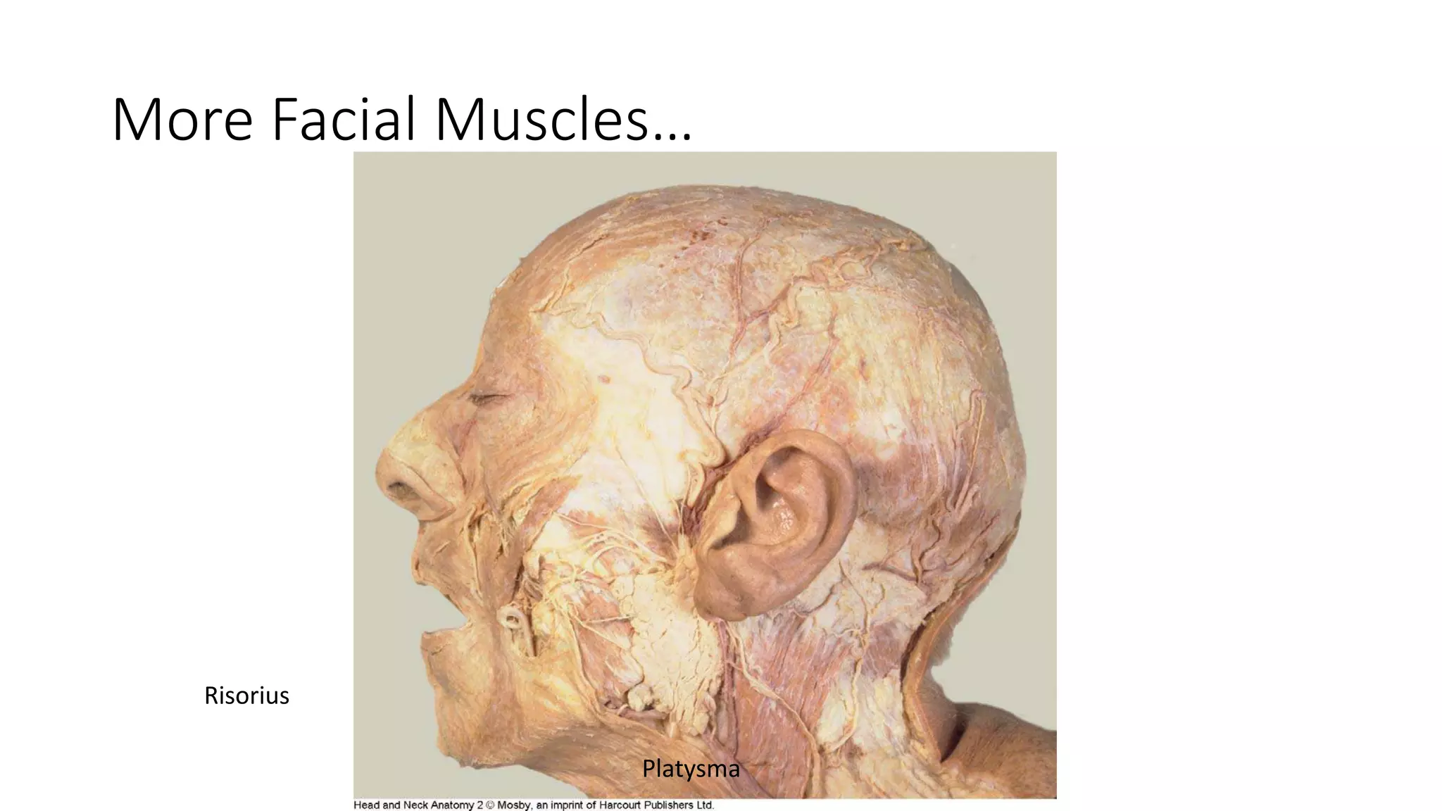
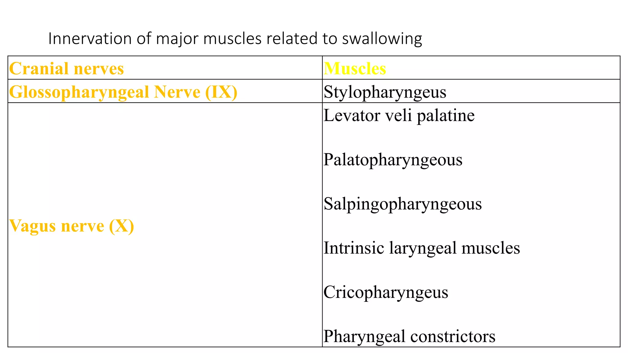
PHYSIOLOGY OF
SWALLOWING
Dr Nadhirah
Supervisor : Prof Madya Dr Marina
PHYSIOLOGY
OF
SWALLOWING
Dr Jamal Nasser bin Salleh
Supervisor : Prof Madya Dr Marina bt Baki
INTRODUCTION
• Deglutition involves co-ordinated activity of muscles of oral cavity,
pharynx, larynx and esophagus
• Definition : Passage of a food bolus or liquid from oral cavity to the
stomach via the pharynx and esophagus
• Phases :
• Oral – voluntary
• Pharyngeal involuntary/autonomic
• Esophageal
ANATOMY
Components of
deglutition :
1) Passage of bolus from
oral cavity to stomach
2) Protection of airway
3) Inhibition of air entry
into stomach
Oral phase
1) Oral preparatory phase
2) Oral phase proper
• Voluntary
• Food prepared for swallowing
• Tongue plays vital role
Oral Preparatory Phase
• Breaking down of food in the oral cavity
• Food chewed & mixed with saliva  Bolus
• The elevators of lower jaw play an important role in bolus preparation
• Tongue (vital role) : intrinsic muscles alter the shape, extrinsic
muscles change its position within the oral cavity – helping in chewing
the food by dental occlusion
• Occlusal action of the lips help in creating an effective seal preventing
bolus from dribbling out of oral cavity
• Action of buccinators muscle helps in pushing the bolus out of the
vestibule into the oral cavity proper
Bolus Formation
• The most important function of preparatory
phase
• Involves repeated transfer of food from oral
cavity to oropharynx surface of tongue
• Bolus accumulates on the oropharyngeal
surface of tongue due to repeated cycles of
upward and downward movement of the
tongue
Oral Phase Proper
• The bolus is moved towards the back of the tongue
• Contraction of soft palate prevents nasal regurgitation
• Soft palate also prevents premature movement of bolus into oropharynx
• Once the bolus is of suitable consistency the transit from mouth to
oropharynx just take a couple of seconds
• Tongue plays a vital role during this phase. Its instrinsic muscles contract
and reduce size, while genioglossus muscle elevates the tongue towards
the plate.
• The elevation of the mandible cause suprahyoid muscles raised the hyoid
bone.
Anatomy of Pharynx
• Midline tube about 15cm long
• Continous with nasal cavities and larynx
above and esophagus below
• The anterior wall of pharynx is
incomplete and is composed of posterior
part of tongue posteriorly and larynx
inferiorly
• Divided into nasopharynx, oropharynx,
laryngopharyx according to those
structures lying anteriorly
• The reminder of pharynx resembles the
gut. It has 4 layers ie areolar, muscular,
sub mucous and inner mucous
membrane (ouside inwards)
Anatomy of Pharynx (cont)
• The muscular layer has 2 component ie longitudinal and circular
muscle layers
• Circular muscles : superior, middle, inferior constrictor muscles
• Inferior constrictor muscle has 2 components : thyropharyngeus and
cricopharyngeus
• Except the cricopharyngeus muscles all the other muscles acting on
the pharynx are paired
• The cricopharynx forms a distinct sphinter at the point where
laryngopharynx enters the esophagus
Pharyngeal Phase
(pumping action of tongue and hypopharyngeal suction)
• This phase is reflexive in nature
• It takes a second for the bolus to transverse the pharynx and reach
the cricopharyngeal area
• Contraction of diaphragm is inhibited making simultaneous breathing
and swallowing impossible
• Soft palate is elevated in order to seal off the nasopharynx (by Tensor
palatine and Levator palatine muscles)
• Vocal cords adduct protecting the airway
• As the bolus passes the palatoglossal and palatopharyngeal folds the
act of swallowing become reflexive
Functions of trigger points in oropharynx
• Stimulation of trigger points present in the oropharynx starts off the
pharyngeal reflexive stage of swallowing
• Trigger points are at the the faucial arches and mucosa of the posterior
pharyngeal wall
• These trigger points are innervated by CN IX
• Stimulation of these trigger points causes dilatation of pharynx due to
relaxation of the constrictors, and elevation of pharynx & larynx due to
contraction of longitudinal muscles
• The pharynx constrict behind the bolus thereby propelling it
• Contraction of the inferior constrictor muscle moves the bolus towards
esophagus
Importance of laryngeal elevation during
pharyngeal stage
• Narrows the laryngeal inlet
• Ensures better sealing of the laryngeal inlet by the downturned
epiglottis
• Laryngeal elevation also contributes to dilatation of pharynx
• The laryngeal inlet is closed due to the actions of interarythenoid,
aryepiglottic and thryoepiglottic muscles.
Role of epiglottis in the pharyngeal phase
• The movement of epiglottis occurs in 2 stages
• The epiglottis moves from vertical – horizontal position
• The upper third of epiglottis moves below the horizontal to a slightly
lower level to cover the narrowed laryngeal inlet
• The bolus during the course of the swallow impinges on the epiglottis
and gets diverted to the lateral food channels
• Solid food tends to go behind the epiglottis into the postcricoid area
• Liquid food gets split by the epiglottis and passes via the pyriform
fossa
Anatomy of esophagus
Esophageal phase
• Purely reflexive
• Begins by relaxing the
cricopharyngeal sphinter
• Time taken : 10 – 15 secs
• Primary/secondary/tertiary
peristaltic waves play active
roles in this phase
• peristaltic movements of
esophagus takes the bolus
down to the stomach
Regurgitation of food back into esophagus is
prevented by :
• Tone of gastro-esophageal sphinter
• Negative intra thoracic pressure
• Pinch-cock effect of diaphragm
• Mucosal folds
• Esophagogastric angle
• Slight positive intra abdominal pressure
Neurological Representation of Swallowing
• Complex sensorimotor activity
• Involving organized interactions between cortical, cerebellar, bulbar and
peripheral systems
• Cranial nerves involve :
- V trigeminal
- VII facial
- IX Glossopharyngeal
- X Vagus
- XI spinal accessory
- XII Hypoglossal
Innervation of major muscles related to swallowing
Cranial nerves Muscles
Trigeminal Nerve (V)
Masticatory muscles
Mylohyoid
Tensor veli palatini
Anterior belly of digastrics
Facial nerve (VII)
Facial muscle
Stylohyoid
Posterior belly of digastrics
V - Trigeminal Nerve
Trigeminal
Ganglion
Mandibular
Branch
Buccal
Nerve
Ophthalmic
Branch
Maxillary
Branch
Inferior
Alveolar
Nerve
Lingual
Nervenn
Mastication Muscles
Temporalis
Masseter
Lateral
Pterygoid
Medial
Pterygoid
VII - Facial Nerve Motor Branches
􀂄 General Motor – branches
• Buccal – orbicularis oris, buccinator (masticator), risorius (lip
retractor)
• Mandibular – orbicularis oris, mentalis (lip protruder)
• Cervical – platysma (mandibular depressor)
􀂄 Visceral Motor
• Salivary glands (hitchhike with CN V)
• Palatal & nasal mucosal membranes
VII – Facial Nerve Sensory Branches
Chorda
Tympani
Facial Muscles- Important for Swallow
Orbicularis
Oris
Buccinator
Mentalis
More Facial Muscles…
Risorius
Platysma
Cranial nerves Muscles
Glossopharyngeal Nerve (IX) Stylopharyngeus
Vagus nerve (X)
Levator veli palatine
Palatopharyngeous
Salpingopharyngeous
Intrinsic laryngeal muscles
Cricopharyngeus
Pharyngeal constrictors
Innervation of major muscles related to swallowing
Cranial nerves Muscles
Hypoglossal nerve (XII)
Intrinsic tongue muscles
Hyoglossus
Geniohyoid
Genioglossus
Styloglossus
Thyrohyoid
Innervation of major muscles related to swallowing
Sensory afferents
I. Trigeminal
• Teeth and gums, palate and roof of the pharynx
II. Glossopharyngeal
• Posterior third of the tongue, pharynx, area of the tonsil
III. Vagus
• Pharynx, larynx
Trigeminal Nerve (V)
• Sensory nerve of the face and neck
• Branches
• Maxillary (sensory) : upper lip, maxillary teeth and palate, small area of
pharynx
• Mandibular (sensory + motor)
- Sensory : anterior 2/3 tongue (not taste) , TMJ, mucous membrane of mouth & buccal,
gums and mandibular teeth
- Motor : mastication muscles (temporalis, masseter, pterygoids), tensor muscles (tensor
veli palatini), suprahyoid muscles ( myolohyoid & anterior belly digastric muscle)
VII – Facial Nerve
• Sensory + Motor Components
• Innervates superficial face and neck muscles
• Contains special sensory and visceral motor components
• Special Sensory- Chorda Tympani: taste for anterior 2/3 of tongue &
other sensory for hard/soft palate
Brainstem centers
• Central pattern generators – Dorsomedial & Ventrolateral
• Mainly involves nucleus of the tractus solitarius and the nucleus
ambiguous
• Programs directing the sequential movements of the various muscles
involved
Brainstem centers
• Activated by
• Descending pathways from the motor cortex (cortico bulbar tracts)
• Ascending pathways from sensory structures within the oropharynx and
esophagus
Nucleus of the tractus solitarius receives the descending and peripheral
afferent influences
Dorsomedial pattern generator
• Resides in the medial reticular formation of the rostral medulla and
the reticulum adjacent to the nucleus tractus solitarius
• Initiation and organization of the swallowing sequence
Ventrolateral pattern generator
• Lies near the nucleus ambiguous and its surrounding reticular
formation
• Serves primarily as a connecting pathway to motor nuclei such as the
nucleus ambiguus and the dorsal motor nucleus of the vagus, which
directly control motor output to the pharyngeal musculature and
proximal esophagus
Cortical control to brainstem structures
Inferior precentral gyrus
• Cortical representations of tongue and face
• Bilaterally symmetrical activation
Other cortical areas involved in swallowing
Supplementary motor area
• Preparation for volitional swallowing
Anterior cingulate cortex
• Monitoring autonomic and vegetative functions.
Other cortical areas involved in swallowing
Anterior insula, particularly on the right
• Activation during volitional swallowing
• Allows gustatory and other intraoral sensations to modulate
swallowing.
• Lesions increase the swallowing threshold and delay the pharyngeal
phase of swallowing
Other higher centers in swallowing
Cerebellum
• Esp left-sided activation
• Coordination, timing, and sequencing of swallowing
• controls output for the motor nuclei of cranial nerves V, VII, and XII
Putamen
• Activated during volitional swallowing
Thank you

Physiology of swallowing

  • 1.
    PHYSIOLOGY OF SWALLOWING Dr Nadhirah Supervisor: Prof Madya Dr Marina PHYSIOLOGY OF SWALLOWING Dr Jamal Nasser bin Salleh Supervisor : Prof Madya Dr Marina bt Baki
  • 2.
    INTRODUCTION • Deglutition involvesco-ordinated activity of muscles of oral cavity, pharynx, larynx and esophagus • Definition : Passage of a food bolus or liquid from oral cavity to the stomach via the pharynx and esophagus • Phases : • Oral – voluntary • Pharyngeal involuntary/autonomic • Esophageal
  • 3.
    ANATOMY Components of deglutition : 1)Passage of bolus from oral cavity to stomach 2) Protection of airway 3) Inhibition of air entry into stomach
  • 4.
    Oral phase 1) Oralpreparatory phase 2) Oral phase proper • Voluntary • Food prepared for swallowing • Tongue plays vital role
  • 5.
    Oral Preparatory Phase •Breaking down of food in the oral cavity • Food chewed & mixed with saliva  Bolus • The elevators of lower jaw play an important role in bolus preparation • Tongue (vital role) : intrinsic muscles alter the shape, extrinsic muscles change its position within the oral cavity – helping in chewing the food by dental occlusion • Occlusal action of the lips help in creating an effective seal preventing bolus from dribbling out of oral cavity • Action of buccinators muscle helps in pushing the bolus out of the vestibule into the oral cavity proper
  • 6.
    Bolus Formation • Themost important function of preparatory phase • Involves repeated transfer of food from oral cavity to oropharynx surface of tongue • Bolus accumulates on the oropharyngeal surface of tongue due to repeated cycles of upward and downward movement of the tongue
  • 7.
    Oral Phase Proper •The bolus is moved towards the back of the tongue • Contraction of soft palate prevents nasal regurgitation • Soft palate also prevents premature movement of bolus into oropharynx • Once the bolus is of suitable consistency the transit from mouth to oropharynx just take a couple of seconds • Tongue plays a vital role during this phase. Its instrinsic muscles contract and reduce size, while genioglossus muscle elevates the tongue towards the plate. • The elevation of the mandible cause suprahyoid muscles raised the hyoid bone.
  • 8.
    Anatomy of Pharynx •Midline tube about 15cm long • Continous with nasal cavities and larynx above and esophagus below • The anterior wall of pharynx is incomplete and is composed of posterior part of tongue posteriorly and larynx inferiorly • Divided into nasopharynx, oropharynx, laryngopharyx according to those structures lying anteriorly • The reminder of pharynx resembles the gut. It has 4 layers ie areolar, muscular, sub mucous and inner mucous membrane (ouside inwards)
  • 9.
    Anatomy of Pharynx(cont) • The muscular layer has 2 component ie longitudinal and circular muscle layers • Circular muscles : superior, middle, inferior constrictor muscles • Inferior constrictor muscle has 2 components : thyropharyngeus and cricopharyngeus • Except the cricopharyngeus muscles all the other muscles acting on the pharynx are paired • The cricopharynx forms a distinct sphinter at the point where laryngopharynx enters the esophagus
  • 10.
    Pharyngeal Phase (pumping actionof tongue and hypopharyngeal suction) • This phase is reflexive in nature • It takes a second for the bolus to transverse the pharynx and reach the cricopharyngeal area • Contraction of diaphragm is inhibited making simultaneous breathing and swallowing impossible • Soft palate is elevated in order to seal off the nasopharynx (by Tensor palatine and Levator palatine muscles) • Vocal cords adduct protecting the airway • As the bolus passes the palatoglossal and palatopharyngeal folds the act of swallowing become reflexive
  • 12.
    Functions of triggerpoints in oropharynx • Stimulation of trigger points present in the oropharynx starts off the pharyngeal reflexive stage of swallowing • Trigger points are at the the faucial arches and mucosa of the posterior pharyngeal wall • These trigger points are innervated by CN IX • Stimulation of these trigger points causes dilatation of pharynx due to relaxation of the constrictors, and elevation of pharynx & larynx due to contraction of longitudinal muscles • The pharynx constrict behind the bolus thereby propelling it • Contraction of the inferior constrictor muscle moves the bolus towards esophagus
  • 14.
    Importance of laryngealelevation during pharyngeal stage • Narrows the laryngeal inlet • Ensures better sealing of the laryngeal inlet by the downturned epiglottis • Laryngeal elevation also contributes to dilatation of pharynx • The laryngeal inlet is closed due to the actions of interarythenoid, aryepiglottic and thryoepiglottic muscles.
  • 15.
    Role of epiglottisin the pharyngeal phase • The movement of epiglottis occurs in 2 stages • The epiglottis moves from vertical – horizontal position • The upper third of epiglottis moves below the horizontal to a slightly lower level to cover the narrowed laryngeal inlet • The bolus during the course of the swallow impinges on the epiglottis and gets diverted to the lateral food channels • Solid food tends to go behind the epiglottis into the postcricoid area • Liquid food gets split by the epiglottis and passes via the pyriform fossa
  • 16.
  • 17.
    Esophageal phase • Purelyreflexive • Begins by relaxing the cricopharyngeal sphinter • Time taken : 10 – 15 secs • Primary/secondary/tertiary peristaltic waves play active roles in this phase • peristaltic movements of esophagus takes the bolus down to the stomach
  • 18.
    Regurgitation of foodback into esophagus is prevented by : • Tone of gastro-esophageal sphinter • Negative intra thoracic pressure • Pinch-cock effect of diaphragm • Mucosal folds • Esophagogastric angle • Slight positive intra abdominal pressure
  • 19.
    Neurological Representation ofSwallowing • Complex sensorimotor activity • Involving organized interactions between cortical, cerebellar, bulbar and peripheral systems • Cranial nerves involve : - V trigeminal - VII facial - IX Glossopharyngeal - X Vagus - XI spinal accessory - XII Hypoglossal
  • 20.
    Innervation of majormuscles related to swallowing Cranial nerves Muscles Trigeminal Nerve (V) Masticatory muscles Mylohyoid Tensor veli palatini Anterior belly of digastrics Facial nerve (VII) Facial muscle Stylohyoid Posterior belly of digastrics
  • 21.
    V - TrigeminalNerve Trigeminal Ganglion Mandibular Branch Buccal Nerve Ophthalmic Branch Maxillary Branch Inferior Alveolar Nerve Lingual Nervenn
  • 22.
  • 23.
    VII - FacialNerve Motor Branches 􀂄 General Motor – branches • Buccal – orbicularis oris, buccinator (masticator), risorius (lip retractor) • Mandibular – orbicularis oris, mentalis (lip protruder) • Cervical – platysma (mandibular depressor) 􀂄 Visceral Motor • Salivary glands (hitchhike with CN V) • Palatal & nasal mucosal membranes
  • 24.
    VII – FacialNerve Sensory Branches Chorda Tympani
  • 25.
    Facial Muscles- Importantfor Swallow Orbicularis Oris Buccinator Mentalis
  • 26.
  • 27.
    Cranial nerves Muscles GlossopharyngealNerve (IX) Stylopharyngeus Vagus nerve (X) Levator veli palatine Palatopharyngeous Salpingopharyngeous Intrinsic laryngeal muscles Cricopharyngeus Pharyngeal constrictors Innervation of major muscles related to swallowing
  • 28.
    Cranial nerves Muscles Hypoglossalnerve (XII) Intrinsic tongue muscles Hyoglossus Geniohyoid Genioglossus Styloglossus Thyrohyoid Innervation of major muscles related to swallowing
  • 29.
    Sensory afferents I. Trigeminal •Teeth and gums, palate and roof of the pharynx II. Glossopharyngeal • Posterior third of the tongue, pharynx, area of the tonsil III. Vagus • Pharynx, larynx
  • 30.
    Trigeminal Nerve (V) •Sensory nerve of the face and neck • Branches • Maxillary (sensory) : upper lip, maxillary teeth and palate, small area of pharynx • Mandibular (sensory + motor) - Sensory : anterior 2/3 tongue (not taste) , TMJ, mucous membrane of mouth & buccal, gums and mandibular teeth - Motor : mastication muscles (temporalis, masseter, pterygoids), tensor muscles (tensor veli palatini), suprahyoid muscles ( myolohyoid & anterior belly digastric muscle)
  • 31.
    VII – FacialNerve • Sensory + Motor Components • Innervates superficial face and neck muscles • Contains special sensory and visceral motor components • Special Sensory- Chorda Tympani: taste for anterior 2/3 of tongue & other sensory for hard/soft palate
  • 32.
    Brainstem centers • Centralpattern generators – Dorsomedial & Ventrolateral • Mainly involves nucleus of the tractus solitarius and the nucleus ambiguous • Programs directing the sequential movements of the various muscles involved
  • 33.
    Brainstem centers • Activatedby • Descending pathways from the motor cortex (cortico bulbar tracts) • Ascending pathways from sensory structures within the oropharynx and esophagus Nucleus of the tractus solitarius receives the descending and peripheral afferent influences
  • 34.
    Dorsomedial pattern generator •Resides in the medial reticular formation of the rostral medulla and the reticulum adjacent to the nucleus tractus solitarius • Initiation and organization of the swallowing sequence
  • 35.
    Ventrolateral pattern generator •Lies near the nucleus ambiguous and its surrounding reticular formation • Serves primarily as a connecting pathway to motor nuclei such as the nucleus ambiguus and the dorsal motor nucleus of the vagus, which directly control motor output to the pharyngeal musculature and proximal esophagus
  • 36.
    Cortical control tobrainstem structures Inferior precentral gyrus • Cortical representations of tongue and face • Bilaterally symmetrical activation
  • 37.
    Other cortical areasinvolved in swallowing Supplementary motor area • Preparation for volitional swallowing Anterior cingulate cortex • Monitoring autonomic and vegetative functions.
  • 38.
    Other cortical areasinvolved in swallowing Anterior insula, particularly on the right • Activation during volitional swallowing • Allows gustatory and other intraoral sensations to modulate swallowing. • Lesions increase the swallowing threshold and delay the pharyngeal phase of swallowing
  • 39.
    Other higher centersin swallowing Cerebellum • Esp left-sided activation • Coordination, timing, and sequencing of swallowing • controls output for the motor nuclei of cranial nerves V, VII, and XII Putamen • Activated during volitional swallowing
  • 40.

Editor's Notes

  • #3 Normal deglutition is a smooth coordinated process that involves a complex of series of voluntary and involuntary neuromuscular contractions which can divided into 3 phases.
  • #5 The oral phase may be split into the preparatory phase where the food is taken into the mouth and broken down, and oral phase proper where the bolus is moved to the back of the tongue.
  • #12 The tongue elevated, blocking the oral cavity Soft palate blocks nasopharynx Vc close glottis and larynx moves up Epiglottis covers larynx
  • #18 After the food enters the esophagus, the cricopharyngeal sphinter will close,and peristaltic movements of esophagus takes the bolus down to the stomach. Gastroesophageal sphinter at lower end esophagus relaxes well before peristaltic wave reaches and permit fluid to pass. Bolus of food is passed by contraction of peristaltic waves and then the spinter closes.