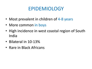
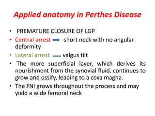
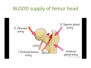
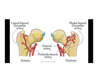
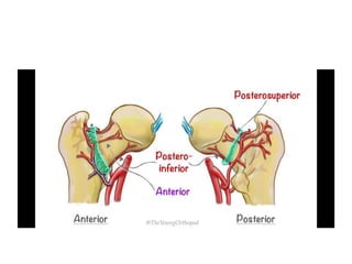
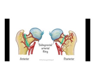
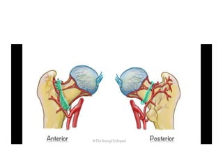
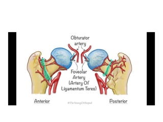
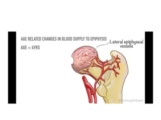
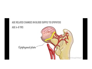
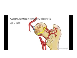
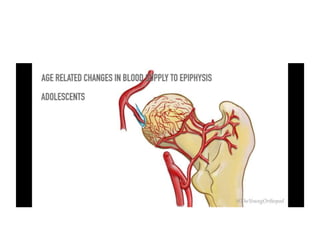
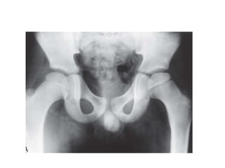
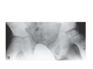
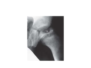
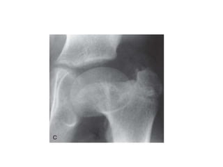
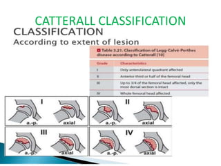
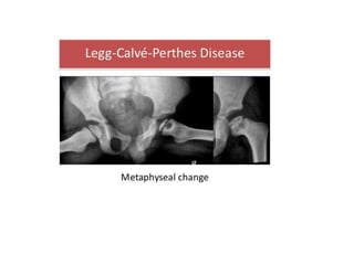
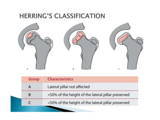
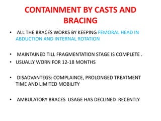
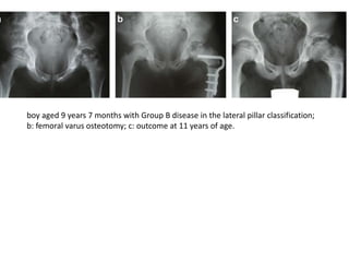
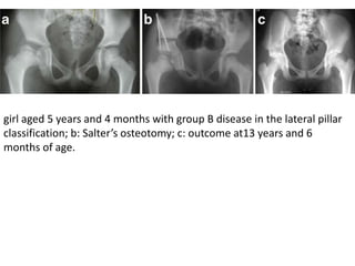
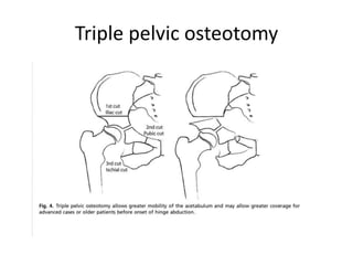
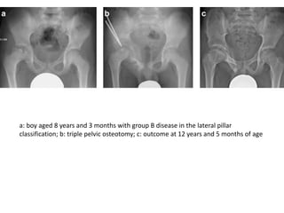
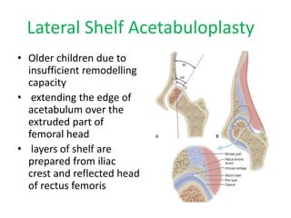
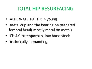
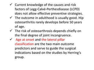
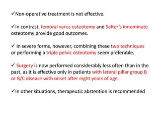
LEGG-CALVE-PERTHES DISEASE
Dr.K.PRASHANTH KUMAR
SEC.DNB RESIDENT
HINDU RAO HOSPITAL
SYNONYMS
1.Legg Calve-perthe’s disease
2. Legg’s stress fracture of femoral head
3. Osteochondritis deformans juvenalis
4. Osteochondrosis of hip joint
5. Pseudocoxalgia
6. Coxa plana
1. What is known about the pathophysiology
and risk factors of LCPD?
2. What is the long-term outcome?
3. What are the main prognostic factors?
4. Which surgical techniques are available?
5. Which patients should receive treatment?
EPIDEMIOLOGY
• Most prevalent in children of 4-8 years
• More common in boys
• High incidence in west coastal region of South
India
• Bilateral in 10-13%
• Rare in Black Africans
DEVELOPMENT OF HIP
• Majority of joint differentitaion completed by 4-8 weeks
• From 8 weeks onwards the limbs and joints undergo
growth and maturation in relative proportions and pre-
established spatial orientations .
• By 7th week cartilaginous model of femur and
acetabulum is complete
• By 11 weeks, all portions of the hip are visible
macroscopically and the infantile configuration of thehip
joint is achieved.
DEVELOPMENT OF HIP
• At 16 weeks, the ossification of the femur is complete up to
the level of the lesser trochanter. The primary centers of
ossification have appeared in the ilium, ischium, and pubis.
• At birth, the acetabular cartilage complex consists of the
saucer-shaped acetabular cartilage laterally and the Y-shaped
triradiate cartilage medially
• Secondary ossification centres of acetabulum develops by 8
years and fuse by 17-18 years.
• Growth in depth and the construction of the final acetabular
shape, however, heavily depends on the interaction with a
spherical femoral head
Applied anatomy in Perthes Disease
• PREMATURE CLOSURE OF LGP
• Central arrest short neck with no angular
deformity
• Lateral arrest valgus tilt
• The more superficial layer, which derives its
nourishment from the synovial fluid, continues to
grow and ossify, leading to a coxa magna.
• The FNI grows throughout the process and may
yield a wide femoral neck
BLOOD supply of femur head
Proposed etiological factors
• Coagulation disorder
• Arterial occlusion
• Disturbance of venous drainage
• Hyperactivity or Attention deficit disorder
• Heriditary influences
• Environmental influences
CURRENT OPINION…
• LCPD develops as a result of proximal femoral
epiphysis ischaemia of unknown aetiology
Pathogenesis and Timing of Femoral
Head Deformation
• The bone necrosis that follows the vascular occlusion
triggers changes in the soft tissue of the hip joint which
include synovitis,articular cartilage hypertrophy and
hypertrophy of the ligamentum teres.
• These soft tissue changes and muscle spasm cause the
femoral head to extrude out laterally of the
acetabulum.
• Stresses of weight-bearing and muscular contraction
pass across the acetabular margin onto the extruded
part of the avascular femoral head.
Unlike normal healthy bone, the avascular bone is not capable of
withstanding these physiological stresses and the trabeculae
collapse.
this results in irreversible femoral head deformation.
Extrusion appears to be a prime factor that predisposes to femoral
head deformation; the greater the extrusion, the greater the
propensity for femoral head deformation.
If more than 20% of the width of the epiphysis extrudes outside the
acetabulum irreversible femoral head deformation is almost
inevitable.
Classification
CHRONOLOGICALLY: Waldenstrom
classification
EXTENT: Catterall/ Salter and
Thompson/ Herring
OUTCOME: Stulberg/ Mose
CATTERALL CLASSIFICATION
CATTERALL’S HEAD AT RISK SIGNS
LATERAL SUBLUXATION GAGE’S SIGN
LATERAL EPIPHYSEAL CALCIFICATION
AND METAPHYSEAL LUCENCIES
Salter and Thompson Classification
HERRING’S classification
CLASSIFICATION BASED ON OUTCOME
• MOSE CLASSIFICATION: Based on fitting of
contour of healed femoral head into template of
concentric circles in both AP & Frog leg lateral
views
• •Good - < 1 mm
• •Fair - < 2 mm
• •Poor - > 2 mm
STULBERG CLASSIFICATION
TREATMENT OF PERTHES DISEASE
• EARLY IN THE COURSE OF THE DISEASE
• LATE IN THE COURSE OF THE DISEASE
• TREATMENT OF SEQUELAE OF THE DISEASE
TREATMENT IN THE EARLY COURSE OF
THE DISEASE
The factors to take into consideration to decide the
treatment include:
1) The age of the child at the onset of symptoms
2) The presence of extrusion of the femoral head
3) The range of motion of the hip
4) The stage of evolution of the disease.
PRINCIPLES OF CONTAINMENT
• BASED ON BIOLOGIC PLASTICITY
• IS THE TERM USED TO DESCRIBE INTERVENTION THAT
ENSURES THAT THE ANTERO-LATERAL PART OF THE FEMORAL
EPIPHYSIS IS POSITIONED WITHIN THE ACETABULUM,THEREBY
PROTECTING THE EPIPHYSIS FROM BEING SUBJECTED TO
DEFORMING STRESSES.
• CAN BE OBTAINED BY CONSERVATIVE OR SURGICAL MEANS
• SHOULD BE DONE BEFORE THE DISEASE EVOLVES TO THE
POINT WHERE THE FEMORAL HEAD IS VULNERABLE FOR
DEFROMATION
CONTAINMENT BY CASTS AND
BRACING
• ALL THE BRACES WORKS BY KEEPING FEMORAL HEAD IN
ABDUCTION AND INTERNAL ROTATION
• MAINTAINED TILL FRAGMENTATION STAGE IS COMPLETE .
• USUALLY WORN FOR 12-18 MONTHS
• DISADVANTEGS: COMPLAINCE, PROLONGED TREATMENT
TIME AND LIMITED MOBILITY
• AMBULATORY BRACES USAGE HAS DECLINED RECENTLY
CONTAINMENT BY SURGICAL
METHODS
• Femoral osteotomies
• pelvic osteotomies
• Combined femoral and pelvic osteotomy
PROXIMAL FEMORAL VARUS OSTEOTOMY
• Axer reported this method in 1965.
• Important to restore reasonable ROM before surgery; reasonable
abduction should be possible (30 degree).
• Preserve a neck shaft angle of 110 degree with desired approx.
115 degree.
• 15-20 degree of Varus angulation is sufficient (Kim et al).
• Greater Varus angulation did not produce better results.
• Rotational: To bring more posterior portion of head under
acetabular coverage.
• Fixation: Blade plate/DCS/Recon plate
• post operatively immobilize in spica for 4-6 weeks
COMPLICATIONS AND DEMERITS
• SHORTENING
• EXCESSIVE POST-OP VARUS
• FAILURE OF VARUS TO REMODEL ( MORE AFTER 9 YEARS AGE)
• PERSISTENT EXT ROTATION
• INCREASED ABDUCTOR LURCH
• TROCHANTERIC OVERGROWTH
• PREMATURE PHYSEAL CLOSURE
• DELAYED UNION AND NON-UNION
• HARDWARE TO BE REMOVED
boy aged 9 years 7 months with Group B disease in the lateral pillar classification;
b: femoral varus osteotomy; c: outcome at 11 years of age.
Pelvic osteotomies
• Salter’s ostetomy
• Triple pelvic osteotomy
• Shelf procedures
1) TRANSVERSE OSTEOTOMY ALONG A LINE FROM SCIATIC NOTCH TO
JUST ABOVE AIIS.
2) ACETABULUM IS THEN ROTATED LATERALLY AND ANTERIORLY USING
THE PUBIC SYMPHYSIS AS A HINGE HENCE INCREASING ANTER0-
LATERAL COVERAGE.
3) Contracture of iliopsoas and adductor muscles must be released.
girl aged 5 years and 4 months with group B disease in the lateral pillar
classification; b: Salter’s osteotomy; c: outcome at13 years and 6
months of age.
Triple pelvic osteotomy
a: boy aged 8 years and 3 months with group B disease in the lateral pillar
classification; b: triple pelvic osteotomy; c: outcome at 12 years and 5 months of age
Lateral Shelf Acetabuloplasty
• Older children due to
insufficient remodelling
capacity
• extending the edge of
acetabulum over the
extruded part of
femoral head
• layers of shelf are
prepared from iliac
crest and reflected head
of rectus femoris
Summary of common osteotomies for
containment
Treatment of Perthes disease :late in
the course of the disease
• HINGE ABDUCTION
• described by Grossbard in 1980
-IMPINGEMENT OF THE OUTER PART OF
FEMORAL HEAD ONTO THE LATERAL LIP OF
ACETABULUM
• sudden deterioration of ROM during follow up
• reducible and irreducible type
• DIAGNOSED by arthrography or dynamic MRI
Arthrography in Hinge abduction
Treatment options
• valgus ostetomy
• shelf procedures
• hip distraction procedures (NOT COMMONLY
USED)
VALGUS OSTEOTOMY
• To improve the joint biomechanics
by restoring the rolling motion of the
femoral head into the acteabulum
and the joint congruity in the weight
bearing position
• Improve the gait pattern, lengthen
the leg improves the abductor lever
arm
• Done in the haealing stage
Hip distraction
• To neutralize muscular weight bearing
forces on the femora epiphysis,induce
neovascularization and prevent femoral
head deformation
• Doesn’t change the anatomy of the
proximal femur
• Can be employed when hip is very stiff
• And other methods of surgical containment
are contraindicated
• 1mm/day
• End point -adequate ossification of lateral
pillar
SEQUELAE OF PERTHES DISEASE
• Femoroacetabular impingement
• Coxa brevis, coxa magna
• osteochondritis dessicans
• secondary arthritis
Coxa magna and FAI
• Coxa magna results due to physeal growth
disturbance caused by ischemia
• Causes FAI
• cam (commonly seen in LCPD) and pincer
types
• groin pain with flexion activities like deep
squatting or sitting in a low set chair
• Positive impingement sign
Managemnt of coxa magna
• Treatment revOlutionalized after Reinhold Ganz
who introduced safe surgical dislocation
method
• Femoral head reduction surgery (FHRO)
• reduced incidence of AVN of femoral head
Coxa brevis
• epiphyseal avascular necrosis causes alteration of longitudinal
growth of neck and relative trochantric overgrowth
• CF: trendelenburg sign positive, limp, loss of ROM, Impingement
• TREATMENT
1. COXA BREVIS WITH FAI- open surgical dislocation+ greater
trochantric advancement
2. COXA BREVIS WITH EQUAL LEG LENGTH : GT advancement
3. COXA BREVIS WITH LLD : MORSCHER PROCEDURE
OSTEOCHONDRITIS DESSICANS
• Appears late in adolescence after
asymptomatic period
• locking or catching type of pain
• Dx by MRI or arthrography
• Mx by rest, NSAIDs, osteochondroplasty
TOTAL HIP RESURFACING
• ALTERNATE TO THR in young
• metal cup and the bearing on prepared
femoral head( mostly metal on metal)
• CI: AKI,osteoporosis, low bone stock
• technically demanding
FUTURE TRENDS IN LCPD
• ANTI RESORPTIVE AGENTS
- RANKL inhibitors
- cathepsin K inhibitors
- oteoclast ATPase and chloride channels
• ANTI INFLAMMATORY
- TNF INHIBITORS
• p MRI
TAKE HOME MESSAGE
 Current knowledge of the causes and risk
factors of Legg-Calvé-Perthesdisease (LCPD)
does not allow effective preventive strategies.
 The outcome in adulthood is usually good. Hip
osteoarthritis rarely develops before 50 years
of age.
 The risk of osteoarthrosis depends chiefly on
the final degree of joint incongruence.
 Age at onset and the lateral pillar
classification are the two main outcome
predictors and serve to guide the surgical
indications based on the studies by Herring’s
group.
Treatment of Perthes disease should aim at
preventing femoral head deformation, thereby
minimizing the risk of secondary degenerative
arthritis.
This is possible in a large proportion of
instances if the hip is contained early in the
course of the disease
Non-operative treatment is not effective.
In contrast, femoral varus osteotomy and Salter’s innominate
osteotomy provide good outcomes.
 In severe forms, however, combining these two techniques
or performing a triple pelvic osteotomy seem preferable.
 Surgery is now performed considerably less often than in the
past, as it is effective only in patients with lateral pillar group B
or B/C disease with onset after eight years of age.
In other situations, therapeutic abstention is recommended
PERHTES DISEASE

PERHTES DISEASE

  • 1.
  • 3.
    SYNONYMS 1.Legg Calve-perthe’s disease 2.Legg’s stress fracture of femoral head 3. Osteochondritis deformans juvenalis 4. Osteochondrosis of hip joint 5. Pseudocoxalgia 6. Coxa plana
  • 4.
    1. What isknown about the pathophysiology and risk factors of LCPD? 2. What is the long-term outcome? 3. What are the main prognostic factors? 4. Which surgical techniques are available? 5. Which patients should receive treatment?
  • 5.
    EPIDEMIOLOGY • Most prevalentin children of 4-8 years • More common in boys • High incidence in west coastal region of South India • Bilateral in 10-13% • Rare in Black Africans
  • 6.
    DEVELOPMENT OF HIP •Majority of joint differentitaion completed by 4-8 weeks • From 8 weeks onwards the limbs and joints undergo growth and maturation in relative proportions and pre- established spatial orientations . • By 7th week cartilaginous model of femur and acetabulum is complete • By 11 weeks, all portions of the hip are visible macroscopically and the infantile configuration of thehip joint is achieved.
  • 7.
    DEVELOPMENT OF HIP •At 16 weeks, the ossification of the femur is complete up to the level of the lesser trochanter. The primary centers of ossification have appeared in the ilium, ischium, and pubis. • At birth, the acetabular cartilage complex consists of the saucer-shaped acetabular cartilage laterally and the Y-shaped triradiate cartilage medially
  • 9.
    • Secondary ossificationcentres of acetabulum develops by 8 years and fuse by 17-18 years. • Growth in depth and the construction of the final acetabular shape, however, heavily depends on the interaction with a spherical femoral head
  • 11.
    Applied anatomy inPerthes Disease • PREMATURE CLOSURE OF LGP • Central arrest short neck with no angular deformity • Lateral arrest valgus tilt • The more superficial layer, which derives its nourishment from the synovial fluid, continues to grow and ossify, leading to a coxa magna. • The FNI grows throughout the process and may yield a wide femoral neck
  • 12.
    BLOOD supply offemur head
  • 22.
    Proposed etiological factors •Coagulation disorder • Arterial occlusion • Disturbance of venous drainage • Hyperactivity or Attention deficit disorder • Heriditary influences • Environmental influences
  • 23.
    CURRENT OPINION… • LCPDdevelops as a result of proximal femoral epiphysis ischaemia of unknown aetiology
  • 24.
    Pathogenesis and Timingof Femoral Head Deformation • The bone necrosis that follows the vascular occlusion triggers changes in the soft tissue of the hip joint which include synovitis,articular cartilage hypertrophy and hypertrophy of the ligamentum teres. • These soft tissue changes and muscle spasm cause the femoral head to extrude out laterally of the acetabulum. • Stresses of weight-bearing and muscular contraction pass across the acetabular margin onto the extruded part of the avascular femoral head.
  • 25.
    Unlike normal healthybone, the avascular bone is not capable of withstanding these physiological stresses and the trabeculae collapse. this results in irreversible femoral head deformation. Extrusion appears to be a prime factor that predisposes to femoral head deformation; the greater the extrusion, the greater the propensity for femoral head deformation. If more than 20% of the width of the epiphysis extrudes outside the acetabulum irreversible femoral head deformation is almost inevitable.
  • 27.
    Classification CHRONOLOGICALLY: Waldenstrom classification EXTENT: Catterall/Salter and Thompson/ Herring OUTCOME: Stulberg/ Mose
  • 34.
  • 35.
  • 36.
  • 37.
  • 39.
    Salter and ThompsonClassification
  • 40.
  • 42.
    CLASSIFICATION BASED ONOUTCOME • MOSE CLASSIFICATION: Based on fitting of contour of healed femoral head into template of concentric circles in both AP & Frog leg lateral views • •Good - < 1 mm • •Fair - < 2 mm • •Poor - > 2 mm
  • 44.
  • 45.
    TREATMENT OF PERTHESDISEASE • EARLY IN THE COURSE OF THE DISEASE • LATE IN THE COURSE OF THE DISEASE • TREATMENT OF SEQUELAE OF THE DISEASE
  • 46.
    TREATMENT IN THEEARLY COURSE OF THE DISEASE
  • 47.
    The factors totake into consideration to decide the treatment include: 1) The age of the child at the onset of symptoms 2) The presence of extrusion of the femoral head 3) The range of motion of the hip 4) The stage of evolution of the disease.
  • 48.
    PRINCIPLES OF CONTAINMENT •BASED ON BIOLOGIC PLASTICITY • IS THE TERM USED TO DESCRIBE INTERVENTION THAT ENSURES THAT THE ANTERO-LATERAL PART OF THE FEMORAL EPIPHYSIS IS POSITIONED WITHIN THE ACETABULUM,THEREBY PROTECTING THE EPIPHYSIS FROM BEING SUBJECTED TO DEFORMING STRESSES. • CAN BE OBTAINED BY CONSERVATIVE OR SURGICAL MEANS • SHOULD BE DONE BEFORE THE DISEASE EVOLVES TO THE POINT WHERE THE FEMORAL HEAD IS VULNERABLE FOR DEFROMATION
  • 51.
    CONTAINMENT BY CASTSAND BRACING • ALL THE BRACES WORKS BY KEEPING FEMORAL HEAD IN ABDUCTION AND INTERNAL ROTATION • MAINTAINED TILL FRAGMENTATION STAGE IS COMPLETE . • USUALLY WORN FOR 12-18 MONTHS • DISADVANTEGS: COMPLAINCE, PROLONGED TREATMENT TIME AND LIMITED MOBILITY • AMBULATORY BRACES USAGE HAS DECLINED RECENTLY
  • 56.
    CONTAINMENT BY SURGICAL METHODS •Femoral osteotomies • pelvic osteotomies • Combined femoral and pelvic osteotomy
  • 57.
    PROXIMAL FEMORAL VARUSOSTEOTOMY • Axer reported this method in 1965. • Important to restore reasonable ROM before surgery; reasonable abduction should be possible (30 degree). • Preserve a neck shaft angle of 110 degree with desired approx. 115 degree. • 15-20 degree of Varus angulation is sufficient (Kim et al). • Greater Varus angulation did not produce better results. • Rotational: To bring more posterior portion of head under acetabular coverage. • Fixation: Blade plate/DCS/Recon plate • post operatively immobilize in spica for 4-6 weeks
  • 59.
    COMPLICATIONS AND DEMERITS •SHORTENING • EXCESSIVE POST-OP VARUS • FAILURE OF VARUS TO REMODEL ( MORE AFTER 9 YEARS AGE) • PERSISTENT EXT ROTATION • INCREASED ABDUCTOR LURCH • TROCHANTERIC OVERGROWTH • PREMATURE PHYSEAL CLOSURE • DELAYED UNION AND NON-UNION • HARDWARE TO BE REMOVED
  • 60.
    boy aged 9years 7 months with Group B disease in the lateral pillar classification; b: femoral varus osteotomy; c: outcome at 11 years of age.
  • 61.
    Pelvic osteotomies • Salter’sostetomy • Triple pelvic osteotomy • Shelf procedures
  • 64.
    1) TRANSVERSE OSTEOTOMYALONG A LINE FROM SCIATIC NOTCH TO JUST ABOVE AIIS. 2) ACETABULUM IS THEN ROTATED LATERALLY AND ANTERIORLY USING THE PUBIC SYMPHYSIS AS A HINGE HENCE INCREASING ANTER0- LATERAL COVERAGE. 3) Contracture of iliopsoas and adductor muscles must be released.
  • 65.
    girl aged 5years and 4 months with group B disease in the lateral pillar classification; b: Salter’s osteotomy; c: outcome at13 years and 6 months of age.
  • 66.
  • 67.
    a: boy aged8 years and 3 months with group B disease in the lateral pillar classification; b: triple pelvic osteotomy; c: outcome at 12 years and 5 months of age
  • 68.
    Lateral Shelf Acetabuloplasty •Older children due to insufficient remodelling capacity • extending the edge of acetabulum over the extruded part of femoral head • layers of shelf are prepared from iliac crest and reflected head of rectus femoris
  • 69.
    Summary of commonosteotomies for containment
  • 70.
    Treatment of Perthesdisease :late in the course of the disease • HINGE ABDUCTION • described by Grossbard in 1980 -IMPINGEMENT OF THE OUTER PART OF FEMORAL HEAD ONTO THE LATERAL LIP OF ACETABULUM • sudden deterioration of ROM during follow up • reducible and irreducible type • DIAGNOSED by arthrography or dynamic MRI
  • 71.
  • 72.
    Treatment options • valgusostetomy • shelf procedures • hip distraction procedures (NOT COMMONLY USED)
  • 73.
    VALGUS OSTEOTOMY • Toimprove the joint biomechanics by restoring the rolling motion of the femoral head into the acteabulum and the joint congruity in the weight bearing position • Improve the gait pattern, lengthen the leg improves the abductor lever arm • Done in the haealing stage
  • 74.
    Hip distraction • Toneutralize muscular weight bearing forces on the femora epiphysis,induce neovascularization and prevent femoral head deformation • Doesn’t change the anatomy of the proximal femur • Can be employed when hip is very stiff • And other methods of surgical containment are contraindicated • 1mm/day • End point -adequate ossification of lateral pillar
  • 79.
    SEQUELAE OF PERTHESDISEASE • Femoroacetabular impingement • Coxa brevis, coxa magna • osteochondritis dessicans • secondary arthritis
  • 80.
    Coxa magna andFAI • Coxa magna results due to physeal growth disturbance caused by ischemia • Causes FAI • cam (commonly seen in LCPD) and pincer types • groin pain with flexion activities like deep squatting or sitting in a low set chair • Positive impingement sign
  • 81.
    Managemnt of coxamagna • Treatment revOlutionalized after Reinhold Ganz who introduced safe surgical dislocation method • Femoral head reduction surgery (FHRO) • reduced incidence of AVN of femoral head
  • 82.
    Coxa brevis • epiphysealavascular necrosis causes alteration of longitudinal growth of neck and relative trochantric overgrowth • CF: trendelenburg sign positive, limp, loss of ROM, Impingement • TREATMENT 1. COXA BREVIS WITH FAI- open surgical dislocation+ greater trochantric advancement 2. COXA BREVIS WITH EQUAL LEG LENGTH : GT advancement 3. COXA BREVIS WITH LLD : MORSCHER PROCEDURE
  • 84.
    OSTEOCHONDRITIS DESSICANS • Appearslate in adolescence after asymptomatic period • locking or catching type of pain • Dx by MRI or arthrography • Mx by rest, NSAIDs, osteochondroplasty
  • 85.
    TOTAL HIP RESURFACING •ALTERNATE TO THR in young • metal cup and the bearing on prepared femoral head( mostly metal on metal) • CI: AKI,osteoporosis, low bone stock • technically demanding
  • 86.
    FUTURE TRENDS INLCPD • ANTI RESORPTIVE AGENTS - RANKL inhibitors - cathepsin K inhibitors - oteoclast ATPase and chloride channels • ANTI INFLAMMATORY - TNF INHIBITORS • p MRI
  • 87.
  • 88.
     Current knowledgeof the causes and risk factors of Legg-Calvé-Perthesdisease (LCPD) does not allow effective preventive strategies.  The outcome in adulthood is usually good. Hip osteoarthritis rarely develops before 50 years of age.  The risk of osteoarthrosis depends chiefly on the final degree of joint incongruence.  Age at onset and the lateral pillar classification are the two main outcome predictors and serve to guide the surgical indications based on the studies by Herring’s group.
  • 89.
    Treatment of Perthesdisease should aim at preventing femoral head deformation, thereby minimizing the risk of secondary degenerative arthritis. This is possible in a large proportion of instances if the hip is contained early in the course of the disease
  • 90.
    Non-operative treatment isnot effective. In contrast, femoral varus osteotomy and Salter’s innominate osteotomy provide good outcomes.  In severe forms, however, combining these two techniques or performing a triple pelvic osteotomy seem preferable.  Surgery is now performed considerably less often than in the past, as it is effective only in patients with lateral pillar group B or B/C disease with onset after eight years of age. In other situations, therapeutic abstention is recommended