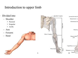
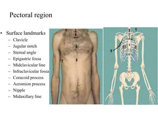
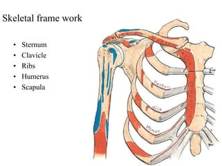
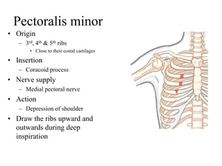
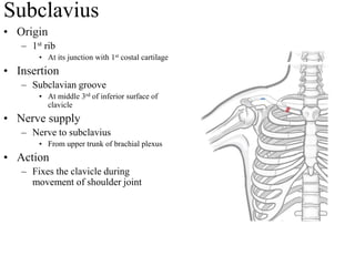
Introduction of upper limb
& Pectoral region
Introduction to upper limb
• Limbs are outgrowth of
anterior part of body wall
• Rotation occurs at 7th week
Introduction to upper limb
• Divided into
– Shoulder
• Pectoral
• Scapular
• Axilla
– Arm
– Forearm
– Hand
Arrangement of muscles
• Arranged in 2 groups
– Anterior compartment
– Posterior compartment
• A few muscles of
posterior compartment
occupy anterolateral
side in forearm
• No muscles in
posterior compartment
Hand
Pectoral Region
Pectoral region
• Surface landmarks
– Clavicle
– Jugular notch
– Sternal angle
– Epigastric fossa
– Midclavicular line
– Infraclavicular fossa
– Coracoid process
– Acromion process
– Nipple
– Midaxillary line
Skin
• Dermatome
– Area of skin supplied by
• An individual spinal nerve
– In trunk dermatome arranged
• Successive Horizontal strip
– Unusual in pectoral region
• In upper part of chest
– Upto sternal angle
• C3 & C4
– But below sternal angle
• T2
• Why this overlap
– Upper ;imb is developed from
C5-T1
Superficial fascia
• Subcutaneous
• Contains
– Fat
– Mammary gland
– Cutaneous nerves
• Supraclavicular nerves (C3,4)
• Anterior and lateral cutaneous
branches of intercostal nerve T2
– T6
– Cutaneous vessels
– Platysma
Skeletal frame work
• Sternum
• Clavicle
• Ribs
• Humerus
• Scapula
Dermatome
Deep fascia
• Pectoral
• Clavipectoral
Pectoral fascia
• Deep fascia of pectoral region
• Covers pectoralis major
• Attachment
– Medially
• Sternum
– Superorlaterally
• Deltoid fascia
– Inferiorly
• Merges with rectus sheath
– Laterally
• Axillary fascia
Muscles
• Pectoralis major
• Pectoralis minor
• Subclavius
• Serratus anterior
Pectoralis major
• Largest muscle of pectoral
region
• Forms anterior axillary fold
• Climbers muscle
Pectoralis major
• Origin
– 2 heads
• Clavicular head
– From medial ½ of the front of the
clavicle.
• Sternocostal head
– Sternum
– Upper 6 costal cartilages.
• Aponeurosis of external oblique muscle
• Insertion
– Lateral lip of bicipital groove
• Upper fibres in lower part
• Lower fibres in upper part
Pectoralis major
• Deltopectoral groove
– Intermuscular groove
– Upper part of Pectoralis major is
near to Deltoid
– A gap between
• Deltoid & pectoralis major
• Structures traversing
– Cephalic vein
– Deltoid branch of
• Thoracoacromial artery
– Deltopectoral lymph node
• Drain lymph from
– Superficial tissue of upper limb along
cephalic vein (including thumb)
Pectoralis major
• Nerve supply
– Lateral pectoral
• Pierces clavipectoral fascia
– Medial pectoral nerve
• Pierces pectoralis major muscle
• Action
– Medial rotation & adduction of arm
Pectoralis minor
• Origin
– 3rd, 4th & 5th ribs
• Close to their costal cartilages
• Insertion
– Coracoid process
• Nerve supply
– Medial pectoral nerve
• Action
– Depression of shoulder
• Draw the ribs upward and
outwards during deep
inspiration
Relations
• Anteriorly
– Pectoralis major
– Interpectoral nodes
• Posteriorly
– Axillary artery (2nd part)
– Axillary vein
– Cords of brachial plexus
• Lower border
– Lateral thoracic vessels
– Anterior group of axillary
lymph nodes
• Tail of breast
Subclavius
• Origin
– 1st rib
• At its junction with 1st costal cartilage
• Insertion
– Subclavian groove
• At middle 3rd of inferior surface of
clavicle
• Nerve supply
– Nerve to subclavius
• From upper trunk of brachial plexus
• Action
– Fixes the clavicle during
movement of shoulder joint
Clavipectoral fascia
• Deep fascia between the
subclavius and pectoralis minor
Clavipectoral fascia
• Location
– In anterior wall of axilla
• Deep to pectoralis major (clavicular
part)
• Vertical extent
– Above
• Clavicle
– Below
• Axillary fascia
• Suspensory ligament of axilla
– Clavipectoral fascia
• Below pectoralis minor
• Maintains concavity of axilla
• Encloses
– 2 muscles
• Subclavius
• Pectoralis minor
Horizontal extent
• Medial
– Costoclavicular ligament
– First 2 external intercostal
membrane
• Lateral
– Coracoid process
– Coracoclavicular ligament
• Costocoracoid ligament
– Upper thickened part
– Extend
• First costochondral junction
• Coracoid process
Structures piercing
• Lateral pectoral nerve
– From lateral cord
• Thoraco- acromial artery
– Branch from 2nd part of axillary
• Cephalic vein
– Into axillary vein
• Few lymph vessels
– From
• Breast to apical group of axillary
lymph node
Serratus anterior
Serratus anterior
• Origin
– Upper eight ribs
• Insertion
– Medial border and inferior angle of
scapula
• Anterior aspect
• Nerve supply
– Long thoracic nerve
• Action
– Draws the scapula forward
(protrusion, in boxing)
8
6
4
2
Long thoracic nerve
• Otherwise
– Nerve of Bell
• Arises from
– Roots of brachial plexus
• Root value
– C5, C6 & C7
• Protected by
– Deep fascia
– Not injured during surgery at
axilla
Winging of scapula
• Paralysis of serratus anterior
• Medial margin of scapula
becomes prominent
• Why
– Protraction of scapula is by
serratus anterior
• In the absence of action of serratus
• Rhomboideus retract
• So medial margin prominent
Mammary gland
• Parts
• Architecture
• Relations
• Blood supply
• Lymphatic drainage
Mammary gland - Extent
• Vertical
• 2nd to 6th rib in
midclavicular plane
• Horizontal
• Lateral border of sternum
to mid axillary plane - 4th
rib
• Axillary tail of Spence
• Projection from upper
outer quadrant
Mammary gland - Parts
• Nipple
-Erectile projection
-15 – 20 Lactiferous ducts
open
- 4th intercostal space
• Areola
- pigmented circular area
-Contains modified
sebaceous glands
Mammary gland - Architecture
• Glandular
-Modified sweat gland
-15 – 20 pyramidal lobes -
lactiferous ducts
• Fibrofatty tissue
-Fibrous tissue forms septa
– ligament of Cooper
* Fatty tissue gives shape
Mammary gland - Relations
• Mammary bed
• Medial 2/3rd – pectoralis
major
• Lateral 1/3rd – serratus
anterior
• Inferomedial - external
oblique
• Retro mammary spaceRetromammary
Space
Mammary gland – Blood supply
• Arteries
• Perforating branches –
internal thoracic
• Lateral thoracic –
axillary
• Intercostal arteries
• Veins
• Corresponding to
arteries
Mammary gland – Lymphatic
drainage
• Parenchyma (including
nipple & areola)
• Skin
Nerve supply
• 4th, 5th and 6th intercostal nerves
• Peau d’ orange –
obsruction in
superficial lymphatics
leads to appearance
of orange peel
Supernumerary breast - polythelia
Age changes
• Appearance of milk ridge – 7th week of IUL
• Nipple appears just before birth
• Lactiferous ducts without alveoli –till
puberty
• Deposition of fibrofatty tissue – puberty
• Proliferation of parenchyma –pregnancy
• Alveoli shrink – after lactation
• Atrophy of skin & fibrous tissue – old age

Pectoral region.ppt

  • 1.
    Introduction of upperlimb & Pectoral region
  • 3.
    Introduction to upperlimb • Limbs are outgrowth of anterior part of body wall • Rotation occurs at 7th week
  • 4.
    Introduction to upperlimb • Divided into – Shoulder • Pectoral • Scapular • Axilla – Arm – Forearm – Hand
  • 5.
    Arrangement of muscles •Arranged in 2 groups – Anterior compartment – Posterior compartment • A few muscles of posterior compartment occupy anterolateral side in forearm • No muscles in posterior compartment Hand
  • 6.
  • 7.
    Pectoral region • Surfacelandmarks – Clavicle – Jugular notch – Sternal angle – Epigastric fossa – Midclavicular line – Infraclavicular fossa – Coracoid process – Acromion process – Nipple – Midaxillary line
  • 8.
    Skin • Dermatome – Areaof skin supplied by • An individual spinal nerve – In trunk dermatome arranged • Successive Horizontal strip – Unusual in pectoral region • In upper part of chest – Upto sternal angle • C3 & C4 – But below sternal angle • T2 • Why this overlap – Upper ;imb is developed from C5-T1
  • 9.
    Superficial fascia • Subcutaneous •Contains – Fat – Mammary gland – Cutaneous nerves • Supraclavicular nerves (C3,4) • Anterior and lateral cutaneous branches of intercostal nerve T2 – T6 – Cutaneous vessels – Platysma
  • 10.
    Skeletal frame work •Sternum • Clavicle • Ribs • Humerus • Scapula
  • 11.
  • 12.
  • 13.
    Pectoral fascia • Deepfascia of pectoral region • Covers pectoralis major • Attachment – Medially • Sternum – Superorlaterally • Deltoid fascia – Inferiorly • Merges with rectus sheath – Laterally • Axillary fascia
  • 14.
    Muscles • Pectoralis major •Pectoralis minor • Subclavius • Serratus anterior
  • 15.
    Pectoralis major • Largestmuscle of pectoral region • Forms anterior axillary fold • Climbers muscle
  • 16.
    Pectoralis major • Origin –2 heads • Clavicular head – From medial ½ of the front of the clavicle. • Sternocostal head – Sternum – Upper 6 costal cartilages. • Aponeurosis of external oblique muscle • Insertion – Lateral lip of bicipital groove • Upper fibres in lower part • Lower fibres in upper part
  • 17.
    Pectoralis major • Deltopectoralgroove – Intermuscular groove – Upper part of Pectoralis major is near to Deltoid – A gap between • Deltoid & pectoralis major • Structures traversing – Cephalic vein – Deltoid branch of • Thoracoacromial artery – Deltopectoral lymph node • Drain lymph from – Superficial tissue of upper limb along cephalic vein (including thumb)
  • 18.
    Pectoralis major • Nervesupply – Lateral pectoral • Pierces clavipectoral fascia – Medial pectoral nerve • Pierces pectoralis major muscle • Action – Medial rotation & adduction of arm
  • 20.
    Pectoralis minor • Origin –3rd, 4th & 5th ribs • Close to their costal cartilages • Insertion – Coracoid process • Nerve supply – Medial pectoral nerve • Action – Depression of shoulder • Draw the ribs upward and outwards during deep inspiration
  • 21.
    Relations • Anteriorly – Pectoralismajor – Interpectoral nodes • Posteriorly – Axillary artery (2nd part) – Axillary vein – Cords of brachial plexus • Lower border – Lateral thoracic vessels – Anterior group of axillary lymph nodes • Tail of breast
  • 22.
    Subclavius • Origin – 1strib • At its junction with 1st costal cartilage • Insertion – Subclavian groove • At middle 3rd of inferior surface of clavicle • Nerve supply – Nerve to subclavius • From upper trunk of brachial plexus • Action – Fixes the clavicle during movement of shoulder joint
  • 23.
    Clavipectoral fascia • Deepfascia between the subclavius and pectoralis minor
  • 24.
    Clavipectoral fascia • Location –In anterior wall of axilla • Deep to pectoralis major (clavicular part) • Vertical extent – Above • Clavicle – Below • Axillary fascia • Suspensory ligament of axilla – Clavipectoral fascia • Below pectoralis minor • Maintains concavity of axilla • Encloses – 2 muscles • Subclavius • Pectoralis minor
  • 26.
    Horizontal extent • Medial –Costoclavicular ligament – First 2 external intercostal membrane • Lateral – Coracoid process – Coracoclavicular ligament • Costocoracoid ligament – Upper thickened part – Extend • First costochondral junction • Coracoid process
  • 27.
    Structures piercing • Lateralpectoral nerve – From lateral cord • Thoraco- acromial artery – Branch from 2nd part of axillary • Cephalic vein – Into axillary vein • Few lymph vessels – From • Breast to apical group of axillary lymph node
  • 28.
  • 29.
    Serratus anterior • Origin –Upper eight ribs • Insertion – Medial border and inferior angle of scapula • Anterior aspect • Nerve supply – Long thoracic nerve • Action – Draws the scapula forward (protrusion, in boxing) 8 6 4 2
  • 30.
    Long thoracic nerve •Otherwise – Nerve of Bell • Arises from – Roots of brachial plexus • Root value – C5, C6 & C7 • Protected by – Deep fascia – Not injured during surgery at axilla
  • 31.
    Winging of scapula •Paralysis of serratus anterior • Medial margin of scapula becomes prominent • Why – Protraction of scapula is by serratus anterior • In the absence of action of serratus • Rhomboideus retract • So medial margin prominent
  • 32.
    Mammary gland • Parts •Architecture • Relations • Blood supply • Lymphatic drainage
  • 33.
    Mammary gland -Extent • Vertical • 2nd to 6th rib in midclavicular plane • Horizontal • Lateral border of sternum to mid axillary plane - 4th rib • Axillary tail of Spence • Projection from upper outer quadrant
  • 34.
    Mammary gland -Parts • Nipple -Erectile projection -15 – 20 Lactiferous ducts open - 4th intercostal space • Areola - pigmented circular area -Contains modified sebaceous glands
  • 35.
    Mammary gland -Architecture • Glandular -Modified sweat gland -15 – 20 pyramidal lobes - lactiferous ducts • Fibrofatty tissue -Fibrous tissue forms septa – ligament of Cooper * Fatty tissue gives shape
  • 36.
    Mammary gland -Relations • Mammary bed • Medial 2/3rd – pectoralis major • Lateral 1/3rd – serratus anterior • Inferomedial - external oblique • Retro mammary spaceRetromammary Space
  • 37.
    Mammary gland –Blood supply • Arteries • Perforating branches – internal thoracic • Lateral thoracic – axillary • Intercostal arteries • Veins • Corresponding to arteries
  • 38.
    Mammary gland –Lymphatic drainage • Parenchyma (including nipple & areola) • Skin
  • 40.
    Nerve supply • 4th,5th and 6th intercostal nerves
  • 41.
    • Peau d’orange – obsruction in superficial lymphatics leads to appearance of orange peel
  • 42.
  • 43.
    Age changes • Appearanceof milk ridge – 7th week of IUL • Nipple appears just before birth • Lactiferous ducts without alveoli –till puberty • Deposition of fibrofatty tissue – puberty • Proliferation of parenchyma –pregnancy • Alveoli shrink – after lactation • Atrophy of skin & fibrous tissue – old age