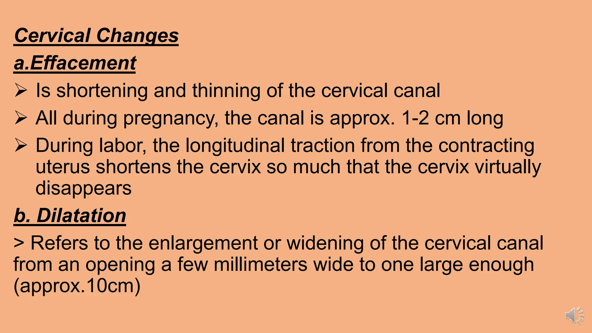
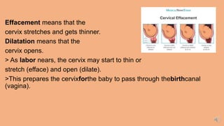
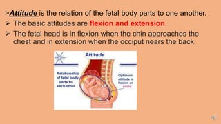
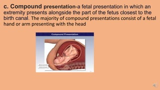
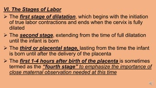
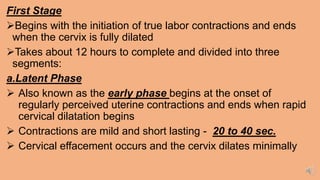
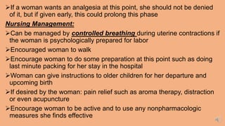
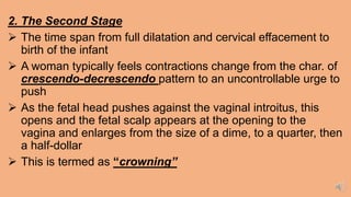
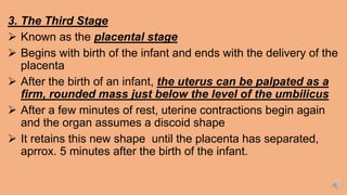
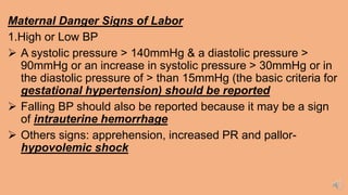
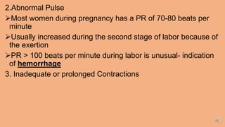
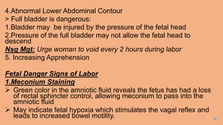
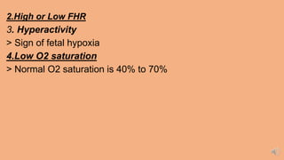
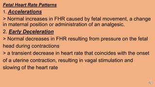
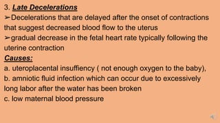
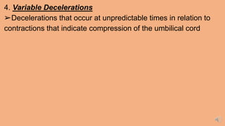
Cervical Changes
a.Effacement
 Is shortening and thinning of the cervical canal
 All during pregnancy, the canal is approx. 1-2 cm long
 During labor, the longitudinal traction from the contracting
uterus shortens the cervix so much that the cervix virtually
disappears
b. Dilatation
> Refers to the enlargement or widening of the cervical canal
from an opening a few millimeters wide to one large enough
(approx.10cm)
Effacement means that the
cervix stretches and gets thinner.
Dilatation means that the
cervix opens.
> As labor nears, the cervix may start to thin or
stretch (efface) and open (dilate).
>This prepares the cervixforthe baby to pass through thebirthcanal
(vagina).
>Attitude is the relation of the fetal body parts to one another.
 The basic attitudes are flexion and extension.
 The fetal head is in flexion when the chin approaches the
chest and in extension when the occiput nears the back.
> Fetal Attitude: Normal: Flexion of head, flexion of arms onto
chest, flexion of legs into abdomen
>Lie is the relation of the long axis (spine) of the fetus to the
long axis (spine) of the mother.
 Presentation refers to the part of the fetus that enters the
pelvic inlet first and leads through the birth canal during labor
at term.
 The presentation can roughly be divided into the
following classifications: cephalic, breech, shoulder,
and compound.
a.Cephalic presentation- the baby is positioned head-down,
facing the mother’s back, with the chin tucked to its chest and
the back of the head ready to enter the pelvis
b.Breech presentation-the fetus in the longitudinal lie
with the buttocks or lower extremity entering the pelvis
first.
>The three types of breech presentation include frank
breech, complete breech, and incomplete breech
c. Shoulder presentation- is a malpresentation at childbirth
where the baby is in a transverse lie (its vertebral column is
perpendicular to that of the mother), thus the leading part (the part
that first enters the birth canal) is an arm, a shoulder, or the trunk.
c. Compound presentation-a fetal presentation in which an
extremity presents alongside the part of the fetus closest to the
birth canal. The majority of compound presentations consist of a fetal
hand or arm presenting with the head
 Position is the relation of the presenting part to the four
quadrants of the mother's pelvis.)
 Position is the relationship
of the presenting part
(occiput, sacrum, mentum [chin],
or sinciput [deflexed vertex])
to the four quadrants of the
mother's pelvis
VI. The Stages of Labor
 The first stage of dilatation, which begins with the initiation
of true labor contractions and ends when the cervix is fully
dilated
 The second stage, extending from the time of full dilatation
until the infant is born
 The third or placental stage, lasting from the time the infant
is born until after the delivery of the placenta
 The first 1-4 hours after birth of the placenta is sometimes
termed as the “fourth stage” to emphasize the importance of
close maternal observation needed at this time
First Stage
Begins with the initiation of true labor contractions and ends
when the cervix is fully dilated
Takes about 12 hours to complete and divided into three
segments:
a.Latent Phase
 Also known as the early phase begins at the onset of
regularly perceived uterine contractions and ends when rapid
cervical dilatation begins
 Contractions are mild and short lasting - 20 to 40 sec.
 Cervical effacement occurs and the cervix dilates minimally
If a woman wants an analgesia at this point, she should not be denied
of it, but if given early, this could prolong this phase
Nursing Management:
Can be managed by controlled breathing during uterine contractions if
the woman is psychologically prepared for labor
Encouraged woman to walk
Encourage woman to do some preparation at this point such as doing
last minute packing for her stay in the hospital
Woman can give instructions to older children for her departure and
upcoming birth
If desired by the woman: pain relief such as aroma therapy, distraction
or even acupuncture
Encourage woman to be active and to use any nonpharmacologic
measures she finds effective
b. The Active Phase
 Cervical dilatation occurs more rapidly
 Contractions grow stronger, lasting 40-60 sec and occur
approx. every 3 to 5 minutes
 Show (increased vaginal secretions) and spontaneous
rupture of the membranes may occur
c. The Transition Phase
> Contractions reach their peak of intensity, occurring every 2 to
3 mins. With a duration of 60 to 70 sec.
Nursing Management:
Encourage woman to be active participant by keeping active and
assuming whatever position is most comfortable for her during this time
Lying flat on her back should be avoided during this time
c. The Transition Phase
Contraction reach their peak of intensity; every 2-3 minutes with a
duration of 60 to 70 seconds and a maximum cervical dilatation of 8 to
10 cm
Woman is experiencing an intense discomfort that is so strong
May accompanied with nausea and vomiting
With loss of control, anxiety, panic or irritability
The irresistible urge to push usually begins
2. The Second Stage
 The time span from full dilatation and cervical effacement to
birth of the infant
 A woman typically feels contractions change from the char. of
crescendo-decrescendo pattern to an uncontrollable urge to
push
 As the fetal head pushes against the vaginal introitus, this
opens and the fetal scalp appears at the opening to the
vagina and enlarges from the size of a dime, to a quarter, then
a half-dollar
 This is termed as “crowning”
3. The Third Stage
 Known as the placental stage
 Begins with birth of the infant and ends with the delivery of the
placenta
 After the birth of an infant, the uterus can be palpated as a
firm, rounded mass just below the level of the umbilicus
 After a few minutes of rest, uterine contractions begin again
and the organ assumes a discoid shape
 It retains this new shape until the placenta has separated,
aprrox. 5 minutes after the birth of the infant.
Maternal Danger Signs of Labor
1.High or Low BP
 A systolic pressure > 140mmHg & a diastolic pressure >
90mmHg or an increase in systolic pressure > 30mmHg or in
the diastolic pressure of > than 15mmHg (the basic criteria for
gestational hypertension) should be reported
 Falling BP should also be reported because it may be a sign
of intrauterine hemorrhage
 Others signs: apprehension, increased PR and pallor-
hypovolemic shock
2.Abnormal Pulse
Most women during pregnancy has a PR of 70-80 beats per
minute
Usually increased during the second stage of labor because of
the exertion
PR > 100 beats per minute during labor is unusual- indication
of hemorrhage
3. Inadequate or prolonged Contractions
4.Abnormal Lower Abdominal Contour
> Full bladder is dangerous:
1.Bladder may be injured by the pressure of the fetal head
2.Pressure of the full bladder may not allow the fetal head to
descend
Nsg Mgt: Urge woman to void every 2 hours during labor
5. Increasing Apprehension
Fetal Danger Signs of Labor
1.Meconium Staining
 Green color in the amniotic fluid reveals the fetus has had a loss
of rectal sphincter control, allowing meconium to pass into the
amniotic fluid
 May indicate fetal hypoxia which stimulates the vagal reflex and
leads to increased bowel motility.
2.High or Low FHR
3. Hyperactivity
> Sign of fetal hypoxia
4.Low O2 saturation
> Normal O2 saturation is 40% to 70%
Fetal Heart Rate Patterns
1. Accelerations
> Normal increases in FHR caused by fetal movement, a change
in maternal position or administration of an analgesic.
2. Early Deceleration
> Normal decreases in FHR resulting from pressure on the fetal
head during contractions
> a transient decrease in heart rate that coincides with the onset
of a uterine contraction, resulting in vagal stimulation and
slowing of the heart rate
3. Late Decelerations
➢Decelerations that are delayed after the onset of contractions
that suggest decreased blood flow to the uterus
➢gradual decrease in the fetal heart rate typically following the
uterine contraction
Causes:
a. uteroplacental insuffiency ( not enough oxygen to the baby),
b. amniotic fluid infection which can occur due to excessively
long labor after the water has been broken
c. low maternal blood pressure
4. Variable Decelerations
➢Decelerations that occur at unpredictable times in relation to
contractions that indicate compression of the umbilical cord

PART 2 WEEK 7 WITH RECORDED DISCUSSION.pptx

  • 1.
    Cervical Changes a.Effacement  Isshortening and thinning of the cervical canal  All during pregnancy, the canal is approx. 1-2 cm long  During labor, the longitudinal traction from the contracting uterus shortens the cervix so much that the cervix virtually disappears b. Dilatation > Refers to the enlargement or widening of the cervical canal from an opening a few millimeters wide to one large enough (approx.10cm)
  • 2.
    Effacement means thatthe cervix stretches and gets thinner. Dilatation means that the cervix opens. > As labor nears, the cervix may start to thin or stretch (efface) and open (dilate). >This prepares the cervixforthe baby to pass through thebirthcanal (vagina).
  • 3.
    >Attitude is therelation of the fetal body parts to one another.  The basic attitudes are flexion and extension.  The fetal head is in flexion when the chin approaches the chest and in extension when the occiput nears the back.
  • 4.
    > Fetal Attitude:Normal: Flexion of head, flexion of arms onto chest, flexion of legs into abdomen
  • 5.
    >Lie is therelation of the long axis (spine) of the fetus to the long axis (spine) of the mother.
  • 6.
     Presentation refersto the part of the fetus that enters the pelvic inlet first and leads through the birth canal during labor at term.  The presentation can roughly be divided into the following classifications: cephalic, breech, shoulder, and compound.
  • 7.
    a.Cephalic presentation- thebaby is positioned head-down, facing the mother’s back, with the chin tucked to its chest and the back of the head ready to enter the pelvis
  • 8.
    b.Breech presentation-the fetusin the longitudinal lie with the buttocks or lower extremity entering the pelvis first. >The three types of breech presentation include frank breech, complete breech, and incomplete breech
  • 9.
    c. Shoulder presentation-is a malpresentation at childbirth where the baby is in a transverse lie (its vertebral column is perpendicular to that of the mother), thus the leading part (the part that first enters the birth canal) is an arm, a shoulder, or the trunk.
  • 10.
    c. Compound presentation-afetal presentation in which an extremity presents alongside the part of the fetus closest to the birth canal. The majority of compound presentations consist of a fetal hand or arm presenting with the head
  • 11.
     Position isthe relation of the presenting part to the four quadrants of the mother's pelvis.)  Position is the relationship of the presenting part (occiput, sacrum, mentum [chin], or sinciput [deflexed vertex]) to the four quadrants of the mother's pelvis
  • 12.
    VI. The Stagesof Labor  The first stage of dilatation, which begins with the initiation of true labor contractions and ends when the cervix is fully dilated  The second stage, extending from the time of full dilatation until the infant is born  The third or placental stage, lasting from the time the infant is born until after the delivery of the placenta  The first 1-4 hours after birth of the placenta is sometimes termed as the “fourth stage” to emphasize the importance of close maternal observation needed at this time
  • 13.
    First Stage Begins withthe initiation of true labor contractions and ends when the cervix is fully dilated Takes about 12 hours to complete and divided into three segments: a.Latent Phase  Also known as the early phase begins at the onset of regularly perceived uterine contractions and ends when rapid cervical dilatation begins  Contractions are mild and short lasting - 20 to 40 sec.  Cervical effacement occurs and the cervix dilates minimally
  • 14.
    If a womanwants an analgesia at this point, she should not be denied of it, but if given early, this could prolong this phase Nursing Management: Can be managed by controlled breathing during uterine contractions if the woman is psychologically prepared for labor Encouraged woman to walk Encourage woman to do some preparation at this point such as doing last minute packing for her stay in the hospital Woman can give instructions to older children for her departure and upcoming birth If desired by the woman: pain relief such as aroma therapy, distraction or even acupuncture Encourage woman to be active and to use any nonpharmacologic measures she finds effective
  • 15.
    b. The ActivePhase  Cervical dilatation occurs more rapidly  Contractions grow stronger, lasting 40-60 sec and occur approx. every 3 to 5 minutes  Show (increased vaginal secretions) and spontaneous rupture of the membranes may occur c. The Transition Phase > Contractions reach their peak of intensity, occurring every 2 to 3 mins. With a duration of 60 to 70 sec.
  • 16.
    Nursing Management: Encourage womanto be active participant by keeping active and assuming whatever position is most comfortable for her during this time Lying flat on her back should be avoided during this time c. The Transition Phase Contraction reach their peak of intensity; every 2-3 minutes with a duration of 60 to 70 seconds and a maximum cervical dilatation of 8 to 10 cm Woman is experiencing an intense discomfort that is so strong May accompanied with nausea and vomiting With loss of control, anxiety, panic or irritability The irresistible urge to push usually begins
  • 17.
    2. The SecondStage  The time span from full dilatation and cervical effacement to birth of the infant  A woman typically feels contractions change from the char. of crescendo-decrescendo pattern to an uncontrollable urge to push  As the fetal head pushes against the vaginal introitus, this opens and the fetal scalp appears at the opening to the vagina and enlarges from the size of a dime, to a quarter, then a half-dollar  This is termed as “crowning”
  • 18.
    3. The ThirdStage  Known as the placental stage  Begins with birth of the infant and ends with the delivery of the placenta  After the birth of an infant, the uterus can be palpated as a firm, rounded mass just below the level of the umbilicus  After a few minutes of rest, uterine contractions begin again and the organ assumes a discoid shape  It retains this new shape until the placenta has separated, aprrox. 5 minutes after the birth of the infant.
  • 19.
    Maternal Danger Signsof Labor 1.High or Low BP  A systolic pressure > 140mmHg & a diastolic pressure > 90mmHg or an increase in systolic pressure > 30mmHg or in the diastolic pressure of > than 15mmHg (the basic criteria for gestational hypertension) should be reported  Falling BP should also be reported because it may be a sign of intrauterine hemorrhage  Others signs: apprehension, increased PR and pallor- hypovolemic shock
  • 20.
    2.Abnormal Pulse Most womenduring pregnancy has a PR of 70-80 beats per minute Usually increased during the second stage of labor because of the exertion PR > 100 beats per minute during labor is unusual- indication of hemorrhage 3. Inadequate or prolonged Contractions
  • 21.
    4.Abnormal Lower AbdominalContour > Full bladder is dangerous: 1.Bladder may be injured by the pressure of the fetal head 2.Pressure of the full bladder may not allow the fetal head to descend Nsg Mgt: Urge woman to void every 2 hours during labor 5. Increasing Apprehension Fetal Danger Signs of Labor 1.Meconium Staining  Green color in the amniotic fluid reveals the fetus has had a loss of rectal sphincter control, allowing meconium to pass into the amniotic fluid  May indicate fetal hypoxia which stimulates the vagal reflex and leads to increased bowel motility.
  • 22.
    2.High or LowFHR 3. Hyperactivity > Sign of fetal hypoxia 4.Low O2 saturation > Normal O2 saturation is 40% to 70%
  • 23.
    Fetal Heart RatePatterns 1. Accelerations > Normal increases in FHR caused by fetal movement, a change in maternal position or administration of an analgesic. 2. Early Deceleration > Normal decreases in FHR resulting from pressure on the fetal head during contractions > a transient decrease in heart rate that coincides with the onset of a uterine contraction, resulting in vagal stimulation and slowing of the heart rate
  • 24.
    3. Late Decelerations ➢Decelerationsthat are delayed after the onset of contractions that suggest decreased blood flow to the uterus ➢gradual decrease in the fetal heart rate typically following the uterine contraction Causes: a. uteroplacental insuffiency ( not enough oxygen to the baby), b. amniotic fluid infection which can occur due to excessively long labor after the water has been broken c. low maternal blood pressure
  • 25.
    4. Variable Decelerations ➢Decelerationsthat occur at unpredictable times in relation to contractions that indicate compression of the umbilical cord