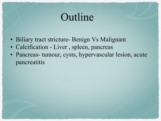
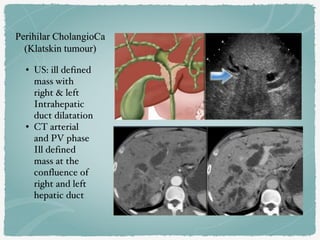
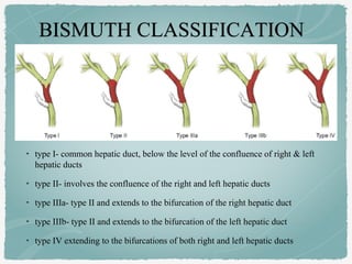
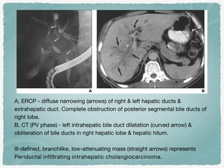
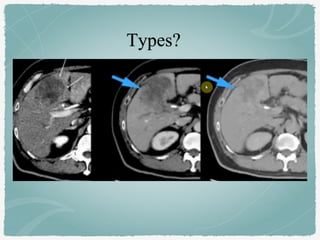
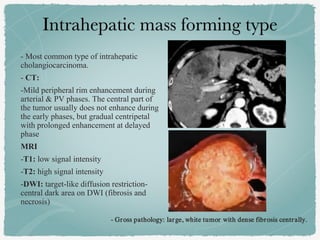
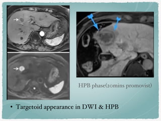
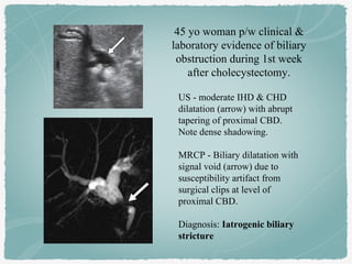
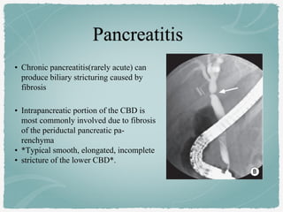
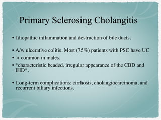
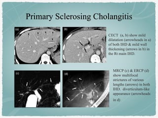
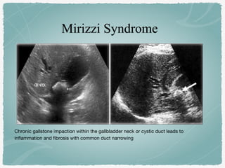
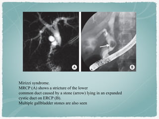
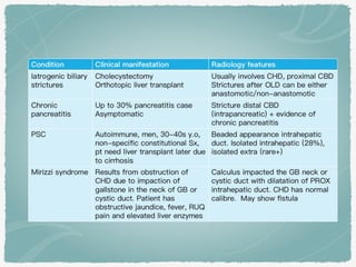
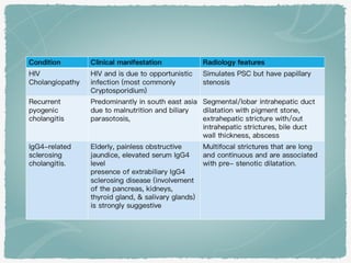
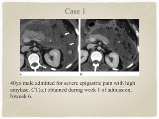
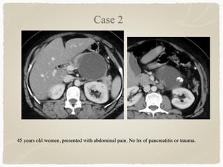
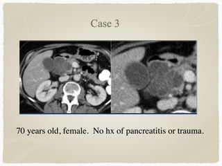
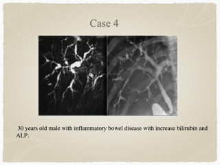
SlideShare a Scribd company logo
Tan Wen Nian
Supervisor: A/Prof Ouzreiah
• Biliary tract stricture- Benign Vs Malignant
• Calcification - Liver , spleen, pancreas
• Pancreas- tumour, cysts, hypervascular lesion, acute
pancreatitis
• Risk factors: Pre-existing bile duct disease (PSC, choledocolithiasis),
IBD (ulcerative colitis), Toxins (Thorotrast exposure)
• Epidemiology:
• 2nd most common primary hepatic tumour
• Gender M:F = 3:2
• Age: peak at 60-70s
• Clinical presentation:
• Painless obstructive jaundice, pain, palpable mass, constitutional Sx
• LFT: increased bilirubin , ALP
• Location
• Intrahepatic
• Perihilar
• Distal
• Growth Pattern
• Mass forming
• Periductal
• Intraductal
Cholangiocarcinoma 2020: the next horizon in mechanisms and management. Banales et.al
Growth pattern
A) Mass‐forming type
- Lesion in the hepatic parenchyma. Mimicker of HCC
B)Periductal‐infiltrating type
- Grows inside the duct wall and spreads longitudinally along the
wall
C) Intraductal
- polypoid or papillary tumour growing towards the duct lumen.
- often resectable surgically, more favorable prognosis than other.
• US: ill defined
mass with
right & left
Intrahepatic
duct dilatation
• CT arterial
and PV phase
Ill defined
mass at the
confluence of
right and left
hepatic duct
• type I- common hepatic duct, below the level of the confluence of right & left
hepatic ducts
• type II- involves the confluence of the right and left hepatic ducts
• type IIIa- type II and extends to the bifurcation of the right hepatic duct
• type IIIb- type II and extends to the bifurcation of the left hepatic duct
• type IV extending to the bifurcations of both right and left hepatic ducts
Intrabiliary mass with biliary dilatation peripheral to the mass.
A, ERCP - diffuse narrowing (arrows) of right & left hepatic ducts &
extrahepatic duct. Complete obstruction of posterior segmental bile ducts of
right lobe.
B, CT (PV phase) - left intrahepatic bile duct dilatation (curved arrow) &
obliteration of bile ducts in right hepatic lobe & hepatic hilum.
Ill-defined, branchlike, low-attenuating mass (straight arrows) represents
Periductal infiltrating intrahepatic cholangiocarcinoma.
- Most common type of intrahepatic
cholangiocarcinoma.
- CT:
-Mild peripheral rim enhancement during
arterial & PV phases. The central part of
the tumor usually does not enhance during
the early phases, but gradual centripetal
with prolonged enhancement at delayed
phase
MRI
-T1: low signal intensity
-T2: high signal intensity
-DWI: target-like diffusion restriction-
central dark area on DWI (fibrosis and
necrosis)
- Gross pathology: large, white tumor with dense fibrosis centrally.
• Targetoid appearance in DWI & HPB
HPB phase(20mins promovist)
ICC HCC
Risk Factors:: Fluke infestation,
recurrent pyogenic infection, Hep
B&C, PSC
Risk factors: Hep B&C, alcohol,
NASH
Peripheral arterial enhancement Mosaic enhancement
Progressive internal enhancement Portal venous washout
Desmoplastic: capsular retraction No capsular retraction, but capsular
expansion
Targetoid appearance on DWI,
HPB
No targetoid appearance
Vascular encasement Vascular thrombosis
Kim et. al 2007
A and B- Coronal MRCP (A)
and axial T1W post contrast
(B) images show long
segment distal CBD stricture
with irregular outline (arrow,
A) & enhancing wall
(arrowhead, B).
Malignant stricture-
cholangiocarcinoma
C and D- Coronal MRCP (C)
and axial T1W post contrast (D)
show smooth and relatively
short stricture (arrow, C) without
enhancing wall (arrowhead,D).
Benign distal CBD stricture
A
B
C
D
• Iatrogenic cause-
Most common cause
(80%–90%) of a
benign stricture
previous hepatobiliary
surgery (80%–90%)
• Chronic pancreatitis-
10%
US - moderate IHD & CHD
dilatation (arrow) with abrupt
tapering of proximal CBD.
Note dense shadowing.
MRCP - Biliary dilatation with
signal void (arrow) due to
susceptibility artifact from
surgical clips at level of
proximal CBD.
Diagnosis: Iatrogenic biliary
stricture
45 yo woman p/w clinical &
laboratory evidence of biliary
obstruction during 1st week
after cholecystectomy.
• Chronic pancreatitis(rarely acute) can
produce biliary stricturing caused by
fibrosis
• Intrapancreatic portion of the CBD is
most commonly involved due to fibrosis
of the periductal pancreatic pa-
renchyma
• *Typical smooth, elongated, incomplete
• stricture of the lower CBD*.
• Idiopathic inflammation and destruction of bile ducts.
• A/w ulcerative colitis. Most (75%) patients with PSC have UC
• > common in males.
• *characteristic beaded, irregular appearance of the CBD and
IHD*.
• Long-term complications: cirrhosis, cholangiocarcinoma, and
recurrent biliary infections.
CECT (a, b) show mild
dilatation (arrowheads in a)
of both IHD & mild wall
thickening (arrows in b) in
the Rt main IHD.
MRCP (c) & ERCP (d)
show multifocal
strictures of various
lengths (arrows) in both
IHD. diverticulum-like
appearance (arrowheads
in d)
Chronic gallstone impaction within the gallbladder neck or cystic duct leads to
inflammation and fibrosis with common duct narrowing
Mirizzi syndrome.
MRCP (A) shows a stricture of the lower
common duct caused by a stone (arrow) lying in an expanded
cystic duct on ERCP (B).
Multiple gallbladder stones are also seen
A 45-year-old male
with history of alcohol
abuse
a)Curvilinear
• Splenic artery atherosclerosis/
aneurysm
• Cyst – hydatid or post
traumatic, simple cyst,
dermoid, epidermoid cyst
• Primary tumours of spleen eg.
Histiocytoma
b)Multiple Small Nodular
• TB, Histoplasmosis, Brucellosis
• Phleboliths
• Haemangioma
• Sickle-cell anaemia
• Lymphoma
c)Diffuse
Homogeneous or
finely granular
• Sickle-cell anaemia
• Pneumocystic carinii
infection
• Lymphoma
d)Solitary > 1cm
• Healed infarct or
haematoma
• Healed abscess
• TB
Splenic artery calcification
- Serpiginous calcifications
Calcified splenic granulomas
2 calcific densities in
the left upper quadrant.
- common incidental
finding- most
commonly resulting
from TB or
histoplasmosis
25-year-old man with HIV and tuberculosis.
Axial contrast-enhanced CT shows hypodense right
hepatic lesion with peripheral curvilinear
calcification (arrow), representing tuberculoma
• Solid tumours
• Cystic tumours
• Endocrine tumours
• 80-90% of all pancreatic tumors.
• > 60 YO, male.
• Risk factors :smoking, alcohol, and chronic pancreatitis.
• Most common location - Pancreatic head.
• Classic appearance
• hypodense (CT)
• T1 hypointense (MR),
• Ill-defined, hypovascular mass causing ductal obstruction and atrophy of the pancreatic tail.
• Double duct signs : Dilation of both the pancreatic duct and CBD.
CT pancreas protocol
- Plain
- Late arterial phase- Normal enhancing pancreas with hypodense lesion
- Portal venous
• Rare, aggressive variant of pancreatic adenocarcinoma,
• Elderly males.
• Clinical triad of lipase hypersecretion syndrome
• Subcutaneous fat necrosis
• bone infarcts causing polyarthralgias
• eosinophilia.
No classical findings- tends to be large, well circumscribed hypervascular lesion
with haemorrhage, cystic change and necrosis
Radiology Assistant
• Benign tumor 
• ‘Grandma' tumour- 60-
70yo
• *Microcystic with
central scar and
calcifications*
• No communication with
pancreatic duct.
Lobulated hypodense lesion with central calcification in the
head of the pancreas.
On T2WI the lesion is multicystic. Note the central low
signal due to the central scar with calcifications.
• 'Mother' tumour - median age:
40-50 yo
• Premalignant tumor
• *Macrocystic with thick wall
septations, capsule, peripheral
calcifications(specific)*
• 95% - tail & body
• Most are symptomatic, p/w
non specific abd pain
T2 hyperintense encapsulated pancreatic tail mass with few
septations inferiorly. intrinsic T1 hyperintensity inferiorly in
keeping with wall calcification seen in CT.
• ‘Daughter’ tumour: mean age
28yo
• Rare
• Large mass with
heterogeneous solid and cystic
areas.
• *Capsule, haemorrhage*
• Low malignant potential.
Large mass in the tail of pancreas with solid & cystic
components. Enhancing rim/capsule.
No internal calcification. No pancreatic duct dilatation.
• Mucin producing tumour from duct epithelium
• ‘Grandfather’ tumour - elderly male
• Main branch, side branch or mixed
• CT -cystic lesion is visible in the
pancreatic head, seemingly in
connection with the moderately
dilated main pancreatic duct
• MRCP - direct connection with the
main duct
• Main duct type IPMN
Coronal MRCP with MIP
reconstruction - 2 discrete side
branch IPMNs and their direct
connection with the adjacent
normal sized pancreatic duct.
Axial CT venous phase
• Small unilocular cystic mass in
the pancreatic body (arrow).
• No pancreatic duct dilatation.
Mucinous cystic neoplasm
• Cyst in the pancreatic tail
• 36 year old woman, incidental findings
in US.
• thick irregular rim and contains solid
components.
Pseudocyst
• Unilocular cyst without solid
components, central scar /
calcification.
• Debris within a cystic lesion
• History of pancreatitis or
abdominal trauma.
• Cysts develop in 4-6 weeks
• Found in any part of the pancreas
Core Radiology
• Hyperfunctioning tumors come to clinical attention due to symptoms of
endocrine excess.
• Non-hyperfunctioning tumors tend to be larger at diagnosis
• undergo cystic change (DDX cystic pancreatic neoplasm)
• Central necrosis and calcification
• Pancreatic endocrine tumors tend to be hypervascular and are best seen in the
late arterial phase. Most are solid unless large.
• MRI: low signal in T1-weighed sequences, and intermediate to high signal in
T2-weighted sequences
Axial CT Plain (A), arterial phase (B) and portal phase (C)
A small, solid lesion is seen on the pancreatic tail (white arrow), with
homogeneous arterial phase hyperenhancement, persisting through the portal
phase (B).
This lesion was a insulinoma.
A larger heterogeneous lesion in the pancreatic head is shown, also with arterial phase
enhancement, with low signal intensity on T1-weighted sequences (E) and high signal
intensity on T2-weighted sequences (F).
Areas of cystic degeneration can also be seen (yellow arrow).
There is also a duodenal hyperenhancing lesion.
Both these lesions were non-functioning neuroendocrine tumors.
P. Pereira, Pancreatic hypervascular lesions: Neuroendocrine tumors and beyond. ECR 2015
Prasad et.al. Hypervascular pancreatic ‘‘lesions’’
Diagnosis: require 2 out of 3 features
1. Typical abdominal pain
2. Serum lipase / amylase >3x upper limit normal
3. Characteristic CT findings
Initial imaging - most useful when performed 5–7 days after hospital
admission, when local complications have developed and pancreatic
necrosis (if present) should be clearly distinguishable.
Revised Atlanta Classification for Acute Pancreatitis 2012
A)
B)
C)
D)
Acute interstitial pancreastitis: Normal enhancement
of the entire pancreas with mild surrounding fatty
infiltration.
ANC. Necrosis of the pancreas.
Inhomogeneous collection in the peripancreatic
tissue. No wall.
Acute necrotizing pancreatitis.
The pancreas do not enhance.
APFC: fluid collection at tail of
pancreas with no wall
40yo male admitted for severe epigastric pain with high
amylase. CT(a.) obtained during week 1 of admission,
b)week 6.
45 years old women, presented with abdominal pain. No hx of pancreatitis or trauma.
70 years old, female. No hx of pancreatitis or trauma.
30 years old male with inflammatory bowel disease with increase bilirubin and
ALP.
1. Radiology Assistant
2. Kim JY, Lee JM, Han JK, et al. Contrast-enhanced MRI combined with MR
cholangiopancreatography for the evaluation of patients with biliary strictures:
differentiation of malignant from benign bile duct strictures. J Magn Reson Imaging
2007;26(2): 304–312.
3. Radiology Survival Course 2020. HCC Vs ICC
4. Banales, J.M., Marin, J.J.G., Lamarca, A. et al. Cholangiocarcinoma 2020: the next horizon
in mechanisms and management. Nat Rev Gastroenterol Hepatol 17, 557–588 (2020).
5. Pancreatic hypervascular lesions: Neuroendocrine tumors and beyond. P. Pereira, R. Gil,
ECR 2015
6. Shankar PR, Wasnik AP, Al-Hawary MM, Francis IR, Kaza RK. Hypervascular pancreatic
"lesions": a pattern-based approach to differentiation. Abdom Radiol (NY). 2018
Apr;43(4):1013-1028.
7. Bryan R. Foster. Revised Atlanta Classification for Acute Pancreatitis: A Pictorial Essay.
Radiographic 2016
Pancreas and spleen 04022021

More Related Content

What's hot

Pediatric chest
Pediatric chestPediatric chest
Pediatric chest
Anish Choudhary
 
Pediatric chest part 2
Pediatric chest part 2Pediatric chest part 2
Pediatric chest part 2
Anish Choudhary
 
Abdominal wall mass
Abdominal wall mass Abdominal wall mass
Abdominal wall mass
Hisham Khatib
 
Acute abdomen
Acute abdomenAcute abdomen
Acute abdomen
AvinashDahatre
 
Bowel Wall Thickening CT Pattern
Bowel Wall Thickening CT PatternBowel Wall Thickening CT Pattern
Bowel Wall Thickening CT Pattern
DrBhishm Sevendra
 
Imaging of benign focal hepatic lesions
Imaging of benign focal hepatic lesionsImaging of benign focal hepatic lesions
Imaging of benign focal hepatic lesions
Dev Lakhera
 
Diseases of ureters
Diseases of uretersDiseases of ureters
Diseases of ureters
Milan Silwal
 
Liver lesions SYMPOSIUM RADIOLOGY
Liver lesions SYMPOSIUM RADIOLOGYLiver lesions SYMPOSIUM RADIOLOGY
Liver lesions SYMPOSIUM RADIOLOGY
Satyendra Raghuwanshi
 
Imaging features of hepatobiliary and pancretic ds
Imaging features of hepatobiliary and pancretic dsImaging features of hepatobiliary and pancretic ds
Imaging features of hepatobiliary and pancretic ds
radiologyoffice
 
Imaging of Non tubercular infections of the urinary tract
Imaging of Non tubercular infections of the urinary tractImaging of Non tubercular infections of the urinary tract
Imaging of Non tubercular infections of the urinary tract
vinothmezoss
 
Urinary tract infections
Urinary tract infectionsUrinary tract infections
Urinary tract infections
Milan Silwal
 
Biliary pathologies
Biliary pathologiesBiliary pathologies
Biliary pathologies
Navdeep Shah
 
Spleen; Imaging Anatomy, Investigations and Pathology
Spleen; Imaging Anatomy, Investigations and PathologySpleen; Imaging Anatomy, Investigations and Pathology
Spleen; Imaging Anatomy, Investigations and Pathology
Ali Aboelsouad
 
Malignant tumors of esophagus
Malignant tumors of esophagusMalignant tumors of esophagus
Malignant tumors of esophagus
Dev Lakhera
 
KEYS OF RADIOLOGY SPOTTERS GIT
KEYS OF RADIOLOGY SPOTTERS GITKEYS OF RADIOLOGY SPOTTERS GIT
KEYS OF RADIOLOGY SPOTTERS GIT
Anish Choudhary
 
CT Abdomen Case based presentation
CT Abdomen Case based presentationCT Abdomen Case based presentation
CT Abdomen Case based presentation
Dr.Damodhar.M.V MBBS,CSSGB,MBA,CPHQ
 
Usg gb & biliary t
Usg gb & biliary tUsg gb & biliary t
Usg gb & biliary t
Anish Choudhary
 
Diseases of spleen
Diseases of spleenDiseases of spleen
Diseases of spleenairwave12
 
RETROPERITONEAL FIBROSIS
RETROPERITONEAL FIBROSISRETROPERITONEAL FIBROSIS
RETROPERITONEAL FIBROSIS
GAURAV NAHAR
 
Presentation1.pptx urinary system part 11.
Presentation1.pptx urinary system part 11.Presentation1.pptx urinary system part 11.
Presentation1.pptx urinary system part 11.Abdellah Nazeer
 

What's hot (20)

Pediatric chest
Pediatric chestPediatric chest
Pediatric chest
 
Pediatric chest part 2
Pediatric chest part 2Pediatric chest part 2
Pediatric chest part 2
 
Abdominal wall mass
Abdominal wall mass Abdominal wall mass
Abdominal wall mass
 
Acute abdomen
Acute abdomenAcute abdomen
Acute abdomen
 
Bowel Wall Thickening CT Pattern
Bowel Wall Thickening CT PatternBowel Wall Thickening CT Pattern
Bowel Wall Thickening CT Pattern
 
Imaging of benign focal hepatic lesions
Imaging of benign focal hepatic lesionsImaging of benign focal hepatic lesions
Imaging of benign focal hepatic lesions
 
Diseases of ureters
Diseases of uretersDiseases of ureters
Diseases of ureters
 
Liver lesions SYMPOSIUM RADIOLOGY
Liver lesions SYMPOSIUM RADIOLOGYLiver lesions SYMPOSIUM RADIOLOGY
Liver lesions SYMPOSIUM RADIOLOGY
 
Imaging features of hepatobiliary and pancretic ds
Imaging features of hepatobiliary and pancretic dsImaging features of hepatobiliary and pancretic ds
Imaging features of hepatobiliary and pancretic ds
 
Imaging of Non tubercular infections of the urinary tract
Imaging of Non tubercular infections of the urinary tractImaging of Non tubercular infections of the urinary tract
Imaging of Non tubercular infections of the urinary tract
 
Urinary tract infections
Urinary tract infectionsUrinary tract infections
Urinary tract infections
 
Biliary pathologies
Biliary pathologiesBiliary pathologies
Biliary pathologies
 
Spleen; Imaging Anatomy, Investigations and Pathology
Spleen; Imaging Anatomy, Investigations and PathologySpleen; Imaging Anatomy, Investigations and Pathology
Spleen; Imaging Anatomy, Investigations and Pathology
 
Malignant tumors of esophagus
Malignant tumors of esophagusMalignant tumors of esophagus
Malignant tumors of esophagus
 
KEYS OF RADIOLOGY SPOTTERS GIT
KEYS OF RADIOLOGY SPOTTERS GITKEYS OF RADIOLOGY SPOTTERS GIT
KEYS OF RADIOLOGY SPOTTERS GIT
 
CT Abdomen Case based presentation
CT Abdomen Case based presentationCT Abdomen Case based presentation
CT Abdomen Case based presentation
 
Usg gb & biliary t
Usg gb & biliary tUsg gb & biliary t
Usg gb & biliary t
 
Diseases of spleen
Diseases of spleenDiseases of spleen
Diseases of spleen
 
RETROPERITONEAL FIBROSIS
RETROPERITONEAL FIBROSISRETROPERITONEAL FIBROSIS
RETROPERITONEAL FIBROSIS
 
Presentation1.pptx urinary system part 11.
Presentation1.pptx urinary system part 11.Presentation1.pptx urinary system part 11.
Presentation1.pptx urinary system part 11.
 

Similar to Pancreas and spleen 04022021

Focal liver lesions- in the eye of a radiologist
Focal liver lesions- in the eye of a radiologistFocal liver lesions- in the eye of a radiologist
Focal liver lesions- in the eye of a radiologist
Dr.Santosh Atreya
 
sandra liver.pptx
sandra liver.pptxsandra liver.pptx
sandra liver.pptx
Sandra710258
 
Pancreatic tumours.ppt.pptx
Pancreatic tumours.ppt.pptxPancreatic tumours.ppt.pptx
Pancreatic tumours.ppt.pptx
Abhi906519
 
Billiary tract
Billiary tractBilliary tract
Billiary tract
RMLIMS
 
Paediatric abdominal masses
Paediatric abdominal massesPaediatric abdominal masses
Paediatric abdominal masses
airwave12
 
Hcc(hepatocellular carcinoma)
Hcc(hepatocellular carcinoma)Hcc(hepatocellular carcinoma)
Hcc(hepatocellular carcinoma)
Dr pradeep Kumar
 
Choledochal cyst
Choledochal cystCholedochal cyst
Choledochal cyst
macshrestha
 
Liver cancer
Liver cancer Liver cancer
Liver cancer
PRANAV TVK
 
Cystic masses of pancreas.pptx
Cystic masses of pancreas.pptxCystic masses of pancreas.pptx
Cystic masses of pancreas.pptx
SadafIbrahim2
 
Liver & billary apparatus
Liver & billary apparatusLiver & billary apparatus
Liver & billary apparatus
Isha Jaiswal
 
Neoplasm of liver.pptx
Neoplasm of liver.pptxNeoplasm of liver.pptx
Neoplasm of liver.pptx
Nabin Paudyal
 
Pediatric GIT Imaging
Pediatric GIT ImagingPediatric GIT Imaging
Pediatric GIT Imaging
Ali Aboelsouad
 
patterns of enhancement in hepatocellular carcinoma
patterns of enhancement in hepatocellular carcinomapatterns of enhancement in hepatocellular carcinoma
patterns of enhancement in hepatocellular carcinoma
Haseeb Manzoor
 
Carcinoma Rectum by Dr.K.AmrithaAnilkumar
Carcinoma Rectum by Dr.K.AmrithaAnilkumarCarcinoma Rectum by Dr.K.AmrithaAnilkumar
Carcinoma Rectum by Dr.K.AmrithaAnilkumar
Dr. Amritha Anilkumar
 
pancreatitis Gi disorder diagnosis management
pancreatitis Gi disorder diagnosis managementpancreatitis Gi disorder diagnosis management
pancreatitis Gi disorder diagnosis management
THaripriya1
 
CHOLANGIOCARCINOMA- BILE DUCT CANCER
CHOLANGIOCARCINOMA- BILE DUCT CANCERCHOLANGIOCARCINOMA- BILE DUCT CANCER
CHOLANGIOCARCINOMA- BILE DUCT CANCER
Dr. Naina Kumar Agarwal
 
cystic neoplasm of pancreas
cystic neoplasm of pancreascystic neoplasm of pancreas
cystic neoplasm of pancreas
MaheshAdhikari19
 
Liver imaging dr Munazza Zafar
Liver imaging dr Munazza ZafarLiver imaging dr Munazza Zafar
Liver imaging dr Munazza Zafar
Tabish Javed
 

Similar to Pancreas and spleen 04022021 (20)

Focal liver lesions- in the eye of a radiologist
Focal liver lesions- in the eye of a radiologistFocal liver lesions- in the eye of a radiologist
Focal liver lesions- in the eye of a radiologist
 
sandra liver.pptx
sandra liver.pptxsandra liver.pptx
sandra liver.pptx
 
Pancreatic tumours.ppt.pptx
Pancreatic tumours.ppt.pptxPancreatic tumours.ppt.pptx
Pancreatic tumours.ppt.pptx
 
Billiary tract
Billiary tractBilliary tract
Billiary tract
 
Paediatric abdominal masses
Paediatric abdominal massesPaediatric abdominal masses
Paediatric abdominal masses
 
Hcc(hepatocellular carcinoma)
Hcc(hepatocellular carcinoma)Hcc(hepatocellular carcinoma)
Hcc(hepatocellular carcinoma)
 
Choledochal cyst
Choledochal cystCholedochal cyst
Choledochal cyst
 
Liver cancer
Liver cancer Liver cancer
Liver cancer
 
Cystic masses of pancreas.pptx
Cystic masses of pancreas.pptxCystic masses of pancreas.pptx
Cystic masses of pancreas.pptx
 
Pancreatitis
PancreatitisPancreatitis
Pancreatitis
 
Liver & billary apparatus
Liver & billary apparatusLiver & billary apparatus
Liver & billary apparatus
 
Neoplasm of liver.pptx
Neoplasm of liver.pptxNeoplasm of liver.pptx
Neoplasm of liver.pptx
 
Pediatric GIT Imaging
Pediatric GIT ImagingPediatric GIT Imaging
Pediatric GIT Imaging
 
patterns of enhancement in hepatocellular carcinoma
patterns of enhancement in hepatocellular carcinomapatterns of enhancement in hepatocellular carcinoma
patterns of enhancement in hepatocellular carcinoma
 
Carcinoma Rectum by Dr.K.AmrithaAnilkumar
Carcinoma Rectum by Dr.K.AmrithaAnilkumarCarcinoma Rectum by Dr.K.AmrithaAnilkumar
Carcinoma Rectum by Dr.K.AmrithaAnilkumar
 
pancreatitis Gi disorder diagnosis management
pancreatitis Gi disorder diagnosis managementpancreatitis Gi disorder diagnosis management
pancreatitis Gi disorder diagnosis management
 
CHOLANGIOCARCINOMA- BILE DUCT CANCER
CHOLANGIOCARCINOMA- BILE DUCT CANCERCHOLANGIOCARCINOMA- BILE DUCT CANCER
CHOLANGIOCARCINOMA- BILE DUCT CANCER
 
Diseases of the liver
Diseases of the liverDiseases of the liver
Diseases of the liver
 
cystic neoplasm of pancreas
cystic neoplasm of pancreascystic neoplasm of pancreas
cystic neoplasm of pancreas
 
Liver imaging dr Munazza Zafar
Liver imaging dr Munazza ZafarLiver imaging dr Munazza Zafar
Liver imaging dr Munazza Zafar
 

More from FaizahMohdZakiPPUKM

Peadiatric msk 031220
Peadiatric msk 031220Peadiatric msk 031220
Peadiatric msk 031220
FaizahMohdZakiPPUKM
 
Paediatric chest 031220
Paediatric chest 031220Paediatric chest 031220
Paediatric chest 031220
FaizahMohdZakiPPUKM
 
Neuroradiology 1a
Neuroradiology 1a Neuroradiology 1a
Neuroradiology 1a
FaizahMohdZakiPPUKM
 
Benign bone tumor
Benign bone tumorBenign bone tumor
Benign bone tumor
FaizahMohdZakiPPUKM
 
Radiology Malignant bone tumors
Radiology Malignant bone tumorsRadiology Malignant bone tumors
Radiology Malignant bone tumors
FaizahMohdZakiPPUKM
 
Cardiac disease
Cardiac diseaseCardiac disease
Cardiac disease
FaizahMohdZakiPPUKM
 
Chest diseases
Chest diseasesChest diseases
Chest diseases
FaizahMohdZakiPPUKM
 

More from FaizahMohdZakiPPUKM (7)

Peadiatric msk 031220
Peadiatric msk 031220Peadiatric msk 031220
Peadiatric msk 031220
 
Paediatric chest 031220
Paediatric chest 031220Paediatric chest 031220
Paediatric chest 031220
 
Neuroradiology 1a
Neuroradiology 1a Neuroradiology 1a
Neuroradiology 1a
 
Benign bone tumor
Benign bone tumorBenign bone tumor
Benign bone tumor
 
Radiology Malignant bone tumors
Radiology Malignant bone tumorsRadiology Malignant bone tumors
Radiology Malignant bone tumors
 
Cardiac disease
Cardiac diseaseCardiac disease
Cardiac disease
 
Chest diseases
Chest diseasesChest diseases
Chest diseases
 

Recently uploaded

Prix Galien International 2024 Forum Program
Prix Galien International 2024 Forum ProgramPrix Galien International 2024 Forum Program
Prix Galien International 2024 Forum Program
Levi Shapiro
 
Charaka Samhita Sutra Sthana 9 Chapter khuddakachatuspadadhyaya
Charaka Samhita Sutra Sthana 9 Chapter khuddakachatuspadadhyayaCharaka Samhita Sutra Sthana 9 Chapter khuddakachatuspadadhyaya
Charaka Samhita Sutra Sthana 9 Chapter khuddakachatuspadadhyaya
Dr KHALID B.M
 
Physiology of Chemical Sensation of smell.pdf
Physiology of Chemical Sensation of smell.pdfPhysiology of Chemical Sensation of smell.pdf
Physiology of Chemical Sensation of smell.pdf
MedicoseAcademics
 
ARTHROLOGY PPT NCISM SYLLABUS AYURVEDA STUDENTS
ARTHROLOGY PPT NCISM SYLLABUS AYURVEDA STUDENTSARTHROLOGY PPT NCISM SYLLABUS AYURVEDA STUDENTS
ARTHROLOGY PPT NCISM SYLLABUS AYURVEDA STUDENTS
Dr. Vinay Pareek
 
For Better Surat #ℂall #Girl Service ❤85270-49040❤ Surat #ℂall #Girls
For Better Surat #ℂall #Girl Service ❤85270-49040❤ Surat #ℂall #GirlsFor Better Surat #ℂall #Girl Service ❤85270-49040❤ Surat #ℂall #Girls
For Better Surat #ℂall #Girl Service ❤85270-49040❤ Surat #ℂall #Girls
Savita Shen $i11
 
Lung Cancer: Artificial Intelligence, Synergetics, Complex System Analysis, S...
Lung Cancer: Artificial Intelligence, Synergetics, Complex System Analysis, S...Lung Cancer: Artificial Intelligence, Synergetics, Complex System Analysis, S...
Lung Cancer: Artificial Intelligence, Synergetics, Complex System Analysis, S...
Oleg Kshivets
 
KDIGO 2024 guidelines for diabetologists
KDIGO 2024 guidelines for diabetologistsKDIGO 2024 guidelines for diabetologists
KDIGO 2024 guidelines for diabetologists
د.محمود نجيب
 
BRACHYTHERAPY OVERVIEW AND APPLICATORS
BRACHYTHERAPY OVERVIEW  AND  APPLICATORSBRACHYTHERAPY OVERVIEW  AND  APPLICATORS
BRACHYTHERAPY OVERVIEW AND APPLICATORS
Krishan Murari
 
Cervical & Brachial Plexus By Dr. RIG.pptx
Cervical & Brachial Plexus By Dr. RIG.pptxCervical & Brachial Plexus By Dr. RIG.pptx
Cervical & Brachial Plexus By Dr. RIG.pptx
Dr. Rabia Inam Gandapore
 
The hemodynamic and autonomic determinants of elevated blood pressure in obes...
The hemodynamic and autonomic determinants of elevated blood pressure in obes...The hemodynamic and autonomic determinants of elevated blood pressure in obes...
The hemodynamic and autonomic determinants of elevated blood pressure in obes...
Catherine Liao
 
Pulmonary Thromboembolism - etilogy, types, medical- Surgical and nursing man...
Pulmonary Thromboembolism - etilogy, types, medical- Surgical and nursing man...Pulmonary Thromboembolism - etilogy, types, medical- Surgical and nursing man...
Pulmonary Thromboembolism - etilogy, types, medical- Surgical and nursing man...
VarunMahajani
 
Tom Selleck Health: A Comprehensive Look at the Iconic Actor’s Wellness Journey
Tom Selleck Health: A Comprehensive Look at the Iconic Actor’s Wellness JourneyTom Selleck Health: A Comprehensive Look at the Iconic Actor’s Wellness Journey
Tom Selleck Health: A Comprehensive Look at the Iconic Actor’s Wellness Journey
greendigital
 
ARTIFICIAL INTELLIGENCE IN HEALTHCARE.pdf
ARTIFICIAL INTELLIGENCE IN  HEALTHCARE.pdfARTIFICIAL INTELLIGENCE IN  HEALTHCARE.pdf
ARTIFICIAL INTELLIGENCE IN HEALTHCARE.pdf
Anujkumaranit
 
Report Back from SGO 2024: What’s the Latest in Cervical Cancer?
Report Back from SGO 2024: What’s the Latest in Cervical Cancer?Report Back from SGO 2024: What’s the Latest in Cervical Cancer?
Report Back from SGO 2024: What’s the Latest in Cervical Cancer?
bkling
 
THOA 2.ppt Human Organ Transplantation Act
THOA 2.ppt Human Organ Transplantation ActTHOA 2.ppt Human Organ Transplantation Act
THOA 2.ppt Human Organ Transplantation Act
DrSathishMS1
 
ANATOMY AND PHYSIOLOGY OF URINARY SYSTEM.pptx
ANATOMY AND PHYSIOLOGY OF URINARY SYSTEM.pptxANATOMY AND PHYSIOLOGY OF URINARY SYSTEM.pptx
ANATOMY AND PHYSIOLOGY OF URINARY SYSTEM.pptx
Swetaba Besh
 
Ophthalmology Clinical Tests for OSCE exam
Ophthalmology Clinical Tests for OSCE examOphthalmology Clinical Tests for OSCE exam
Ophthalmology Clinical Tests for OSCE exam
KafrELShiekh University
 
Hemodialysis: Chapter 3, Dialysis Water Unit - Dr.Gawad
Hemodialysis: Chapter 3, Dialysis Water Unit - Dr.GawadHemodialysis: Chapter 3, Dialysis Water Unit - Dr.Gawad
Hemodialysis: Chapter 3, Dialysis Water Unit - Dr.Gawad
NephroTube - Dr.Gawad
 
New Directions in Targeted Therapeutic Approaches for Older Adults With Mantl...
New Directions in Targeted Therapeutic Approaches for Older Adults With Mantl...New Directions in Targeted Therapeutic Approaches for Older Adults With Mantl...
New Directions in Targeted Therapeutic Approaches for Older Adults With Mantl...
i3 Health
 
How to Give Better Lectures: Some Tips for Doctors
How to Give Better Lectures: Some Tips for DoctorsHow to Give Better Lectures: Some Tips for Doctors
How to Give Better Lectures: Some Tips for Doctors
LanceCatedral
 

Recently uploaded (20)

Prix Galien International 2024 Forum Program
Prix Galien International 2024 Forum ProgramPrix Galien International 2024 Forum Program
Prix Galien International 2024 Forum Program
 
Charaka Samhita Sutra Sthana 9 Chapter khuddakachatuspadadhyaya
Charaka Samhita Sutra Sthana 9 Chapter khuddakachatuspadadhyayaCharaka Samhita Sutra Sthana 9 Chapter khuddakachatuspadadhyaya
Charaka Samhita Sutra Sthana 9 Chapter khuddakachatuspadadhyaya
 
Physiology of Chemical Sensation of smell.pdf
Physiology of Chemical Sensation of smell.pdfPhysiology of Chemical Sensation of smell.pdf
Physiology of Chemical Sensation of smell.pdf
 
ARTHROLOGY PPT NCISM SYLLABUS AYURVEDA STUDENTS
ARTHROLOGY PPT NCISM SYLLABUS AYURVEDA STUDENTSARTHROLOGY PPT NCISM SYLLABUS AYURVEDA STUDENTS
ARTHROLOGY PPT NCISM SYLLABUS AYURVEDA STUDENTS
 
For Better Surat #ℂall #Girl Service ❤85270-49040❤ Surat #ℂall #Girls
For Better Surat #ℂall #Girl Service ❤85270-49040❤ Surat #ℂall #GirlsFor Better Surat #ℂall #Girl Service ❤85270-49040❤ Surat #ℂall #Girls
For Better Surat #ℂall #Girl Service ❤85270-49040❤ Surat #ℂall #Girls
 
Lung Cancer: Artificial Intelligence, Synergetics, Complex System Analysis, S...
Lung Cancer: Artificial Intelligence, Synergetics, Complex System Analysis, S...Lung Cancer: Artificial Intelligence, Synergetics, Complex System Analysis, S...
Lung Cancer: Artificial Intelligence, Synergetics, Complex System Analysis, S...
 
KDIGO 2024 guidelines for diabetologists
KDIGO 2024 guidelines for diabetologistsKDIGO 2024 guidelines for diabetologists
KDIGO 2024 guidelines for diabetologists
 
BRACHYTHERAPY OVERVIEW AND APPLICATORS
BRACHYTHERAPY OVERVIEW  AND  APPLICATORSBRACHYTHERAPY OVERVIEW  AND  APPLICATORS
BRACHYTHERAPY OVERVIEW AND APPLICATORS
 
Cervical & Brachial Plexus By Dr. RIG.pptx
Cervical & Brachial Plexus By Dr. RIG.pptxCervical & Brachial Plexus By Dr. RIG.pptx
Cervical & Brachial Plexus By Dr. RIG.pptx
 
The hemodynamic and autonomic determinants of elevated blood pressure in obes...
The hemodynamic and autonomic determinants of elevated blood pressure in obes...The hemodynamic and autonomic determinants of elevated blood pressure in obes...
The hemodynamic and autonomic determinants of elevated blood pressure in obes...
 
Pulmonary Thromboembolism - etilogy, types, medical- Surgical and nursing man...
Pulmonary Thromboembolism - etilogy, types, medical- Surgical and nursing man...Pulmonary Thromboembolism - etilogy, types, medical- Surgical and nursing man...
Pulmonary Thromboembolism - etilogy, types, medical- Surgical and nursing man...
 
Tom Selleck Health: A Comprehensive Look at the Iconic Actor’s Wellness Journey
Tom Selleck Health: A Comprehensive Look at the Iconic Actor’s Wellness JourneyTom Selleck Health: A Comprehensive Look at the Iconic Actor’s Wellness Journey
Tom Selleck Health: A Comprehensive Look at the Iconic Actor’s Wellness Journey
 
ARTIFICIAL INTELLIGENCE IN HEALTHCARE.pdf
ARTIFICIAL INTELLIGENCE IN  HEALTHCARE.pdfARTIFICIAL INTELLIGENCE IN  HEALTHCARE.pdf
ARTIFICIAL INTELLIGENCE IN HEALTHCARE.pdf
 
Report Back from SGO 2024: What’s the Latest in Cervical Cancer?
Report Back from SGO 2024: What’s the Latest in Cervical Cancer?Report Back from SGO 2024: What’s the Latest in Cervical Cancer?
Report Back from SGO 2024: What’s the Latest in Cervical Cancer?
 
THOA 2.ppt Human Organ Transplantation Act
THOA 2.ppt Human Organ Transplantation ActTHOA 2.ppt Human Organ Transplantation Act
THOA 2.ppt Human Organ Transplantation Act
 
ANATOMY AND PHYSIOLOGY OF URINARY SYSTEM.pptx
ANATOMY AND PHYSIOLOGY OF URINARY SYSTEM.pptxANATOMY AND PHYSIOLOGY OF URINARY SYSTEM.pptx
ANATOMY AND PHYSIOLOGY OF URINARY SYSTEM.pptx
 
Ophthalmology Clinical Tests for OSCE exam
Ophthalmology Clinical Tests for OSCE examOphthalmology Clinical Tests for OSCE exam
Ophthalmology Clinical Tests for OSCE exam
 
Hemodialysis: Chapter 3, Dialysis Water Unit - Dr.Gawad
Hemodialysis: Chapter 3, Dialysis Water Unit - Dr.GawadHemodialysis: Chapter 3, Dialysis Water Unit - Dr.Gawad
Hemodialysis: Chapter 3, Dialysis Water Unit - Dr.Gawad
 
New Directions in Targeted Therapeutic Approaches for Older Adults With Mantl...
New Directions in Targeted Therapeutic Approaches for Older Adults With Mantl...New Directions in Targeted Therapeutic Approaches for Older Adults With Mantl...
New Directions in Targeted Therapeutic Approaches for Older Adults With Mantl...
 
How to Give Better Lectures: Some Tips for Doctors
How to Give Better Lectures: Some Tips for DoctorsHow to Give Better Lectures: Some Tips for Doctors
How to Give Better Lectures: Some Tips for Doctors
 

Pancreas and spleen 04022021

  • 1. Tan Wen Nian Supervisor: A/Prof Ouzreiah
  • 2. • Biliary tract stricture- Benign Vs Malignant • Calcification - Liver , spleen, pancreas • Pancreas- tumour, cysts, hypervascular lesion, acute pancreatitis
  • 3.
  • 4. • Risk factors: Pre-existing bile duct disease (PSC, choledocolithiasis), IBD (ulcerative colitis), Toxins (Thorotrast exposure) • Epidemiology: • 2nd most common primary hepatic tumour • Gender M:F = 3:2 • Age: peak at 60-70s • Clinical presentation: • Painless obstructive jaundice, pain, palpable mass, constitutional Sx • LFT: increased bilirubin , ALP
  • 5. • Location • Intrahepatic • Perihilar • Distal • Growth Pattern • Mass forming • Periductal • Intraductal Cholangiocarcinoma 2020: the next horizon in mechanisms and management. Banales et.al
  • 6. Growth pattern A) Mass‐forming type - Lesion in the hepatic parenchyma. Mimicker of HCC B)Periductal‐infiltrating type - Grows inside the duct wall and spreads longitudinally along the wall C) Intraductal - polypoid or papillary tumour growing towards the duct lumen. - often resectable surgically, more favorable prognosis than other.
  • 7.
  • 8. • US: ill defined mass with right & left Intrahepatic duct dilatation • CT arterial and PV phase Ill defined mass at the confluence of right and left hepatic duct
  • 9. • type I- common hepatic duct, below the level of the confluence of right & left hepatic ducts • type II- involves the confluence of the right and left hepatic ducts • type IIIa- type II and extends to the bifurcation of the right hepatic duct • type IIIb- type II and extends to the bifurcation of the left hepatic duct • type IV extending to the bifurcations of both right and left hepatic ducts
  • 10.
  • 11. Intrabiliary mass with biliary dilatation peripheral to the mass.
  • 12.
  • 13. A, ERCP - diffuse narrowing (arrows) of right & left hepatic ducts & extrahepatic duct. Complete obstruction of posterior segmental bile ducts of right lobe. B, CT (PV phase) - left intrahepatic bile duct dilatation (curved arrow) & obliteration of bile ducts in right hepatic lobe & hepatic hilum. Ill-defined, branchlike, low-attenuating mass (straight arrows) represents Periductal infiltrating intrahepatic cholangiocarcinoma.
  • 14.
  • 15. - Most common type of intrahepatic cholangiocarcinoma. - CT: -Mild peripheral rim enhancement during arterial & PV phases. The central part of the tumor usually does not enhance during the early phases, but gradual centripetal with prolonged enhancement at delayed phase MRI -T1: low signal intensity -T2: high signal intensity -DWI: target-like diffusion restriction- central dark area on DWI (fibrosis and necrosis) - Gross pathology: large, white tumor with dense fibrosis centrally.
  • 16. • Targetoid appearance in DWI & HPB HPB phase(20mins promovist)
  • 17. ICC HCC Risk Factors:: Fluke infestation, recurrent pyogenic infection, Hep B&C, PSC Risk factors: Hep B&C, alcohol, NASH Peripheral arterial enhancement Mosaic enhancement Progressive internal enhancement Portal venous washout Desmoplastic: capsular retraction No capsular retraction, but capsular expansion Targetoid appearance on DWI, HPB No targetoid appearance Vascular encasement Vascular thrombosis
  • 18. Kim et. al 2007
  • 19. A and B- Coronal MRCP (A) and axial T1W post contrast (B) images show long segment distal CBD stricture with irregular outline (arrow, A) & enhancing wall (arrowhead, B). Malignant stricture- cholangiocarcinoma C and D- Coronal MRCP (C) and axial T1W post contrast (D) show smooth and relatively short stricture (arrow, C) without enhancing wall (arrowhead,D). Benign distal CBD stricture A B C D
  • 20. • Iatrogenic cause- Most common cause (80%–90%) of a benign stricture previous hepatobiliary surgery (80%–90%) • Chronic pancreatitis- 10%
  • 21. US - moderate IHD & CHD dilatation (arrow) with abrupt tapering of proximal CBD. Note dense shadowing. MRCP - Biliary dilatation with signal void (arrow) due to susceptibility artifact from surgical clips at level of proximal CBD. Diagnosis: Iatrogenic biliary stricture 45 yo woman p/w clinical & laboratory evidence of biliary obstruction during 1st week after cholecystectomy.
  • 22. • Chronic pancreatitis(rarely acute) can produce biliary stricturing caused by fibrosis • Intrapancreatic portion of the CBD is most commonly involved due to fibrosis of the periductal pancreatic pa- renchyma • *Typical smooth, elongated, incomplete • stricture of the lower CBD*.
  • 23. • Idiopathic inflammation and destruction of bile ducts. • A/w ulcerative colitis. Most (75%) patients with PSC have UC • > common in males. • *characteristic beaded, irregular appearance of the CBD and IHD*. • Long-term complications: cirrhosis, cholangiocarcinoma, and recurrent biliary infections.
  • 24. CECT (a, b) show mild dilatation (arrowheads in a) of both IHD & mild wall thickening (arrows in b) in the Rt main IHD. MRCP (c) & ERCP (d) show multifocal strictures of various lengths (arrows) in both IHD. diverticulum-like appearance (arrowheads in d)
  • 25. Chronic gallstone impaction within the gallbladder neck or cystic duct leads to inflammation and fibrosis with common duct narrowing
  • 26. Mirizzi syndrome. MRCP (A) shows a stricture of the lower common duct caused by a stone (arrow) lying in an expanded cystic duct on ERCP (B). Multiple gallbladder stones are also seen
  • 27.
  • 28.
  • 29.
  • 30.
  • 31. A 45-year-old male with history of alcohol abuse
  • 32. a)Curvilinear • Splenic artery atherosclerosis/ aneurysm • Cyst – hydatid or post traumatic, simple cyst, dermoid, epidermoid cyst • Primary tumours of spleen eg. Histiocytoma b)Multiple Small Nodular • TB, Histoplasmosis, Brucellosis • Phleboliths • Haemangioma • Sickle-cell anaemia • Lymphoma c)Diffuse Homogeneous or finely granular • Sickle-cell anaemia • Pneumocystic carinii infection • Lymphoma d)Solitary > 1cm • Healed infarct or haematoma • Healed abscess • TB
  • 33. Splenic artery calcification - Serpiginous calcifications
  • 34. Calcified splenic granulomas 2 calcific densities in the left upper quadrant. - common incidental finding- most commonly resulting from TB or histoplasmosis
  • 35.
  • 36.
  • 37. 25-year-old man with HIV and tuberculosis. Axial contrast-enhanced CT shows hypodense right hepatic lesion with peripheral curvilinear calcification (arrow), representing tuberculoma
  • 38. • Solid tumours • Cystic tumours • Endocrine tumours
  • 39. • 80-90% of all pancreatic tumors. • > 60 YO, male. • Risk factors :smoking, alcohol, and chronic pancreatitis. • Most common location - Pancreatic head. • Classic appearance • hypodense (CT) • T1 hypointense (MR), • Ill-defined, hypovascular mass causing ductal obstruction and atrophy of the pancreatic tail. • Double duct signs : Dilation of both the pancreatic duct and CBD. CT pancreas protocol - Plain - Late arterial phase- Normal enhancing pancreas with hypodense lesion - Portal venous
  • 40.
  • 41. • Rare, aggressive variant of pancreatic adenocarcinoma, • Elderly males. • Clinical triad of lipase hypersecretion syndrome • Subcutaneous fat necrosis • bone infarcts causing polyarthralgias • eosinophilia. No classical findings- tends to be large, well circumscribed hypervascular lesion with haemorrhage, cystic change and necrosis
  • 43.
  • 44. • Benign tumor  • ‘Grandma' tumour- 60- 70yo • *Microcystic with central scar and calcifications* • No communication with pancreatic duct. Lobulated hypodense lesion with central calcification in the head of the pancreas. On T2WI the lesion is multicystic. Note the central low signal due to the central scar with calcifications.
  • 45. • 'Mother' tumour - median age: 40-50 yo • Premalignant tumor • *Macrocystic with thick wall septations, capsule, peripheral calcifications(specific)* • 95% - tail & body • Most are symptomatic, p/w non specific abd pain T2 hyperintense encapsulated pancreatic tail mass with few septations inferiorly. intrinsic T1 hyperintensity inferiorly in keeping with wall calcification seen in CT.
  • 46. • ‘Daughter’ tumour: mean age 28yo • Rare • Large mass with heterogeneous solid and cystic areas. • *Capsule, haemorrhage* • Low malignant potential. Large mass in the tail of pancreas with solid & cystic components. Enhancing rim/capsule. No internal calcification. No pancreatic duct dilatation.
  • 47. • Mucin producing tumour from duct epithelium • ‘Grandfather’ tumour - elderly male • Main branch, side branch or mixed
  • 48. • CT -cystic lesion is visible in the pancreatic head, seemingly in connection with the moderately dilated main pancreatic duct • MRCP - direct connection with the main duct • Main duct type IPMN
  • 49. Coronal MRCP with MIP reconstruction - 2 discrete side branch IPMNs and their direct connection with the adjacent normal sized pancreatic duct. Axial CT venous phase • Small unilocular cystic mass in the pancreatic body (arrow). • No pancreatic duct dilatation.
  • 50. Mucinous cystic neoplasm • Cyst in the pancreatic tail • 36 year old woman, incidental findings in US. • thick irregular rim and contains solid components. Pseudocyst • Unilocular cyst without solid components, central scar / calcification. • Debris within a cystic lesion • History of pancreatitis or abdominal trauma. • Cysts develop in 4-6 weeks • Found in any part of the pancreas
  • 52. • Hyperfunctioning tumors come to clinical attention due to symptoms of endocrine excess. • Non-hyperfunctioning tumors tend to be larger at diagnosis • undergo cystic change (DDX cystic pancreatic neoplasm) • Central necrosis and calcification • Pancreatic endocrine tumors tend to be hypervascular and are best seen in the late arterial phase. Most are solid unless large. • MRI: low signal in T1-weighed sequences, and intermediate to high signal in T2-weighted sequences
  • 53. Axial CT Plain (A), arterial phase (B) and portal phase (C) A small, solid lesion is seen on the pancreatic tail (white arrow), with homogeneous arterial phase hyperenhancement, persisting through the portal phase (B). This lesion was a insulinoma.
  • 54. A larger heterogeneous lesion in the pancreatic head is shown, also with arterial phase enhancement, with low signal intensity on T1-weighted sequences (E) and high signal intensity on T2-weighted sequences (F). Areas of cystic degeneration can also be seen (yellow arrow). There is also a duodenal hyperenhancing lesion. Both these lesions were non-functioning neuroendocrine tumors.
  • 55. P. Pereira, Pancreatic hypervascular lesions: Neuroendocrine tumors and beyond. ECR 2015
  • 56. Prasad et.al. Hypervascular pancreatic ‘‘lesions’’
  • 57. Diagnosis: require 2 out of 3 features 1. Typical abdominal pain 2. Serum lipase / amylase >3x upper limit normal 3. Characteristic CT findings Initial imaging - most useful when performed 5–7 days after hospital admission, when local complications have developed and pancreatic necrosis (if present) should be clearly distinguishable.
  • 58. Revised Atlanta Classification for Acute Pancreatitis 2012
  • 59. A) B) C) D) Acute interstitial pancreastitis: Normal enhancement of the entire pancreas with mild surrounding fatty infiltration. ANC. Necrosis of the pancreas. Inhomogeneous collection in the peripancreatic tissue. No wall. Acute necrotizing pancreatitis. The pancreas do not enhance. APFC: fluid collection at tail of pancreas with no wall
  • 60.
  • 61.
  • 62. 40yo male admitted for severe epigastric pain with high amylase. CT(a.) obtained during week 1 of admission, b)week 6.
  • 63. 45 years old women, presented with abdominal pain. No hx of pancreatitis or trauma.
  • 64. 70 years old, female. No hx of pancreatitis or trauma.
  • 65. 30 years old male with inflammatory bowel disease with increase bilirubin and ALP.
  • 66. 1. Radiology Assistant 2. Kim JY, Lee JM, Han JK, et al. Contrast-enhanced MRI combined with MR cholangiopancreatography for the evaluation of patients with biliary strictures: differentiation of malignant from benign bile duct strictures. J Magn Reson Imaging 2007;26(2): 304–312. 3. Radiology Survival Course 2020. HCC Vs ICC 4. Banales, J.M., Marin, J.J.G., Lamarca, A. et al. Cholangiocarcinoma 2020: the next horizon in mechanisms and management. Nat Rev Gastroenterol Hepatol 17, 557–588 (2020). 5. Pancreatic hypervascular lesions: Neuroendocrine tumors and beyond. P. Pereira, R. Gil, ECR 2015 6. Shankar PR, Wasnik AP, Al-Hawary MM, Francis IR, Kaza RK. Hypervascular pancreatic "lesions": a pattern-based approach to differentiation. Abdom Radiol (NY). 2018 Apr;43(4):1013-1028. 7. Bryan R. Foster. Revised Atlanta Classification for Acute Pancreatitis: A Pictorial Essay. Radiographic 2016