PAGE Gel Electrophoresis Explained
•
16 likes•3,389 views

Polyacrylamide gel electrophoresis (PAGE) allows researchers to separate proteins by size. Proteins are first denatured using heat and detergents to impart a uniform charge. They are then loaded into a discontinuous polyacrylamide gel system, where they are stacked and separated. The stacking gel prepares the proteins into a narrow band before entering the resolving gel, which separates the proteins based on size differences through a process of electrophoresis using buffer systems. After separation, proteins can be visualized on the gel by staining. PAGE is a powerful technique that provides high resolution to analyze protein samples.

Recommended
Recommended
More Related Content
What's hot
What's hot (20)
Similar to PAGE Gel Electrophoresis Explained
Similar to PAGE Gel Electrophoresis Explained (20)
More from Amany Elsayed
More from Amany Elsayed (20)
Recently uploaded
Recently uploaded (20)
PAGE Gel Electrophoresis Explained
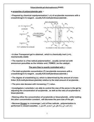
- 1. 1 Polyacrylamide gel electrophoresis (PAGE) ◘ properties of polyacrylamide gels :- • Prepared by chemical copolymerization) (بلمرة of acrylamide monomers with a crosslinking)(تشابك reagent , usually N,N-methylenbisacrylamide . • A clear Transparent gel is obtained , which is chemically inert ),(خامل mechanically stable • The reaction is a free radical polymerization , usually carried out with ammonium persulfate as the initiator and ( TEMED ) as the catalyst . The pore Size is exactly controlled with :- • The total acrylamide concentration (T) (acrylamide monomers with a crosslinking)(تشابك reagent , usually N,N-methylenbisacrylamide ) • The degree of crosslinking (c), which is determined by the amount of cross- linker (N,N-methylenbisacrylamide) relative to the total amount of acrylamide . • The pore size decease with increasing ( T ) value . • Investigators ( scientists ) are able to control the size of the pores in the gel by adjusting the concentration of acrylamide , as well as the ratio of acrylamide to bisacrylamide . • Raising either the concentration of acrylamide or bisacrylamide , while holding the other concentration constant , will decrease the pore size of the gel . • Because Oxygen is a scavenger ) (يلتهم of free radicals , polymerization is performed in closed cassettes . ) لالكسجين يتعرض ال حتي مغلق وسط في البلمره تتم (
- 2. 2 Chemistry of acrylamide polymerization • Polymerization occurs because of free oxygen radicals that react with the vinyl groups in acrylamide and bisacrylamide . • The Oxygen radicals are generated from the catalyst , ammonium persulfate ( APS ) , when it reacts with a second catalyst ( TEMED ) .
- 3. 3 Proteins are denatured prior to electrophoresis • Because proteins are so diverse with respect to either surface charges and geometries • The molecular weight of folded proteins cannot be simply determined by their migration rate in electric field . • Positively and Negatively charged proteins would migrate in different directions ! • To resolve the proteins in a sample according to their size :- - investigators must convert the proteins to uniform geometry and impart a uniform (charge / mass ratio ) to the proteins . ألن الموجب الكاثود إلي السالب األنود من يتجه عشان سالبه لشحنه هتكونا وهنا موحده شحنه ويكتسب يتكسر البروتين (الزم هتروح واجزاء الموجب القطب ناحيه هتروح واجزاء تشتت يحصل وبكداه سالبه وشحنه موجبه شحنه ليه االساس في البروتين ) السالب القطب ناحية - In SDSPAGE , The solution is denature the proteins by boiling Them with the Anionic detergent , Sodium dodecyl sulfate ( SDS) and 2-mercaptoethanol .
- 4. 4 • The combination of heat and detergent is sufficient to break the many non-covalent bonds that stabilize proteins folds . • and 2- mercaptoethanol breaks any covalent bonds between cysteine residues . • SDS consisting of a hydrophobic 12-carbon chain and hydrophilic sulfate group . • The SDS Hydrocarbon chain permeates ) (يتخلل the protein interior and binds to hydrophobic groups , reducing the protein to a random coil , coated with negatively charged detergent molecule all along its length . • Denatured proteins bind quite a lot of SDS , amounting to 1.4 g SDS/g protein , or one SDS molecule for every two amino acids .
- 5. 5 Continuous and discontinuous buffer systems Continuous buffer systems • The identity and concentration of the buffer components are the same in both the gel and the tank . • Although continuous buffer system are easy to prepared and give adequate resolution for some applications , bands tend to be broader and resolution consequently poorer in these gels . • These buffer systems are used for most forms of DNA ( eg : Agarose gel electrophoresis )
- 6. 6 Discontinuous buffer systems • Employ different buffers for tank and gel , and often two different buffers within the gel . • Discontinuous systems concentrate or “ stack “ the samples into a very narrow zone prior to separation . • Which results in improved band sharpness and resolution . ◘ The gel is divided into :- - an upper (stacking) gel of low percentage of acrylamide and low pH (6.8) and a (separation=running=resolving) gel with a pH of 8.8 and much smaller pores ( Higher percentage of acrylamide ) - The Stacking gel prevents any high-molecular-weight DNA present in the sample from clogging )(انسداد the pores at the top of the running gel before low molecular-weight DNA has entered . - both , the stacking and separating gels , contain only chloride as the mobile anion , while the tank buffer contains glycine as its anion , at a pH of 8.8 .
- 7. 7 The stacking and running gels underlie the resolving power of the SDS-PAGE gels SDS-PAGE system can be considered a 3-component system :- • The stacking and running ( resolving ) gels have different pore sizes , ionic strengths and pHs . • The third component is the electrophoresis buffer , which contains large amounts of glycine . • The Ionization state of glycine is critical to the separation . • The sample is usually dissolved in glycine chloride buffer ( pH 8.9 ) before loading on the gel . • The glycine exists primarily in two forms i.e Zwitter ions at low pH ( 6.9 – stacking gel ) and anion at high pH ( 8.9- resolving gel )
- 8. 8 Stacking Gel • The sample pH is greater than the pH of stacking gel . • After loading the sample on the well of the gel , the protein molecules present in the sample are in anionic form . • When electric fields is applied on the gel , the glycine-chloride buffer ions and sample move in the stacking gel which has pH of 6.9 . • in this pH , the glycine ion is in the form of zwitter ion with net charge zero and no electrophoretic mobility . • But the chloride ion and sample are in anionic form at pH 6.9 and act as mobile ions . • The sample will tend to accumulated and form a thin , concentrated band sandwiched between chloride and glycinate . •The chloride ion and protein carry the most of the current . The Resolving gel • The ionic from of concentrated band reaches the resolving gel with pH 8.9 . • The zwitter ionic glycine is changed into anionic form . • In Resloving gel anionic glycine and chloride carry most of the current . • The proteins present in the sample encounter with high pH and smaller pore size .
- 9. 9 The sample buffer • contain a tracking dye , bromophenole blue ( BPB ) , which will migrate with the leading edge of the proteins being separated on the gel . • Also , contains glycerol , which allows the protein samples to settle into the bottom of the gel wells . • Once a voltage is applied , the chloride ions in the sample buffer and stacking gel move rapidly toward the positive pole , forming the leading edge of a moving ion front . Sodium Dodecyl Sulphate – Polyacrylamide ( SDS-PAGE) • SDS binds to hydrophobic regions of denaturated protein chain in constant ratio of about 1.4 g of SDS per gram of protein. • The bound SDS molecules carrying negative charges mask the native charge of the protein. • The SDS binds to the unfolded proteins giving all proteins a similar shape ( i.e random ciol or extend conformation ) and a uniform charge to mass ratio .
- 10. 10 ◘ • ◘ A common way to detect proteins after electrophoresis is to stain the gel with Coomassie blue , a gye that binds proteins . - Gel are usually “ Fixed “ before staining with an acetic acid and methanol solution which precipitates proteins into the acrylamide matrix . Calculation of Relative mobility • (RF) the distance the protein migrated is compared to the length of the gel or :- Rf = Distance protein migrated / gel length
- 11. 11 PAGE The lecture is Done ♥ ☺ Q.A