2. (similar to Fas inhibition the adenoviral protein E3) (32). At
the DISC, the apoptotic pathway may be inhibited by cFLIP
protein that is capable of blocking processing and activation of
caspase 8 (33, 34). Downstream of DISC, IAP proteins may
specifically inhibit the executor caspases 3 and 7 (35). In those
cells that require mitochondria to stimulate apoptosis, the sig-
nal may be inhibited by Bcl-2/Bcl-XL types of proteins that
prevent the release of proapoptotic factors from the mitochon-
dria (30).
In the present study, we tested the cytotoxic effects of TRAIL
on six human prostate cancer cell lines, demonstrating variable
responses, with some cell lines being extremely sensitive and
others highly resistant. The highly resistant cell line LNCaP
was further investigated to examine mechanisms that protect
it from TRAIL-mediated apoptosis. We find that the TRAIL-
induced death signal in LNCaP cells is negatively regulated by
a high constitutive activity of protein kinase Akt. Furthermore,
the antiapoptotic block occurs downstream of caspase 8 activa-
tion at the level of BID protein cleavage. This study is the first
demonstration that the PI 3-kinase/Akt pathway may interfere
with an apoptotic signal by inhibiting processing of BID.
EXPERIMENTAL PROCEDURES
Antibodies—Antibodies were obtained from the following sources:
anti-phospho-Akt (New England Biolabs, Beverly, MA); anti-cyto-
chrome c and anti-BID (Zymed Laboratories Inc.); anti-Akt and anti-
XIAP (Transduction Laboratories, Lexington, KY); anti-HA1 tag
(Babco, Richmond, CA); anti-caspase 8 (Upstate Biotechnology, Inc.,
Lake Placid, NY); anti-caspase 7 (PharMingen, San Diego, CA); anti-
caspase 9 (Oncogene Research Products, Boston, MA); anti-FLIPL (Af-
finity BioReagents, Golden, CO); anti-FLIP␥/␦ (Calbiochem).
Cell Culture—Prostate cancer cell lines LNCaP, PC-3, DU 145, TSU-
Pr1, JCA-1, and ALVA-31 were passaged in RPMI 1640 with 10% fetal
calf serum, 50 units/ml penicillin, and 50 units/ml streptomycin. The
sources for these cell lines, their characterization, and use in our labo-
ratories have been described previously (36). LNCaP cells overexpress-
ing Bcl-2 (37) were kindly provided by Dr. R. Buttyan (Columbia Pres-
byterian Medical Center, New York, NY) and grown in medium
supplemented with 400 g/ml of G418.
Expression of Recombinant TRAIL in Yeast Pichia pastoris—A cDNA
encoding for soluble human TRAIL (residues 114–281) was amplified
by polymerase chain reaction from the expressed sequence tag clone
117926 (GenBankTM
accession number T90422) in frame with the N-t-
erminal hexahistidine tag using oligonucleotides 5Ј-AGTCATGAATTC-
CATCACCATCACCATCACGTGAGAGAAAGAGGTCCTCAGAGAGT-
AG-3Ј and 5Ј-AGTCATGGTACCTTAGCCAACTAAAAAGGCCCCGAA-
AAA-3Ј. This cDNA was then cloned into the EcoRI/KpnI sites of pPIC-
Z␣A vector (Invitrogen, Carlsbad, CA) in frame with the cleavable sec-
retion signal from yeast ␣ factor. All manipulations of yeast were
performed in general as outlined in the Invitrogen manual. Briefly, the
expression vector was linearized and transformed by electroporation
into P. pastoris strain SMD1168 (38). Transformants were selected on
500 g/ml of Zeocin, and secretion of TRAIL was tested by Western
blotting. For large scale production, yeast were grown for 24 h in 10
liters of complex medium containing glycerol and antifoam 289 (Sigma,
St. Louis, MO) and buffered with 100 mM potassium phosphate buffer,
pH 6.0, at constant aeration and mixing to A600 of 15. To induce TRAIL
production, cells were pelleted by centrifugation, resuspended in
complex medium containing 0.5% methanol, and grown for 24 h. The
supernatant was concentrated using tangential flow Prep/Scale-TFF
cartridge (Millipore Corp., Bedford, MA) and recombinant TRAIL
purified by nickel-chelate chromatography on a Ni2ϩ
-nitrilotriacetic
acid-agarose column (Qiagen, Valencia, CA). This procedure yielded
about 2 mg of pure protein from 1 liter of yeast supernatant.
Cytotoxicity Assays—Cell viability was determined spectrophoto-
metrically using an Aqueous One tetrazolium-based assay (Promega,
Madison, WI). Absorbance was measured at 490 nm, and data from
duplicate determinations were plotted as percentage of untreated con-
trol cells. Quantitative analysis of DNA fragmentation was done using
a Cell Death Detection ELISAplus
kit (Roche Diagnostics Corp., Indian-
apolis, IN) by measuring relative amounts of DNA-histone complexes
released into the cytoplasm. Data from triplicate determinations were
plotted as percentage of control of untreated cells. A TUNEL assay was
performed using the FragELTM
DNA fragmentation detection kit (On-
cogene Research Products, Cambridge, MA).
Measurement of Cytochrome c Release from Mitochondria—Cytosolic
extracts from LNCaP cells were prepared by the hypotonic lysis proce-
dure originally described by Bossy-Wetzel et al. (39) and modified by
Carson et al. (40). LNCaP cells grown on 15-cm plates to 50% confluence
were placed on ice and then scraped directly into growth medium and
centrifuged for 2 min at 200 ϫ g. Cell pellets were then washed once
with ice-cold phosphate-buffered saline and resuspended in 300 l of
hypotonic lysis buffer (220 mM mannitol, 68 mM sucrose, 50 mM PIPES-
KOH (pH 7.4), 50 mM KCl, 5 mM EDTA, 2 mM MgCl2, 1 mM dithiothre-
itol) containing protease inhibitors, including Complete Mixture (Roche
Molecular Biochemicals, Germany), 1 mM phenylmethylsulfonyl fluo-
ride, 10 g/ml leupeptin, and 2 g/ml aprotinin. Cells were incubated on
ice for 45 min and homogenized by pipetting (10 passes up and down).
Supernatants were cleared by 10-min centrifugation at 1000 ϫ g, fol-
lowed by 30 min at 100,000 ϫ g and analyzed by Western blotting with
the anti-cytochrome c antibody.
Construction of Adenoviral Vectors Expressing myr-Akt—The full-
length coding sequence of human Akt1 was fused in frame with the
myristoylation signal from the human Src protein in the N terminus
and HA tag in the C terminus (myr-Akt). Kinase-dead construct was
created by mutating lysine 179 for alanine, destroying in that way an
ATP-binding site (myr-Akt(KϪ)). Recombinant adenoviruses were con-
structed by the method described by Crouzet et al. (41). Briefly, cDNAs
of interest were subcloned into the expression cassette in plasmid vector
pXL2996 under the control of the cytomegalovirus promoter. Each
expression cassette was subcloned into the shuttle vector pXL3474. The
resulting shuttle plasmids were introduced into Escherichia coli JM83
cells by electroporation. After double homologous recombinations, plas-
mid DNA for recombinant virus was purified by CsCl density gradient
centrifugation. This DNA was linearized and transfected into 293 cells.
2–3 weeks after transfections, recombinant adenovirus was harvested
from the conditioned medium and amplified in 293 cells.
RESULTS
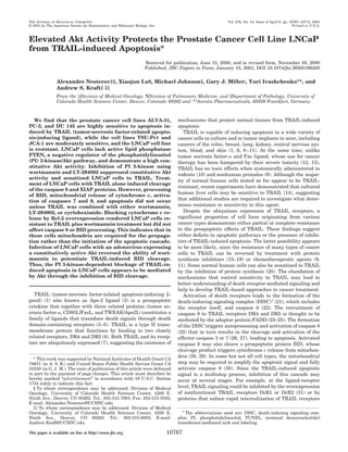
Effect of Soluble TRAIL on Six Prostate Cancer Cell Lines—
Recombinant human TRAIL (residues 114–281) was produced
in methylotrophic yeast P. pastoris as a fusion protein contain-
ing an N-terminal hexahistidine tag and a cleavable secretion
signal from yeast ␣ factor. These features allowed quick one-
step purification of secreted 20-kDa TRAIL by nickel-chelate
chromatography from yeast supernatant yielding ϳ2 mg of
pure protein from each liter of yeast culture medium (Fig. 1A).
The cytotoxic effects of TRAIL were tested on a panel of six
prostate cancer cell lines (Fig. 1B). Cell viability assays dem-
onstrated that three of these cell lines, ALVA-31, DU 145, and
PC-3 were very sensitive to TRAIL, JCA-1, and TSU-Pr1 re-
vealed moderate sensitivity, whereas LNCaP cells were resist-
ant to as high as 4 g/ml of TRAIL. Internuclosomal fragmen-
tation (DNA laddering) confirmed that cell death occurred by
apoptosis (data not shown).
To investigate the mechanisms controlling the resistance of
LNCaP cells to the cytotoxic effect of TRAIL, a series of West-
ern and Northern blot experiments were done to compare the
expression of various components of the TRAIL signaling path-
way among the six prostate cancer cell lines. However, no
correlation was found between the sensitivity of cells to TRAIL
and the expression of TRAIL receptors DR4 and DR5, decoy
receptors for TRAIL DcR1 and DcR2, initiator caspase 8, and
apoptosis inhibitory protein cFLIP (data not shown). LNCaP
cells contain a deactivating frameshift mutation in the gene
encoding the tumor suppresser PTEN (42). This dual specificity
phosphatase cleaves D3 phosphate of second messenger lipid
phosphatidylinositol (PI) 3,4,5-trisphosphate (43). PI 3,4,5-
trisphosphate produced by PI 3-kinase activates protein kinase
Akt, and therefore, the lack of negative regulation by PTEN
results in the constitutive activation of Akt in LNCaP cells (40).
Immunoblot analysis with an antibody that specifically recog-
nizes the phosphorylated/activated form of Akt (Ser473
) dem-
onstrates that LNCaP cells possess the highest Akt activity
among the six prostate cancer cell lines (Fig. 2A). Treating cells
with the inhibitor of PI 3-kinase, wortmannin (200 nM), for 6 h
PI 3-Kinase Blocks TRAIL-Induced Apoptosis and BID Cleavage10768
3. reverses the high constitutive activity of Akt (Fig. 2B).
Inhibition of PI 3-Kinase Activity or Protein Synthesis Ren-
ders LNCaP Cells Sensitive to TRAIL—To test whether the
high constitutive activity of Akt in LNCaP cells results in their
resistance to TRAIL, we first examined how PI 3-kinase inhib-
itors wortmannin (200 nM) and LY-294002 (20 M) effect
TRAIL cytotoxicity. Wortmannin acts at nanomolar concentra-
tions by covalently modifying PI 3-kinase (44) but is unstable in
aqueous solutions (45), making it possible that some PI 3-ki-
nase activity can be restored by de novo synthesis in the course
of the experiment. LY-294002 does not bind the enzyme co-
valently and has an IC50 value for PI 3-kinase about 500-fold
higher than that of wortmannin (46) but is much more stable in
culture medium. We have found that both substances signifi-
cantly enhanced the proapoptotic activity of TRAIL in LNCaP
cells as judged by apoptotic morphology (Fig. 3A) and DNA
fragmentation (Fig. 3B), quantitated by measuring the relative
amounts of DNA-histone complexes released into cytoplasm.
Since wortmannin and LY-294002 inhibit PI 3-kinase by dif-
ferent mechanisms, this result confirms that sensitization of
cells to TRAIL occurs through the inhibition of the PI 3-kinase
pathway. Inhibition of protein synthesis with cycloheximide
also sensitized LNCaP cells to TRAIL (Fig. 3, A and B). The
DNA fragmentation induced by TRAIL in combination with
wortmannin, LY-294002, or cycloheximide was greater than
that triggered by the potassium ionophore valinomycin (Fig.
3B), a potent inducer of apoptosis (47). Thus, the resistance of
LNCaP cells to TRAIL results from the blockage of the TRAIL-
induced apoptotic signal transduction cascade rather than the
defects in apoptotic machinery. These data demonstrate that
the inhibition of TRAIL-mediated apoptosis in LNCaP cells
requires PI 3-kinase activity and involves some short lived
protein component(s).
TRAIL-mediated Cytochrome c Release Is Blocked in LNCaP
Cells—Depending on the cell type, apoptotic signaling medi-
ated by CD95/Fas may or may not require the release of pro-
apoptotic factors (cytochrome c and apoptosis-inducing factor)
from mitochondria. In type II, but not in type I cells, inhibition
of mitochondrial apoptogenic activities by overexpression of
Bcl-2 protein blocks Fas-mediated apoptosis (30). To examine
whether the apoptogenic activity of mitochondria is required
for the transduction of the TRAIL-induced death signal in
LNCaP cells, the cytotoxic effects of TRAIL alone or in combi-
nation with wortmannin were studied in an LNCaP cell line
overexpressing Bcl-2 (37). Quantitation of apoptotic nuclei by
the TUNEL technique clearly demonstrates that Bcl-2 overex-
pression impairs the cytotoxic effect of TRAIL (Fig. 4A), indi-
cating that mitochondria play an important role in TRAIL-
induced apoptosis of LNCaP cells. If the resistance of LNCaP
cells to TRAIL results from the high constitutive activity of
Akt, this enzyme may block apoptosis either upstream (48, 49)
or downstream (50) of mitochondrial cytochrome c release. To
discriminate between these two possibilities, experiments were
done to examine whether TRAIL-induced cytochrome c release
is inhibited in LNCaP cells. LNCaP cells were incubated for 6 h
with TRAIL alone or TRAIL in combination with cycloheximide
or wortmannin. Cytosolic extracts were then prepared under
conditions that keep mitochondria intact (39), and cytochrome
c released to the cytosolic fraction was then detected by immu-
noblotting (Fig. 4B). This experiment demonstrated that in
LNCaP cells TRAIL alone does not trigger the release of cyto-
chrome c from the mitochondria, but it does so in combination
with wortmannin and, to a lesser extent, cycloheximide. Thus,
TRAIL-induced apoptotic signaling in LNCaP cells is blocked
upstream of the mitochondria.
TRAIL-induced Apoptotic Signaling in LNCaP Cells Is
Blocked at the Level of BID Cleavage—To understand at what
biochemical step the TRAIL-mediated apoptotic cascade is
blocked in LNCaP cells, a series of immunoblotting experi-
ments were carried out using antibodies to proteins involved in
this cascade. Our results demonstrate that processing of initi-
ator caspase 8 is induced by TRAIL alone as efficiently as when
FIG. 1. Sensitivity of human prostate cancer cell lines to solu-
ble human TRAIL. A, purification of recombinant TRAIL from P.
pastoris supernatant by nickel-chelate chromatography. B, relative vi-
ability of six prostate cancer cell lines treated for 24 h with TRAIL, as
measured by the tetrazolium conversion assay. Data are expressed as
the means for duplicate determinations.
FIG. 2. Constitutive activity of Akt in prostate cancer cells
determined by immunoblot with anti-phospho-Akt antibody
(Ser473
). A, cell lysates prepared from six prostate cancer cell lines were
probed by immunoblotting with anti-phospho-Akt antibody (top panel)
or anti-Akt antibody (bottom panel). B, LNCaP cells were treated with
wortmannin (200 nM) or cycloheximide (10 M) for 6 h, and cell lysates
were immunoblotted with anti-phospho-Akt antibody (top panel) or
anti-Akt/PKB␣ antibody (bottom panel).
PI 3-Kinase Blocks TRAIL-Induced Apoptosis and BID Cleavage 10769
4. TRAIL is combined with cycloheximide and wortmannin (Fig.
5A). Similarly, these two compounds did not enhance TRAIL-
induced cleavage of the apoptosis inhibitory protein XIAP, a
substrate for several caspases including caspase 8 (51). These
results suggest that the antiapoptotic block in LNCaP occurs
downstream of caspase 8 activation. In contrast, proteolytic
cleavage of the caspase 8 substrate BID was not detected in
TRAIL-treated cells unless TRAIL was administered in combi-
nation with cycloheximide or wortmannin. Caspase 8-mediated
cleavage of BID generates a proteolytic fragment, tBID, that is
capable of inducing mitochondrial cytochrome c release and
providing a functional link between death receptors and the
mitochondria (28, 29). The lack of BID cleavage is thus consist-
ent with the observation that TRAIL alone is not capable of
inducing cytochrome c release. TRAIL-mediated processing of
cytochrome c-dependent caspase 9 and effector caspase 7 were
also detected only if TRAIL was combined with wortmannin or
cycloheximide. The involvement of PI 3-kinase in the blockage
of TRAIL-induced BID cleavage was further confirmed by the
experiment with another PI 3-kinase inhibitor, LY-294002.
Fig. 5B demonstrates that treatment of LNCaP cells with LY-
294002 in combination with TRAIL results in the decreasing of
cellular BID level. Thus, the PI 3-kinase- and protein synthesis-
dependent antiapoptotic block in LNCaP cells occurs down-
stream of caspase 8, at the level of BID cleavage.
Alternatively, it is possible that the lack of BID cleavage may
result from an inhibition of mitochondrial function. By analogy
with the CD95/Fas system, LNCaP cells may be classified as
type II cells, since mitochondrial function appears to be neces-
sary for apoptosis. In type II cells, mitochondrial cytochrome c
release serves as an amplification loop that potentiates the
activation of caspase 8. If a similar mitochondria-dependent
amplification loop is involved in TRAIL signaling in LNCaP
cells, its disruption may affect caspase 8-mediated BID cleav-
age. To test whether or not cleavage of BID in LNCaP cells
depends on mitochondrial function, the processing of BID in
Bcl-2 overexpressor LNCaP cells versus parental cells was ex-
amined. Immunoblot analysis (Fig. 5C) demonstrates that after
6 h of treatment with TRAIL plus wortmannin or TRAIL plus
cycloheximide, BID is processed equally well in parental and
Bcl-2-overexpressing LNCaP cells. In addition, caspase 8 was
processed efficiently in both cell lines as judged by the TRAIL-
induced appearance of a cleavage product that corresponds to
the 20-kDa active subunit of caspase 8. Thus, apoptogenic
activity of mitochondria is not required for TRAIL-induced
cleavage of BID and caspase 8.
Our results demonstrate that the blockage of TRAIL-induced
apoptosis at the level of BID cleavage can be removed by
cycloheximide treatment, suggesting the possibility that this
inhibition may be mediated by a short lived protein. It has been
hypothesized that inhibition of protein synthesis sensitizes
cells to death-inducing ligands by down-regulating antiapop-
totic cFLIP proteins (15, 19, 52). To determine whether this is
the case for LNCaP cells, cell lysates from a previous experi-
ment (Fig. 5A) were immunoblotted with antibodies that rec-
ognize different splice variants of cFLIP proteins: FLIPL,
FLIP␥, and FLIP␦ (53). In contrast to published data, treat-
ment of LNCaP cells for up to 16 h with cycloheximide or
wortmannin had no effect on the level of cFLIP proteins (Fig.
5C), suggesting that they are unlikely to be involved in the
FIG. 3. Inhibitors of PI 3-kinase or
protein synthesis potentiate the cyto-
toxic activity of TRAIL. A, LNCaP cells
were treated for 24 h with 1 g/ml TRAIL,
200 nM wortmannin, 20 M LY-294002, or
10 M cycloheximide alone or in combina-
tions. The cells were visualized by light
microscopy. B, LNCaP cells were treated
for 6 h with 1 g/ml TRAIL, 200 nM wort-
mannin (WM), 20 M LY-294002, 10 M
cycloheximide (CHX), or 100 M valino-
mycin alone or in combinations. DNA
fragmentation was quantitated by meas-
uring the relative amounts of DNA-his-
tone complexes released into the cyto-
plasm using a Cell Death Detection
ELISAplus
kit.
FIG. 4. The role of mitochondrial cy-
tochrome c release for TRAIL-in-
duced apoptosis in LNCaP cells. A,
parental LNCaP cells or LNCaP cells
overexpressing Bcl-2 were treated as de-
scribed in the legend to Fig. 3B, and ap-
optotic nuclei were scored by TUNEL
staining. Several randomly chosen micro-
scopic fields were visualized, and both
normal and TUNEL-positive cells were
counted. The numbers of TUNEL-positive
versus total numbers of counted cells are
represented as ratios above the bar
graphs. B, LNCaP cells were treated with
TRAIL, wortmannin, or cycloheximide as
described above. Cells were lysed in hypo-
tonic buffer, and cytochrome c in the cy-
tosolic fraction was measured by immu-
noblotting with cytochrome c-specific
antibodies.
PI 3-Kinase Blocks TRAIL-Induced Apoptosis and BID Cleavage10770
5. inhibition of TRAIL signaling in LNCaP cells.
Constitutively Active Akt Blocks TRAIL/Wortmannin-in-
duced BID Cleavage—The potentiating effect of wortmannin on
TRAIL-induced BID cleavage suggests that Akt may be in-
volved in the inhibition of TRAIL signaling in LNCaP cells. To
confirm this hypothesis, a constitutively active Akt, con-
structed by fusing Akt to the myristoylation signal of Src pro-
tein (myr-Akt) was introduced into LNCaP cells by adenovirus-
mediated gene transfer. If Akt is the sole target of the
wortmannin effect, then this infection would be expected to
counteract the ability of wortmannin to sensitize LNCaP cells
to TRAIL-induced BID cleavage. As a control, an adenovirus
containing kinase-inactive Akt (myr-Akt(KϪ) was used. LN-
CaP cells infected with adenoviral constructs 16 h prior to the
experiment were treated for an additional 6 h with TRAIL or
TRAIL plus wortmannin, and BID cleavage was examined by
immunoblotting. Our results demonstrate (Fig. 6A) that the
infection of LNCaP cells with myr-Akt, but not with the kinase-
inactive Akt, inhibits processing of BID induced by TRAIL plus
wortmannin treatment. TRAIL-mediated cell death was also
inhibited in myr-Akt-infected cells as judged by cell morphol-
ogy (data not shown). Thus, activated Akt is capable of rescuing
LNCaP cells from the apoptogenic action of TRAIL plus wort-
mannin treatment, supporting the hypothesis that the resist-
ance of LNCaP cells to TRAIL results from high constitutive
activity of Akt.
We next tested whether activated Akt can also inhibit cleav-
age of BID induced by TRAIL plus cycloheximide treatment.
However, no rescue was observed even when the adenovirus
titer was 16 times higher than that sufficient to inhibit pro-
apoptotic effects of TRAIL plus wortmannin treatment (Fig.
6B). These results suggest that the protective effects of Akt on
BID cleavage may require Akt-induced protein synthesis.
Our results (Figs. 1B and 2A) indicate the existence of
TRAIL-sensitive cell lines that possess an elevated Akt activ-
ity, albeit at a much lower level than that found in LNCaP
cells. This result raises the question of whether the protective
effect of Akt is cell type-specific or it occurs only when the level
of Akt activity is above a certain threshold. To examine these
possibilities, we overexpressed myristoylated Akt in various
TRAIL-sensitive cell lines: DU 145 and ALVA-31 prostate can-
cer cells, A498 renal cancer cells, and HeLa cervical cancer
cells. Of them, only ALVA-31 cells acquired significant resist-
ance to TRAIL upon myr-Akt overexpression (Fig. 6C). Thus,
the protective effect of Akt appears to be cell type-specific.
DISCUSSION
We have developed a novel approach to obtaining prepara-
tive amounts of proapoptotic ligand TRAIL and tested the
effects of this reagent on a panel of six prostate cancer cell
lines. Soluble TRAIL was produced by a methylotrophic yeast
P. pastoris, secreted into the medium, and then purified to
homogeneity by one-step chromatography on a nickel-chelate
column. Cytotoxicity assays demonstrated that three cell lines,
ALVA-31, DU 145, and PC-3, were very sensitive to TRAIL,
while in comparison JCA-1 and TSU-Pr1 revealed moderate
sensitivity, and LNCaP cells were resistant to as high as 4
g/ml TRAIL. Comparing these results with the data published
on Fas ligand-induced apoptosis indicates that prostate cancer
cells differ in their responses to these two apoptotic stimuli.
Whereas cells believed to be derived from primary prostate
cancer tumors (ALVA-31 and JCA-1) were reported to be sen-
sitive to Fas ligand-induced apoptosis, cells originating from
distant metastasis (DU 145, PC-3, TSU-Pr1, and LNCaP) ap-
peared to be Fas-resistant despite the expression of Fas anti-
gen on the cell surface (36, 54). In contrast, among the above
listed cell lines, only LNCaP cells were resistant to TRAIL-
induced apoptosis, indicating that TRAIL has a greater poten-
tial as an agent to treat metastatic prostate cancer. These data
also suggest that despite the similarity of CD95/Fas and TRAIL
receptors, TRAIL and Fas ligand-mediated apoptosis may em-
ploy different signal transduction pathways or be negatively
regulated by different mechanisms in these prostate cancer
cells.
We found that among six prostate cancer cell lines examined,
the LNCaP cells, which are the most highly resistant to TRAIL-
induced apoptosis, have the highest constitutive activity of the
Akt protein kinase. This result is consistent with the lack of the
functional tumor suppressor PTEN, a negative regulator of the
PI 3-kinase/Akt pathway in these cells (42). Because the Akt
FIG. 5. Block of TRAIL-mediated ap-
optotic signal in LNCaP cells occurs at
the level of BID cleavage. A, LNCaP cells
were treated for 6 or 16 h with 1 g/ml
TRAIL, 200 nM wortmannin (WM), or 10 M
cycloheximide (CHX) alone or in combina-
tions. Cell lysates were electrophoresed and
consecutively immunoblotted with antibod-
ies specific to caspase 8, XIAP, BID, caspase
9, and caspase 7. The arrows on the left
indicate cleavage products. B, LNCaP cells
were treated for 6 h with 1 g/ml TRAIL or
20 M LY-294002 alone or in combinations.
Cell lysates were electrophoresed and con-
secutively immunoblotted with antibodies
specific to BID or the phosphorylated form of
Akt (Ser473
). C, parental LNCaP cells and
LNCaP cells overexpressing Bcl-2 were
treated for 6 h with 1 g/ml TRAIL and 200
nM wortmannin alone or in combination.
Cleavage of caspase 8 and BID was analyzed
by immunoblotting with the corresponding
antibodies. Blots were processed by ECL,
and two different exposures were taken to
visualize holocaspase 8 (short exposure) and
its 20-kDa proteolytic fragment (long expo-
sure). The arrow indicates caspase 8 cleav-
age product. D, cell lysates from the experi-
ment described for A were immunoblotted
with antibodies that specifically recognize
different splice variants of cFLIP protein:
FLIPL, FLIP␥, and FLIP␦.
PI 3-Kinase Blocks TRAIL-Induced Apoptosis and BID Cleavage 10771
6. protein kinase is known to block apoptosis (55), we tested
whether inhibition of this pathway affects the sensitivity of
LNCaP cells to TRAIL. We found that treatment with the PI
3-kinase inhibitors wortmannin and LY-294002 or the protein
synthesis inhibitor cycloheximide renders them sensitive to
TRAIL-induced apoptosis. Thus, the resistance of LNCaP cells
to TRAIL results not from defects in apoptotic machinery, but
from PI 3-kinase-dependent inhibition of the TRAIL-mediated
apoptotic signaling pathway.
It has been reported that apoptosis induced by triggering of
CD95/Fas (56, 57) is counteracted by the PI 3-kinase/Akt path-
way, but the molecular mechanisms that cause apoptosis re-
sistance remain unclear. To identify which step of the TRAIL-
mediated apoptotic pathway is blocked in LNCaP cells, we first
tested whether the release of proapoptotic factors from mito-
chondria is essential for TRAIL-induced death of these cells.
The involvement of mitochondria in apoptosis induced by death
receptors remains controversial. Scaffidi et al. (30) have pro-
posed that two types of cells exist that differ with respect to
their requirement for mitochondria during Fas-mediated apop-
tosis. In type I cells, caspase 8 is activated without involvement
of mitochondria to a level sufficient to process the effector
caspase 3. In contrast, in type II cells a mitochondria-depend-
ent amplification loop is required to fully activate caspase 8
and transduce an apoptotic signal. This model has recently
been questioned by Huang et al. (58), who argue that the
difference between type I and type II cells is an artifact of using
agonistic anti-Fas antibodies to trigger Fas signaling instead of
Fas ligand. To clarify the role of mitochondria in TRAIL-in-
duced apoptosis in LNCaP cells, we used Bcl-2-overexpressing
LNCaP cells, which were shown to exhibit an impaired cyto-
chrome c release in response to various apoptotic stimuli (37).
Our results demonstrate that these cells are much more resist-
ant to TRAIL plus wortmannin-induced apoptosis compared
with the parental cells. In these experiments, apoptosis was
triggered by soluble death receptor ligand and not agonistic
antibody, supporting the notion that in some cells mitochon-
drial function is indeed essential for death receptor-mediated
apoptosis.
Using a cell fractionation approach, we have found that
TRAIL-induced cytochrome c release was blocked in LNCaP
cells, but both wortmannin and cycloheximide are capable of
overcoming this block. Release of mitochondrial cytochrome c
by death receptors is triggered by a multistep mechanism. The
formation of the DISC results in autoprocessing and activation
of the initiator caspase 8 followed by cleavage of the proapop-
totic protein BID (28, 29). A proteolytic fragment of BID trans-
locates to the mitochondria as an integral membrane protein
and triggers the release of mitochondrial cytochrome c (59).
Using immunoblot analysis, we found that cleavage of caspase
8 and one of its substrates, the antiapoptotic protein XIAP (51)
were induced by TRAIL alone as efficiently as when TRAIL was
combined with either wortmannin or cycloheximide. This im-
portant result indicates that DISC formation or caspase 8
activation was not blocked in LNCaP cells. In contrast, wort-
mannin and cycloheximide were required for TRAIL-induced
cleavage of BID, the release of cytochrome c, and processing of
caspases 9 and 7. Thus, the PI 3-kinase-dependent block of
TRAIL-induced apoptosis in LNCaP cells occurs at the level of
BID cleavage.
The requirement for mitochondrial apoptogenic activity in
TRAIL-induced death suggests that LNCaP cells are similar to
type II cells. If so, the lack of BID cleavage could, in principle,
be explained by the disruption of a mitochondria-dependent
FIG. 6. Constitutively active Akt inhibits proapoptotic effects of TRAIL. A, LNCaP cells were infected with adenoviral constructs
expressing myristoylated Akt (Adeno-myr-Akt) or kinase-inactive myristoylated Akt (Adeno-myr-Akt(KϪ)) at a titer of 3 ϫ 106
TCID50/ml. Control
cells were not infected with adenoviruses. 16 h postinfection, the cells were treated for 6 h with 1 g/ml TRAIL and 200 nM wortmannin alone or
in combination. Cell lysates were consecutively probed with BID-specific antibody and anti-HA1 antibody that recognizes hemagglutinin-tagged
myr-Akt. B, LNCaP cells were infected where indicated with adenoviral constructs expressing myristoylated Akt (Adeno-myr-Akt) at a titer
increasing from 6 to 48 ϫ 106
TCID50/ml. Control cells were not infected with adenovirus. 16 h after infection, the cells were treated for 6 h with
1 g/ml of TRAIL and 10 M cycloheximide alone or in combination. Cell lysates were probed as outlined for A. C, ALVA-31 cells were transiently
cotransfected with an expression plasmid encoding the E. coli lacZ gene plus an expression plasmid for myristoylated Akt (myr-Akt) (60) or empty
expression vector (Mock). 24 h after transfection, the cells were incubated with or without 0.1 g/ml TRAIL and scored for apoptosis 24 h later.
Cells positive for -galactosidase activity were checked for morphological changes characteristic of apoptosis, and the percentage of live cells was
quantitated.
PI 3-Kinase Blocks TRAIL-Induced Apoptosis and BID Cleavage10772
7. amplification loop, resulting in only partial activation of
caspase 8. To see whether this hypothesis could be true, we
compared the cleavage of BID and caspase 8 in Bcl-2-overex-
pressing versus parental LNCaP cells and found that these
proteins are processed equally well in both cell lines. These
results demonstrate that although mitochondrial function is
important for TRAIL-induced apoptosis in LNCaP cells, unlike
“typical” type II cells mitochondria are required not to amplify
caspase 8 activation but to transduce apoptotic signal down-
stream of the initiator caspase. Therefore, it may be possible to
classify LNCaP as type III cells where mitochondria are in-
volved in the propagation rather than the initiation of the
apoptotic cascade.
Involvement of PI 3-kinase in the block of apoptosis suggests
that Akt could mediate resistance of LNCaP cells to TRAIL. To
confirm this hypothesis, we tested whether overexpression of
constitutively active Akt could inhibit the proapoptotic effect of
TRAIL plus wortmannin treatment. For this purpose, we used
a myristoylated derivative of Akt, which exhibits kinase activ-
ity independently of PI 3-kinase (60). Both apoptosis (data not
shown) and BID cleavage induced by treatment of LNCaP cells
with TRAIL plus wortmannin were inhibited by overexpression
of myristoylated Akt, indicating that resistance of LNCaP cells
to TRAIL is, at least in part, mediated by Akt.
It has been documented that Akt may inhibit a variety of
apoptotic stimuli in multiple ways (55). These include direct
phosphorylation and modulation of proapoptotic proteins BAD
(48) and caspase 9 (50), activation of antiapoptotic NF-B-
mediated transcriptional pathways (61, 62), or phosphorylation
of the Forkhead family of transcription factors, preventing
them from inducing the transcription of proapoptotic genes
(63). Inhibition of BID cleavage has not been previously re-
ported as a mechanism through which PI 3-kinase and Akt
block apoptotic signals.
Although it remains unclear how the PI 3-kinase/Akt path-
way mediates inhibition of BID cleavage, our data suggest an
indirect mechanism. First, inhibition of protein synthesis by
cycloheximide affected the same step of TRAIL apoptotic cas-
cade as the inhibition of PI 3-kinase. However, even very high
levels of constitutively active Akt did not rescue BID from
cleavage when TRAIL was combined with cycloheximide rather
than wortmannin. These results suggest that a short lived
protein is involved in the PI 3-kinase/Akt-mediated blockage of
BID cleavage, and the synthesis of this hypothetical protein
may be triggered by Akt. Second, the effect of myristoylated
Akt appears to be cell type-specific, since its overexpression did
not rescue HeLa, DU-145, or A498 cells from TRAIL-induced
apoptosis (data not shown) but did rescue LNCaP and ALVA-31
cells. This could reflect either the difference in apoptotic path-
ways employed by different cell types or the absence of certain
factors required for the protective effect of Akt. In particular,
human prostate cancer cell lines have scores of chromosomal
deletions and rearrangements (64), so that LNCaP and PC-3
differ in much more than Akt levels.
It has been reported that short term (3–7-h) treatment of
human keratinocytes (52), HeLa and Kym-1 cells (19) with
cycloheximide significantly reduces the level of cellular cFLIP
protein. Since upon overexpression cFLIP is capable of inhib-
iting Fas-mediated apoptosis (33, 34), it has been suggested
that protein synthesis inhibitors sensitize cells to TRAIL by
down-regulating cFLIP. To examine this hypothesis, we tested
the level of various splice variants of cFLIP (FLIPL, FLIP␥, and
FLIP␦) in LNCaP cells and found that neither cycloheximide
nor wortmannin treatment affected cFLIP levels after as long
as 16 h of treatment. These data are consistent with our obser-
vation on renal carcinoma cells (65) and published results on
Kaposi’s sarcoma cells (17) in which that inhibition of protein
synthesis sensitized cells to TRAIL without affecting the ex-
pression of cFLIP proteins. Thus, mediators of the PI 3-kinase-
dependent blockage of TRAIL-induced BID cleavage and apop-
tosis in LNCaP cells still await identification and
characterization.
Acknowledgments—We thank Dr. Ralph Buttyan (Columbia Presby-
terian Medical Center, New York, NY) for Bcl-2-overexpressing LNCaP
cells, and we thank Dr. Richard A. Roth (Stanford University School of
Medicine, Stanford, CA), Dr. Joseph Biggs, and other members of Kraft
laboratory for helpful discussions. We appreciate the excellent technical
assistance of Sarah Winbourn.
REFERENCES
1. Wiley, S. R., Schooley, K., Smolak, P. J., Din, W. S., Huang, C. P., Nicholl, J. K.,
Sutherland, G. R., Smith, T. D., Rauch, C., Smith, C. A., and Goodwin, R. G.
(1995) Immunity 3, 673–682
2. Pitti, R. M., Marsters, S. A., Ruppert, S., Donahue, C. J., Moore, A., and
Ashkenazi, A. (1996) J. Biol. Chem. 271, 12687–12690
3. Schulze-Osthoff, K., Ferrari, D., Los, M., Wesselborg, S., and Peter, M. E.
(1998) Eur. J. Biochem. 254, 439–459
4. Ashkenazi, A., and Dixit, V. M. (1998) Science 281, 1305–1308
5. Walczak, H., and Krammer, P. H. (2000) Exp. Cell Res. 256, 58–66
6. Griffith, T. S., and Lynch, D. H. (1998) Curr. Opin. Immunol. 10, 559–563
7. Golstein, P. (1997) Curr. Biol. 7, R750–R753
8. Snell, V., Clodi, K., Zhao, S., Goodwin, R., Thomas, E. K., Morris, S. W., Kadin,
M. E., Cabanillas, F., Andreeff, M., and Younes, A. (1997) Br. J. Haematol.
99, 618–624
9. Ashkenazi, A., Pai, R. C., Fong, S., Leung, S., Lawrence, D. A., Marsters, S. A.,
Blackie, C., Chang, L., McMurtrey, A. E., Hebert, A., DeForge, L.,
Koumenis, I. L., Lewis, D., Harris, L., Bussiere, J., Koeppen, H., Shahrokh,
Z., and Schwall, R. H. (1999) J. Clin. Invest. 104, 155–162
10. Walczak, H., Miller, R. E., Ariail, K., Gliniak, B., Griffith, T. S., Kubin, M.,
Chin, W., Jones, J., Woodward, A., Le, T., Smith, C., Smolak, P., Goodwin,
R. G., Rauch, C. T., Schuh, J. C., and Lynch, D. H. (1999) Nat. Med. 5,
157–163
11. Keane, M. M., Ettenberg, S. A., Nau, M. M., Russell, E. K., and Lipkowitz, S.
(1999) Cancer Res. 59, 734–741
12. Vassalli, P. (1992) Annu. Rev. Immunol. 10, 411–452
13. Nagata, S. (1997) Cell 88, 355–365
14. Jo, M., Kim, T. H., Seol, D. W., Esplen, J. E., Dorko, K., Billiar, T. R., and
Strom, S. C. (2000) Nat. Med. 6, 564–567
15. Griffith, T. S., Chin, W. A., Jackson, G. C., Lynch, D. H., and Kubin, M. Z.
(1998) J. Immunol. 161, 2833–2840
16. Muhlenbeck, F., Haas, E., Schwenzer, R., Schubert, G., Grell, M., Smith, C.,
Scheurich, P., and Wajant, H. (1998) J. Biol. Chem. 273, 33091–33098
17. Mori, S., Murakami-Mori, K., Nakamura, S., Ashkenazi, A., and Bonavida, B.
(1999) J. Immunol. 162, 5616–5623
18. Rieger, J., Naumann, U., Glaser, T., Ashkenazi, A., and Weller, M. (1998)
FEBS Lett. 427, 124–128
19. Wajant, H., Haas, E., Schwenzer, R., Muhlenbeck, F., Kreuz, S., Schubert, G.,
Grell, M., Smith, C., and Scheurich, P. (2000) J. Biol. Chem. 275,
24357–24366
20. Bretz, J. D., Rymaszewski, M., Arscott, P. L., Myc, A., Ain, K. B., Thompson,
N. W., and Baker, J. R., Jr. (1999) J. Biol. Chem. 274, 23627–23632
21. Kischkel, F. C., Hellbardt, S., Behrmann, I., Germer, M., Pawlita, M.,
Krammer, P. H., and Peter, M. E. (1995) EMBO J. 14, 5579–5588
22. Medema, J. P., Scaffidi, C., Kischkel, F. C., Shevchenko, A., Mann, M.,
Krammer, P. H., and Peter, M. E. (1997) EMBO J. 16, 2794–2804
23. Chaudhary, P. M., Eby, M., Jasmin, A., Bookwalter, A., Murray, J., and Hood,
L. (1997) Immunity 7, 821–830
24. Schneider, P., Thome, M., Burns, K., Bodmer, J. L., Hofmann, K., Kataoka, T.,
Holler, N., and Tschopp, J. (1997) Immunity 7, 831–836
25. Bodmer, J. L., Holler, N., Reynard, S., Vinciguerra, P., Schneider, P., Juo, P.,
Blenis, J., and Tschopp, J. (2000) Nat. Cell Biol. 2, 241–243
26. Srinivasula, S. M., Ahmad, M., Fernandes-Alnemri, T., Litwack, G., and
Alnemri, E. S. (1996) Proc. Natl. Acad. Sci. U. S. A. 93, 14486–14491
27. Muzio, M., Salvesen, G. S., and Dixit, V. M. (1997) J. Biol. Chem. 272,
2952–2956
28. Luo, X., Budihardjo, I., Zou, H., Slaughter, C., and Wang, X. (1998) Cell 94,
481–490
29. Li, H., Zhu, H., Xu, C. J., and Yuan, J. (1998) Cell 94, 491–501
30. Scaffidi, C., Fulda, S., Srinivasan, A., Friesen, C., Li, F., Tomaselli, K. J.,
Debatin, K. M., Krammer, P. H., and Peter, M. E. (1998) EMBO J. 17,
1675–1687
31. Ashkenazi, A., and Dixit, V. M. (1999) Curr. Opin. Cell Biol. 11, 255–260
32. Tollefson, A. E., Hermiston, T. W., Lichtenstein, D. L., Colle, C. F., Tripp, R. A.,
Dimitrov, T., Toth, K., Wells, C. E., Doherty, P. C., and Wold, W. S. (1998)
Nature 392, 726–730
33. Kataoka, T., Schroter, M., Hahne, M., Schneider, P., Irmler, M., Thome, M.,
Froelich, C. J., and Tschopp, J. (1998) J. Immunol. 161, 3936–3942
34. Scaffidi, C., Schmitz, I., Krammer, P. H., and Peter, M. E. (1999) J. Biol. Chem.
274, 1541–1548
35. Deveraux, Q. L., and Reed, J. C. (1999) Genes Dev. 13, 239–252
36. Hedlund, T. E., Duke, R. C., Schleicher, M. S., and Miller, G. J. (1998) Prostate
36, 92–101
37. Raffo, A. J., Perlman, H., Chen, M. W., Day, M. L., Streitman, J. S., and
Buttyan, R. (1995) Cancer Res. 55, 4438–4445
38. Gleeson, M. A., White, C. E., Meininger, D. P., and Komives, E. A. (1998)
PI 3-Kinase Blocks TRAIL-Induced Apoptosis and BID Cleavage 10773
8. Methods Mol. Biol. 103, 81–94
39. Bossy-Wetzel, E., Newmeyer, D. D., and Green, D. R. (1998) EMBO J. 17,
37–49
40. Carson, J. P., Kulik, G., and Weber, M. J. (1999) Cancer Res. 59, 1449–53
41. Crouzet, J., Naudin, L., Orsini, C., Vigne, E., Ferrero, L., Le Roux, A., Benoit,
P., Latta, M., Torrent, C., Branellec, D., Denefle, P., Mayaux, J. F.,
Perricaudet, M., and Yeh, P. (1997) Proc. Natl. Acad. Sci. U. S. A. 94,
1414–1419
42. Vlietstra, R. J., van Alewijk, D. C., Hermans, K. G., van Steenbrugge, G. J.,
and Trapman, J. (1998) Cancer Res. 58, 2720–2723
43. Di Cristofano, A., and Pandolfi, P. P. (2000) Cell 100, 387–90
44. Wymann, M. P., Bulgarelli-Leva, G., Zvelebil, M. J., Pirola, L.,
Vanhaesebroeck, B., Waterfield, M. D., and Panayotou, G. (1996) Mol. Cell.
Biol. 16, 1722–1733
45. Budavari, S. (ed) (1989) The Merck Index, 11th Ed., p. 1585, Merck, Rahway,
NJ
46. Walker, E. H., Pacold, M. E., Perisic, O., Stephens, L., Hawkins, P. T.,
Wymann, M. P., and Williams, R. L. (2000) Mol. Cell 6, 909–919
47. Duke, R. C., Witter, R. Z., Nash, P. B., Young, J. D., and Ojcius, D. M. (1994)
FASEB J. 8, 237–246
48. Datta, S. R., Dudek, H., Tao, X., Masters, S., Fu, H., Gotoh, Y., and Greenberg,
M. E. (1997) Cell 91, 231–241
49. Kennedy, S. G., Kandel, E. S., Cross, T. K., and Hay, N. (1999) Mol. Cell. Biol.
19, 5800–5810
50. Cardone, M. H., Roy, N., Stennicke, H. R., Salvesen, G. S., Franke, T. F.,
Stanbridge, E., Frisch, S., and Reed, J. C. (1998) Science 282, 1318–1321
51. Deveraux, Q. L., Leo, E., Stennicke, H. R., Welsh, K., Salvesen, G. S., and
Reed, J. C. (1999) EMBO J. 18, 5242–5251
52. Leverkus, M., Neumann, M., Mengling, T., Rauch, C. T., Brocker, E. B.,
Krammer, P. H., and Walczak, H. (2000) Cancer Res. 60, 553–559
53. Tschopp, J., Irmler, M., and Thome, M. (1998) Curr. Opin. Immunol. 10,
552–558
54. Uslu, R., Borsellino, N., Frost, P., Garban, H., Ng, C. P., Mizutani, Y.,
Belldegrun, A., and Bonavida, B. (1997) Clin. Cancer Res. 3, 963–972
55. Datta, S. R., Brunet, A., and Greenberg, M. E. (1999) Genes Dev. 13,
2905–2927
56. Hausler, P., Papoff, G., Eramo, A., Reif, K., Cantrell, D. A., and Ruberti, G.
(1998) Eur. J. Immunol. 28, 57–69
57. Rohn, J. L., Hueber, A. O., McCarthy, N. J., Lyon, D., Navarro, P., Burgering,
B. M., and Evan, G. I. (1998) Oncogene 17, 2811–2818
58. Huang, D. C., Hahne, M., Schroeter, M., Frei, K., Fontana, A., Villunger, A.,
Newton, K., Tschopp, J., and Strasser, A. (1999) Proc. Natl. Acad. Sci.
U. S. A. 96, 14871–14876
59. Gross, A., Yin, X. M., Wang, K., Wei, M. C., Jockel, J., Milliman, C.,
Erdjument-Bromage, H., Tempst, P., and Korsmeyer, S. J. (1999) J. Biol.
Chem. 274, 1156–63
60. Kohn, A. D., Summers, S. A., Birnbaum, M. J., and Roth, R. A. (1996) J. Biol.
Chem. 271, 31372–31378
61. Ozes, O. N., Mayo, L. D., Gustin, J. A., Pfeffer, S. R., Pfeffer, L. M., and Donner,
D. B. (1999) Nature 401, 82–85
62. Romashkova, J. A., and Makarov, S. S. (1999) Nature 401, 86–90
63. Kops, G. J., and Burgering, B. M. (1999) J. Mol. Med. 77, 656–665
64. Webber, M. M., Bello, D., and Quader, S. (1997) Prostate 30, 58–64
65. Pawlowski, J. E., Nesterov, A., Scheinman, R. I., Johnson, T. R., and Kraft,
A. S. (2001) Anticancer Res. 20, 4243–4256
PI 3-Kinase Blocks TRAIL-Induced Apoptosis and BID Cleavage10774