Vitamin D plays an important role in regulating calcium and phosphorus balance and promoting bone growth. It also has immunomodulating, anticancer, and neuromodulating effects. The document reviews vitamin D's structure, metabolism, mechanisms of action, and functions in the body. It then discusses recent research on vitamin D's impact on oral cavity diseases, focusing on its potential role in autoimmune conditions of the oral mucosa like recurrent aphthous stomatitis.
![Lips 2006). The two basic forms of vitamin D: ergocal-
ciferol (vitamin D2, which appears in plants, yeasts and
fungi) and cholecalciferol (vitamin D3, of an animal ori-
gin), differ in the structure of the side chain attached to the
sterol group (Lips 2006; Miller and Portale 2001; Myszka
and Klinger 2014).
Both forms are biologically inactive. The active form in
humans is 1a,25-dihydroxy vitamin D [1,25(OH)2D],
which is derived from provitamin D3 (7-dehydrocholes-
terol). Provitamin D3 is present in epithelial basal and
spinous layers and in fibroblasts of the dermis. On exposure
to sunlight (UVB radiation, 290–315 nm) provitamin D3 is
transformed into cholecalciferol, which undergoes photoi-
somerization at body temperature and is then released into
intracellular space and into the blood. The initial hydrox-
ylation of cholecalciferol occurs in liver at the C25 position
and is catalyzed by a group of 25-hydroxylases, consisting
of cytochromes CYP27A1, CYP3A4 and CYP2R1. This
results in the formation of 25-hydroxycholecalciferol
(25(OH)D3; calcidiol).
Subsequent hydroxylation catalyzed by 1a-hydroxylasis
(CYP27B1) occurs at the C1 and/or C24 position, mainly
in the kidneys and to a lesser degree in bony tissue, lungs,
liver, placenta, parotid glands, keratinocytes, neoplasmatic
cells and macrophages. Hydroxylation at the C1 position
produces the biologically active calcitriol [1,25(OH)2D3].
Hydroxylation of calcidiol at C24 position leads to the
formation of 24,25-dihydroxycholecalciferol, and is cat-
alyzed by CYP24 a hydroxylase commonly found in body
tissues. The key role of this derivative is the metabolism of
the cartilage and bony tissue. The serum levels of both
hydroxylated derivatives are regulated by a feedback
mechanism and are dependent on the 1,25(OH)2D3 con-
centration in the organism together with the indirect
regulatory role of calcium and phosphoric ions, calcitonin,
somatotropin, parathyroid hormone and other hormones
(Grygiel-Go´rniak and Puszczewicz 2014; Hewison 2012b;
Miller and Portale 2001).
Since 1998 normal serum vitamin D (25(OH)D)
concentration for the Central European population have
been established as 30–50 ng/ml. Levels between 21 and
29 ng/ml indicate vitamin D insufficiency, while con-
centrations below 20 ng/ml are defined as vitamin D
deficiency and require medical intervention (Holick et al.
2011, Pludowski et al. 2013a, b, Yin and Agrawal 2014).
A 25(OH)D serum level of 50–100 ng/ml reflects a high
vitamin D supply, which may require some modifications
to the vitamin D intake especially for the upper con-
centrations ([100 ng/ml) at which potentially negative
health outcomes may arise. Serum levels in excess of
200 ng/ml are considered to be toxic (Pludowski et al.
2013a, b).
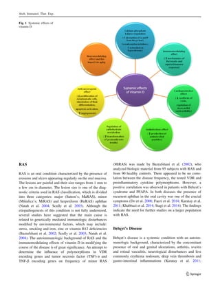
Vitamin D Mechanism of Action
The mechanism of vitamin D activity is both genomic and
extra-genomic (Fernandes de Abreu et al. 2009; Lips 2006;
Pawlak and Doboszyn´ska 2014). The genomic action is
mediated by the VDR which belongs to the nuclear
receptor subfamily (Jurutka et al. 2007; Valdivielso and
Fernandez 2006). It acts as a ligand activated transcription
factor by modifying the transcription on binding the
selected sequences in the target genes promoter regions,
called vitamin D responsive elements. The bonding of
1,25(OH)2D-VDR complex with a specific DNA sequence
is preceded by heterodimerization with the retinoid X
receptor (Fernandes de Abreu et al. 2009; Tuohimaa 2009;
Valdivielso and Fernandez 2006). Vitamin D as calcitriol is
known to modify the expression of more than 200 vitamin
D-responsive genes. Vitamin D receptors maintain the
structure and function of the skeleton and are located in the
tissues and organs responsible for calcium-phosphate
homeostasis, such as bones, kidneys and parotid glands
(Grygiel-Go´rniak and Puszczewicz 2014). Stumpf et al.
(1979), reported the presence of VDRs in other organs,
such as skin, brain and immune cells. That led to further
research focused on the extra-bony effects of vitamin D
(Grygiel-Go´rniak and Puszczewicz 2014; Lips 2006). The
VDR encoding gene is located on the chromosome 12 in
position 12q13.11 and consists of two promoter regions,
eight protein encoding exons, and six untranslated exons
(Martelli et al. 2014). Some specific VDR gene alleles can
influence the action of vitamin D on a cellular level,
including calcium metabolism, transcription, cellular divi-
sions and initiation of the immunologic response (Myszka
and Klinger 2014; Valdivielso and Fernandez 2006). The
role of the selected VDR gene polymorphisms in the
pathogenesis of inflammatory, neurologic and metabolic
conditions is currently an active area of research (Myszka
and Klinger 2014).
Extra-genomic vitamin D effects are mediated by the
receptors of a different structure to the nuclear VDRs and
are classified as membrane-associated protein disulfide
isomerases, family A, member 3. The process involves the
activation of proteases and cell kinases, followed by the
release of prostaglandins. The result is a stimulation of
some intracellular signaling paths (like MAP and Raf
kinase paths) in various cell types, e.g. in enterocytes,
monocytes, vascular smooth muscle cells, osteoblasts and
chondrocytes. The interaction with a second messenger
such as MAP or cyclic AMP involves calcium channels
and leads to increased calcium absorption and osteoclastic
bone resorption, as well as the stimulation of cell differ-
entiation and the modulation of muscle function and insulin
secretion (Lips 2006). While the effects of genomic
Arch. Immunol. Ther. Exp.
123](https://image.slidesharecdn.com/oral-181215232407/85/Oral-2-320.jpg)
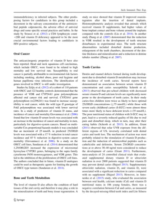
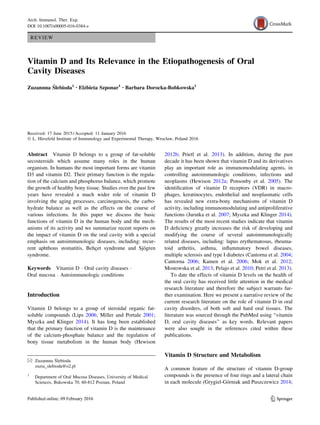
![Table1AsummaryresultsofstudiesontheroleofvitaminDintheoralcavitydiseases
OralcavitydiseasePopulationstudied
(contributingcountry)
SpeciestestedResultsReferences
RASS:95(MiRAS)
C:90
(GreatBritain)
VDRgenepolymorphismsNocorrelationBazrafshanietal.(2002)
Behc¸et’ssyndromeS:32
C:31
(Turkey)
25[OH]D(serum);levelinstudygroupKaratayetal.(2011)
S:48
C:47
(Iran)
25[OH]D(serum);levelinstudygroupKhabbazietal.(2014)
S:112
C:112
(Iran)
25[OH]D(serum);levelinstudygroupFaezietal.(2014)
S:41
C:15
(SouthKorea)
25[OH]D(serum);levelinstudygroupDoetal.(2008))
PFAPAS:25
C:25
(Italy)
25[OH]D(serum);numberoffeverepisodes,;durationof
episodesaftervitaminDsupplementation
Stagietal.(2014)
Sjo¨gren’ssyndromeS:41
C:41
(Danmark)
25[OH]D(serum);levelinstudygroupBangetal.(1999)
S:25
C:15
(Hungary)
25[OH]2D3(serum)NocorrelationSzodorayetal.(2010)
S:35
C:1674
(Danmark)
1a,25[OH]2D3
(serum)
25[OH]D(serum)
Nocorrelation
;levelinstudygroup
Mulleretal.(1990)
S:30
C:46
(Italy)
25[OH]D3(serum)NocorrelationBaldinietal.(2014)
Arch. Immunol. Ther. Exp.
123](https://image.slidesharecdn.com/oral-181215232407/85/Oral-6-320.jpg)
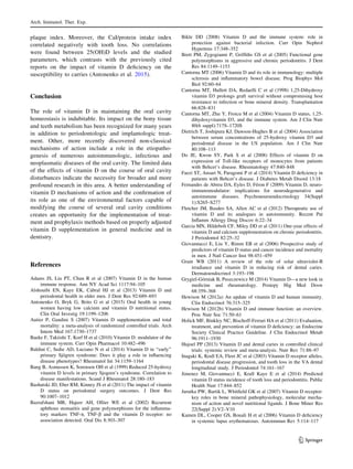
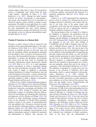
![Table1continued
OralcavitydiseasePopulationstudied
(contributingcountry)
SpeciestestedResultsReferences
PeriodontitisS:24(AP)
37(EOP)
C:37
(Japan)
VDRgenepolymorphisms:TaqI(Tt)frequencyinEOP
NocorrelationinAPandCgroups
Sunetal.(2002)
S:198
(Japan)
VDRgenepolymorphisms:TaqI(TT)frequencyinCPTachietal.(2003)
S:51(AgP)
57(CP)
C:100
(GreatBritain)
VDRgenepolymorphisms:TaqI(TT)frequencyinCPBrettetal.(2005)
S:79(CP)
224(AgP)
C:231
(GreatBritain)
VDRgenepolymorphisms:TaqI(TT)frequencyinsmokerswithCPNibalietal.(2008)
S:107
C:121
(China)
VDRgenepolymorphisms:TaqI(TT)frequencyinCPWangetal.(2009)
S:115(CP)
58(AgP)
C:65
(Italy)
VDRgenepolymorphisms:TaqI(TT)frequencyinCP:TaqI(tt)in
AgP
Martellietal.(2011)
S:99(CP)
63(AgP)
C:126
(Jordan)
VDRgenepolymorphisms:BsmI(bb)andApaI(aa)frequencyinCP,
;inAgP
Karasnehetal.(2013)
S:30
C:30
(Brazil)
VDRgenepolymorphisms:TaqI(Tt)frequencyinCPToniatoBorgesetal.(2009)
S:562
(USA)
TotalvitaminDdailyintakeTotalvitaminDintakeC800IU/day
comparedto400IU/day?;severe
periodontaldiseaseand;rateofmoderate-
to-severeABLforhigherdoses
Alshouibietal.(2013)
S:920
(USA)
25[OH]D(serum);level?
:rateofgingivalbleeding
Millenetal.(2013)
S:23(onvitaminD
supplement)
C:28(nosupplement)
(USA)
VitaminDsupplementation(C400IU/day)Noinfluenceonperiodontaltissuesafter
12monthsofsupplementation
Garciaetal.(2011)
Arch. Immunol. Ther. Exp.
123](https://image.slidesharecdn.com/oral-181215232407/85/Oral-7-320.jpg)
![Khabbazi et al. 2014). As with RAS, the etiopathogenesis
of the syndrome has not been defined. It was found that as a
consequence of the disease the autoimmunologic reaction
leads to vasculitis (Karatay et al. 2011; Khabbazi et al.
2014). Based on the observation of 32 patients with Beh-
c¸et’s disease, Karatay et al. (2011) demonstrated that their
serum 25-hydroxy vitamin D levels were significantly
lower compared to healthy controls. Similar observations
were reported by Khabbazi et al. (2014) who compared
serum levels of vitamin D in 48 subjects with Behc¸et’s
disease and 47 healthy volunteers as a control group. Also
in a study by Faezi et al. (2014) the serum vitamin D
concentrations were found to be significantly lower in
patients with Behc¸et’s syndrome than in controls. Do et al.
(2008) demonstrated that during the active phase of Beh-
c¸et’s disease the expression of TLR-2 and TLR-4 was
found to increase in monocytes, which correlated with a
lower 25-hydroxy vitamin D level compared with a control
group of healthy adults. The modulating effect of vitamin
D on the expression of monocytic TLRs suggests a
potential for therapeutic utilization of vitamin D in patients
with Behc¸et’s disease (Do et al. 2008).
PFAPA Syndrome
Stagi et al. (2014) found that vitamin D deficiency is an
important modifier of immunologic response in PFAPA
syndrome, where aphthous ulcers in the oral cavity are one
of the characteristic symptoms which is accompanied by
the episodes of fever, pharyngitis and cervical lym-
phadenopathy. The disease belongs to the wider group of
periodic fever syndromes (Stagi et al. 2014). To date the
etiopathogenesis of the condition remains undefined, but an
autoimmunologic association has been suggested. It was
found that the supplementation of vitamin D with a dose of
400 IU (international units) per day during the winter
season resulted in an improvement of clinical condition in
25 patients with a fully symptomatic version of the disease.
This was inferred by the reduction in the number and the
duration of fever episodes (Stagi et al. 2014).
Sjo¨gren Syndrome
Sjo¨gren syndrome is a disease with an autoimmunologic
background, the course of which, may be modified by
vitamin D. The primary form of this condition causes
progressive damage to the secretory salivary cells, which
leads to xerostomia together with a dysfunction in tear
secretion, followed by conjunctivitis and keratitis. In its
secondary form the symptoms are accompanied by other
autoimmunologic conditions, for example rheumatoid
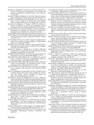
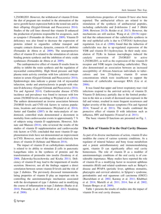
Table1continued
OralcavitydiseasePopulationstudied
(contributingcountry)
SpeciestestedResultsReferences
S:11202
(USA)
25[OH]D(serum);level?
:PALinpeopleC50yearsold
Dietrichetal.(2004)
CandidiasisS:84(HIV?
)
(USA)
25[OH]D(serum);level?
:riskofOC
Sroussietal.(2012)
ToothcariesS:144(S-ECC)
C:122
(Canada)
25[OH]D(serum);levelinstudygroupSchrothetal.(2013)
S:106
(Argentina)
CaI
25[OH]D(serum)
;CaI?:DMFTandPI
Nocorrelation
Antonenkoetal.(2015)
OSCCS:110(OSCC)
C:122
(Serbia)
VDRandCYP24A1genepolymorphismsCYP24A1genepolymorphism(rs2296241)
:susceptibilitytoOSCC;wildtypeff
genotypeofFokIpolymorphism?worse
survival
Zeljicetal.(2012)
RASrecurrentaphthousstomatitis,CPchronicperiodontitis,AgPaggressiveperiodontitis,APadultperiodontitis,EOPearly-onsetperiodontitis,OCoralcandidiasis,S-ECCsevereearly
childhoodcaries,OSCCoralsquamouscellcarcinoma,Sstudygroup,Ccontrolgroup,PALperiodontalattachmentloss,CaIcalciumintake,DMFTdecayed-missing-filledteethindex,PILo¨e
Silnessplaqueindex,PFAPAperiodicfever,aphthousstomatitis,pharyngitisandcervicaladenitis
Arch. Immunol. Ther. Exp.
123](https://image.slidesharecdn.com/oral-181215232407/85/Oral-8-320.jpg)