2. activities. Neurotoxicity is a commonly observed early-onset effect of
acute exposure to RDX in several species. Seizures have been reported
within hours of human acute exposures from prior wartime weapons
production before adoption of modern occupational safety standards
(Stone et al., 1969; Testud et al., 1996; Woody et al., 1986). Intentional
and accidental consumption of C-4 has produced similar effect, with
recovery within days (Davies et al., 2007; Goldberg et al., 1992; Harrell-
Bruder and Hutchins, 1995; Hollander and Colbach, 1969; Kasuske et al.,
2009; Kucukardali et al., 2003). Coincident mild anemia has been less fre-
quently reported (Kucukardali et al., 2003), but for one case with high
dose exposure, persisted for weeks (Stone et al., 1969). Seizures are also
observed shortly after acute exposure to RDX in rats (Burdette et al.,
1988; Schneider et al., 1978; Williams et al., 2010) and dogs (Bruchim
et al., 2005).
Very little is known about the toxicity of RDX degradation prod-
ucts. In our study that directly compared acute toxicity of RDX with
its degradation products in rats, lethality and neurotoxicity of MNX
were comparable to that of RDX and of greater potency than DNX
and TNX (Meyer et al., 2005). Similar results have been reported for
deer mice (Peromyscus maniculatus) (Smith et al., 2007). In addition,
hematotoxic effects of MNX were seen in rats surviving 14 days
after single oral exposure as evidenced by decreased blood hemoglobin
and splenic hemosiderosis (Meyer et al., 2005). Because anemia
resulting from direct chemical destruction of intravascular eryth-
rocytes typically resolves within ~7 days in the rat (Berger, 1985a,
1985b; Harrison and Jollow, 1986), the 14-day persistence led us to
hypothesize that MNX was cytotoxic to bone marrow (BM) progenitor
cells. Incidence of acquired BM failure from non-therapeutic toxicants
has been estimated at ~30% (Montane et al., 2008). Notable environmen-
tal myelosuppressants include benzo(a)pyrene, 7,12-dimethylbenz(a)
anthrecene (DMBA) and benzene (Cronkite et al., 1989; Galvan et al.,
2006; Gasiewicz et al., 2010; Snyder et al., 1980). Toxicant effects on
proliferation, differentiation and apoptosis of hematopoietic stem
and lineage-committed progenitor cells (Wang et al., 2012; Yoon et al.,
2001) result in subsequent loss of their derived mature cells in blood. Re-
ductive activation of C-nitroso 1,2,4-benzotriazine 1,4-dioxide prodrugs
in hypoxic niches of BM associated with hematopoietic stem cell loss
(Parmar et al., 2007) suggests a similar mechanism could apply to
N-nitroso MNX (Uchimiya et al., 2010).
Toxicity of MNX compared to RDX on BM hematopoietic pro-
genitor cells of treated rats with time after single exposure is de-
scribed here. Erythroid and myeloid lineage responses of BM were
evaluated with colony forming assays (Pessina et al., 2003; Rich
and Hall, 2005) and correlated with levels of mature blood cells.
Results demonstrated suppression of BM hematopoiesis by both
RDX and MNX. Loss of both myeloid and erythroid lineages occurred
and RDX was of comparable potency to MNX. Onset of these BM ef-
fects was delayed until after 7 day-post treatment suggesting devel-
opment of preceding events are necessary to drive the suppressive
outcome at the level of the BM that is then apparent at 14 days as
loss of mature blood cells.
Materials and methods
Materials. RDX (>99%) was obtained from Stan Caulder (Naval
Surface Warfare Center, Indianhead, MD) and stored under absolute eth-
anol. MNX was obtained from Dr. Ron Spanggord (SRI Intl., Menlo Park,
CA). Purity of MNX as determined by HPLC with UV detection was greater
than 98.4% with ~1.2% RDX contamination. Both compounds were used
without any additional purification. Assay kits for colony formation
of granulocyte/macrophage-colony forming cells (GM-CFC; catalog no.
K1-GM2-1R, now renamed KCO-GM2-1R), granulocyte–erythrocyte–
monocyte–megakaryocyte-CFCs (CFC-GEMM, KCO-GEMM2-1R) and
burst-forming units-erythroids (BFU-E, KCO-B2-1R) were the
methylcellulose HALO platform (now relabeled CAMEO-96) from
Hemogenix, Inc. (Colorado Springs, CO). Iscove's modified Dulbecco
medium (IMDM) and antibiotic/antimycotic solution were purchased
from Invitrogen (Carlsbad, CA). Bovine serum albumin (BSA) and
Histopaque-1077 were purchased from Sigma (St. Louis, MO).
Antibodies for flow cytometry were mouse anti-rat CD32 (Rat Fc
block [FcγIII/II], mAB D-34-485), phycoerythrin (PE)-conjugated IgG1
κ-isotype control antibody, fluorescein isothiocyanate (FITC)-conjugated
IgG2a κ-isotype control antibody, PE-conjugated antibody to rat Thy1.1
(mAB; OX-7) and FITC-conjugated mouse monoclonal antibody to rat
CD71 (mAB OX-26) purchased from BD Pharmingen (San Jose, CA). All
other reagents were of analytical grade and purchased from commercially
available sources.
Animals and treatment. Female Sprague–Dawley (SD) rats (210–
240 g) were obtained from the in-house breeding colony of University
of Louisiana at Monroe and housed individually with a 12-h light/dark
cycle, controlled temperature (21±1 °C) and humidity (50±10%), and
free access to water and rodent chow (Harlan Teklad rat chow No.7001,
Madison, WI). Rats were allowed to acclimate in polycarbonate cages
for one week prior to study. All animal handling and husbandry were in
accordance with the Guide for Use and Care of Animals (National
Research Council, 2011) and all the protocols were pre-approved by the
Institutional Animal Care and Use Committee. Food was withdrawn the
night before treatments. Treatments were randomly assigned to groups
of rats (n=3–5) and orally administered between 9:00 and 10:00 AM.
Treatments were RDX (0–94 mg/kg) or MNX (0–94 mg/kg) in 5%
DMSO (v/v) in corn oil administered as a single oral dose (10 ml/kg).
High doses were equal to half the RDX and MNX LD50s (Meyer et al.,
2005). Rats were frequently observed for convulsions over the first 8 h
and were euthanized with CO2 if moribund according to OECD criteria
(Organisation for Economic Co-operation and Development (OECD),
2000). Survivors were euthanized with CO2 at different time points rang-
ing from 7 to 14 days and blood was collected by cardiac puncture for
hematological assessment. Both femurs were excised and immediately
processed for BM cell isolation.
Hematology. Blood was collected by cardiac puncture into heparinized
syringes and transferred to EDTA-containing vacutainer tubes (Becton,
Dickinson and Co.). The hematological parameters hemoglobin, erythro-
cyte, leukocyte and platelet counts and leukocyte differentials were
determined with a CELL-DYN Sapphire System (Abbott Laboratories,
Abbott Park, IL) (Fairbanks and Klee, 1986). Hematocrit was derived
from measured red blood cell size and number. Hemoglobin was mea-
sured as absorbance at 540 nm after erythrocyte lysis and conversion
to hemoglobin-hydroxylamine. Granulocyte count was determined by
summing eosinophil, basophil and neutrophil counts.
BM cell isolation. Marrow was extracted from both femurs of each
rat. Bones were disarticulated from the pelvis, excised and the proximal
and distal heads of each were cutoff with bone sheers. Marrow was
flushed by inserting 18 ½-gauge needles with 3 ml ice-cold IMDM
plus 0.2% BSA and antibiotic/antimycotic (1 ml/100 ml medium)
through one end of the bone shaft. Residual fluid in the bones was
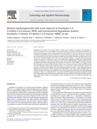
Fig. 1. Structure of hexahydro-1,3,5-trinitro-1,3,5-triazine (RDX) and its environmental
degradation product, hexahydro-1-nitroso-3,5-dinitro-1,3,5-triazine (MNX).
444 S. Jaligama et al. / Toxicology and Applied Pharmacology 266 (2013) 443–451
3. flushed with a 3 ml bolus of air and the same process was repeated for
the second femur. The media was triturated with the same needle and
syringe to produce a single cell suspension, filtered through nylon
mesh and pelleted by centrifugation at 250 ×g for 10 min at room tem-
perature. Cell pellet was suspended in 3.0 ml IMDM medium and cell
number was obtained using a hemocytometer.
Colony forming assays. Mononuclear cells were obtained from BM cell
suspension by density gradient centrifugation using Histopaque-1077
according to supplier's instructions. Briefly, 3 ml of BM cell suspension
was layered on 6 ml of Histopaque-1077 and centrifuged at 400 ×g for
30 min at room temperature. Following centrifugation, the top layer
was discarded and mononuclear cell fraction at the opaque
interface was collected and resuspended in 10 ml IMDM and
centrifuged at 800 ×g for 10 min at room temperature. Cell pellet
was washed in 5 ml IMDM and centrifuged at 250 ×g for 10 min at
room temperature. Supernatant was discarded and mononuclear cell
pellet was suspended in 0.25 ml medium and counted. GM-CFC and
CFC-GEMM assays were performed using HALO kit as per manufacturer's
instructions (Hemogenix, Colorado Springs, CO). In brief, 1.2×10⁵
mono-
nuclear cells/60 μl were added to growth factor mix (rat recombinant
GM-CSF, IL-3 and SCF for GM-CFCs; EPO, GM-CSF, G-CSF, IL-3, IL-6, SCF,
TPO, and Flt3-L for CFU-GEMMs), methylcellulose and serum in propor-
tions of 1:1:4:4. BFU-E assays were performed similarly except that initial
mononuclear cell density was 2.4×10⁵
/60 μl and growth factors were rat
recombinant EPO, IL-3 and CSF. Assay media with cells were plated
(100 μl, n=4) in 96-well plates and incubated for 5 days in a water
jacketed 5% CO2 incubator at 37 °C. After 5 days of incubation, clonal
growth was determined as amount of ATP luminescence produced from
the lysed colonies with HALO kit components. Luminescence was mea-
sured by means of a Chameleon II microplate reader (Hidex, Turku,
Finland). Amount of ATP was quantified from luminescence using an
ATP standard curve run on the same day. Coefficients of variation for
GM-CFC and BFU-E assessments were typically 21% and 25%.
The direct effect of MNX on formation of GM-CFCs was deter-
mined by culturing mononuclear cells in medium containing MNX.
MNX was dissolved in DMSO and added to IMDM medium to attain
final concentrations of 10–2000 μM and 0.1% (v/v) DMSO. Concentra-
tion range exceeded blood levels of ~2 μM that we have seen in rats
24–48 h after a single oral dose of 94 mg/kg MNX (MacMillian, D.,
unpublished observations). Mononuclear cells were isolated from
BM of female SD rats as previously described and were plated in
GM-CFC assay medium plus MNX in the above concentration range.
After 5 days in culture, GM-CFC ATP was measured as before.
Flow cytometry. Total BM cells were analyzed for Thy 1.1 and CD71
(transferrin receptor), markers for rat pluripotential hematopoietic cells
and erythroid-committed precursors, respectively (Goldschneider et al.,
1978; McCarthy et al., 1987; Schomaker et al., 2002). BM cells were isolat-
ed as described earlier for colony forming assays and suspended at 5×10⁶
cells/ml in phosphate buffered saline (PBS) containing 0.1% sodium azide
on ice. The low affinity Fc receptors were blocked by incubating with
mouse anti-rat CD32 [FcγIII/II, mAB D-34-485] at 1:2000 dilution for
10 min. The cells were then incubated with PE-conjugated anti-rat
Thy1.1 (mAB OX-7) at 1:2000 and FITC conjugated mouse monoclo-
nal anti-rat CD71 (mAB OX-26) (BD Pharmingen, San Jose, CA) at
1:200 dilution for 45 min on ice in the dark. After incubation with
antibodies, cells were washed twice at 400 ×g, 4 °C for 10 min and
resuspended in 500 μl PBS. A FACSCalibur flow cytometer (BD Biosci-
ences, San Jose, CA) was setup using isotype control for background
signal and compensation was set with singly labeled cells. Aliquots
containing only single primary antibody (for fluorescence spill over
compensation) and isotype antibodies (negative control) were
processed simultaneously. FL1 (band pass filter, 530±15 nm) and
FL2 (band pass filter, 585±21 nm) detectors were used for FITC
and PE fluorescence, respectively. For each sample, data were
acquired with low flow rate from 20000 cells. Data were analyzed
using Cell Quest Software (BD Biosciences, San Jose, CA).
Data analysis. Data are expressed as mean±SE (n=3–5 animals).
Dose–response data were analyzed by one-way ANOVA with post-hoc
comparisons of treatment means against vehicle control done with
Dunnet's test. Time-course data were analyzed by two-way ANOVA
and Bonferroni's post-tests. Percent data were transformed to square
root with a 3/8 continuity factor before ANOVA. CFC and BFU data
were expressed as mean±SE pmol ATP/well. Results of flow cytometric
analysis for CD 71 and CD 90.1 (Thy 1.1) were expressed as mean±SE
percent of total cells (20,000 cells). Treatment means were considered
as statistically significant if pb0.05. Statistical analysis for all the studies
was carried out with Prism, v. 4 (GraphPad Software, Inc., San Diego,
CA).
Results
Clinical observations
In the 14-day study (Figs. 2–5), one rat of 4 treated with 94 mg/kg
RDX required euthanasia because of onset of multiple tonic–clonic
convulsions. These occurred within 20 min of RDX administration.
No further mortalities or clinical effects requiring euthanasia were
noted during the remaining 13 days for MNX or RDX. Transient,
mild audiogenic convulsions were noted for ~60% of 24 and 47 mg/kg
RDX-treated rats at ~10–20 min of dosing. Body weight gain over the
14 days was not affected by treatment. Similarly, 2 of 5 rats treated
with 94 mg/kg RDX and 1 of 5 treated with 47 mg/kg RDX were lost to
neurotoxicity in the 7-day study (Fig. 6).
Hematology
A significant decrease in the hemoglobin content and hematocrit
levels at 14 days after single oral exposure was observed with MNX
(NOAEL=47 mg/kg) in the previous study (Meyer et al., 2005).
Blood of rats treated with 94 mg/kg RDX exhibited a similar modest
decrease in hemoglobin content (pb0.05). Values for rats treated
with vehicle control and RDX at 47 and 94 mg/kg were 14.8±0.3,
13.8±0.2 and 13.5±0.2 g/dL (mean±SEM, n=5). Fig. 2A summa-
rizes the effect of RDX and MNX on WBCs. A 40% decrease in WBCs
was seen with RDX and MNX (NOAEL=47 mg/kg) at 14-days
post-exposure that included a 60% reduction in granulocyte count
(Fig. 2B). Relative percentages of granulocyte neutrophils, eosinophils
and basophils and of monocytes and lymphocytes in Wright-stained
blood smears were unchanged by RDX or MNX (data not shown).
No effect of RDX or MNX was observed on platelet count, red cell distri-
bution width (RDW), mean corpuscular volume or mean corpuscular
hemoglobin concentration at 14 days nor on these blood parameters or
on RBC count, hematocrit, hemoglobin or WBC count at 7 days after
treatment.
Effect of RDX and MNX on BM cellularity (BMC)
In order to determine whether BM toxicity contributed to the ob-
served decrease in blood hemoglobin and granulocytes, we counted
BM cells as affected by exposure to these compounds. A decline of ap-
proximately 25% in total femoral BMC occurred 14 days after a single
oral administration of RDX or MNX (Fig. 3). Although effect of RDX just
reached statistical significance at 12 mg/kg, monotonic trend was not ev-
ident until ≥47 mg/kg. The LOAEL for MNX was 94 mg/kg. At 7 days
after treatment, femoral BMC of RDX or MNX treated rats did not differ
from that of vehicle-treated rats (~225–270×106
cells).
445
S. Jaligama et al. / Toxicology and Applied Pharmacology 266 (2013) 443–451
4. Effect of RDX and MNX on colony forming cells
Rat GM-CFC colony formation was significantly impacted by acute
RDX and MNX exposure at 14 days (Fig. 4). ATP from GM-CFCs of
RDX- and MNX-treated rats was decreased by half due to both re-
duced density and size of colonies. Significant effects were seen at
47 and 94 mg/kg for both RDX and MNX. Decreases in the myeloid
progenitor cells paralleled loss of peripheral granulocytes at high
RDX and MNX dose at 14 days post exposure (Fig. 2B). Similarly,
BFU-Es were decreased at 14 days after RDX and MNX (Fig. 5), but
only the high dose statistically differed from vehicle control. At the
earlier time of 7 days post-exposure, MNX and RDX were without
effect on either erythroid or myeloid colony formation (Figs. 6A and
B). Likewise, neither chemical affected CFC-GEMMs, progenitors of
BFU-Es and GM-CFCs, at 7 days (Fig. 6C).
Effects of MNX on BM multipotential progenitor cells
CD71(transferrin receptor) is expressed on post-BFU-E erythrocyte
progenitors (Rogers et al., 1996; Schomaker et al., 2002), while Thy1.1
(CD 90.1) is present on early multi-lineage progenitor cells of rat BM
(Goldschneider et al., 1978; McCarthy et al., 1987; Thierfelder, 1977).
Effects of MNX (0–94 mg/kg) 14 days after acute oral exposure on
percentage of BM cells expressing surface markers CD71 and Thy1.1 are
shown in Table 1 and representative histograms are presented in Fig. 7.
Cells positive for CD71 were in the range of ~25–29% of total BM cells
and for Thy1.1 were ~47–57% of mononuclear cells as assessed by flow
cytometry. There was no significant change observed on percent of cells
expressing CD71 or Thy1.1 upon treatment with MNX at any dose.
Time course for MNX BM suppression
In order to identify the time of onset of MNX-induced myelo-
suppression that occurred between 7 and 14 days, we treated rats
with MNX (47 mg/kg) and assessed hematology and colony forma-
tion for GM-CFCs at 10, 12, and 14 days post-exposure. As shown
in Fig. 8A, a small, but significant decrease in blood hemoglobin
was observed at 12, but not 10, days. Hemoglobin at 14 days did
not differ from vehicle controls, as seen before with 47 mg/kg
MNX. WBC and granulocyte counts were unchanged by 47 mg/kg
Fig. 2. Effect of RDX and MNX on WBCs (A) and granulocytes (B) at 14 days post-exposure.
Blood samples were collected by cardiac puncture and analyzed for WBC and granulocyte
counts. Data are presented as mean±SE for WBCs and granulocytes of female SD rats
(n=5/group) dosed with 0, 12, 24, 47 or 94 mg/kg RDX or MNX. Significant differences of
treatment means from vehicle control (0 mg/kg) are indicated (*, pb0.05; **, pb0.01).
Fig. 3. Effect of RDX and MNX on total femoral bone marrow cellularity at 14 days
post-exposure. Bone marrow was flushed from both femurs and total number of bone
marrow cells counted. Data are presented as mean±SE for female SD rats (n=5/group)
dosed with 0, 12, 24, 47 or 94 mg/kg RDX or MNX. Significant differences of treatment
means from vehicle control (0 mg/kg) are indicated (*, pb0.05; **, pb0.01).
Fig. 4. Effect of RDX and MNX at 14 days post-exposure on clonal proliferation of
GM-CFCs. BM mononuclear cells were isolated from female SD rats (n=3–5/group)
at 14 days after single exposure to 0, 12, 24, 47 or 94 mg/kg of RDX or MNX. 20,000
cells/well were grown in methylcellulose in quadruplicate in 96-well plates with
GM-CSF, IL-3 and SCF (Halo kit, Hemogenix Inc., Colorado Springs, CO). After 5 days in-
cubation at 37 °C in a 5% CO2 incubator, ATP was quantified against a standard curve.
Data are mean±SE of pmol ATP/well. Significant differences of treatment means com-
pared to vehicle control are indicated (*, pb0.05). Phase contrast micrographs of
GM-CFC colonies from rats treated with vehicle (left) and 94 mg/kg MNX (right) are
shown below the graph.
Fig. 5. Effect of RDX and MNX at 14 days post-exposure on clonal proliferation of BFU-Es. BM
mononuclear cells were isolated from female SD rats (n=3–5/group) at 14 days after single
exposure to 0, 12, 24, 47 or 94 mg/kg of RDX or MNX. 40,000 cells/well were grown in meth-
ylcellulose in quadruplicate in 96-well plates with EPO, IL-3 and CSF (Halo kit, Hemogenix
Inc., Colorado Springs, CO). After 5 days incubation at 37 °C in a 5% CO2 incubator, ATP was
quantified against a standard curve. Data are mean±SE of pmol ATP/well. Significant differ-
ences of treatment means compared to vehicle control are indicated (*, pb0.05).
446 S. Jaligama et al. / Toxicology and Applied Pharmacology 266 (2013) 443–451
5. MNX at any time, consistent with previously observed 14-day MNX
NOAEL of 47 mg/kg (Fig. 2). For GM-CFCs, MNX suppression was
observed at all the time points with maximal effect at 12 days
(Fig. 8B).
Effect of MNX in culture on GM-CFC formation
To test whether MNX directly inhibited development of BM cells
into GM-CFCs in response to GM-CSF, IL-3 and SCF, MNX was added
directly to the methylcellulose culture medium. MNX concentration
range tested exceeded that we previously observed in blood of rats
after oral treatment with 94 mg/kg MNX. MNX did not inhibit forma-
tion of GM-CFCs when present in the culture medium from 10 to
1000 μM, but rather increased colonies at 100 and 500 μM (Fig. 9).
Cloudiness was evident in medium with 2000 μM MNX suggesting
insolubility.
Discussion
In an earlier study to compare lethality of acute exposure of environ-
mental degradation product MNX to parent RDX, we observed a mild,
but statistically significant drop in blood hemoglobin and hematocrit
at 14 days after a single dose of 94 mg/kg MNX to rats (Meyer et al.,
2005). Since anemia from single-dose treatment with hematotoxicants
that cause intravascular hemolysis resolves in about 7 days in rats
(Berger, 1985a, 1985b; Harrison and Jollow, 1986), we suspected that
MNX acted through a different mechanism. Here we asked whether
loss of peripheral erythrocytes was a consequence of MNX suppression
of BM precursor cells. We did find loss of hematopoietic erythroid pre-
cursors (BFU-Es) from BM of MNX-treated rats at 14 days, but not
7 days, after dosing. Precursor cells of the myeloid lineage (GM-CFCs)
were likewise affected after 14 days. Such a delayed-onset effect on
hematopoiesis could result from a time-dependent accumulation of
BM MNX that reached cytotoxic levels at 14 days. However, several ob-
servations from our work with MNX discount this mechanism. First, we
have shown the MNX is not directly toxic to GM-CFCs at concentrations
exceeding those we have measured in blood of MNX-treated rats
(Fig. 9). Second, we had previously seen that MNX was not detect-
able in liver, kidney, brain, or spleen of rats at 14 days post-dose
(MacMillian, D., unpublished observations), so presumably it had
also cleared other tissues.
We also continued parallel assessment of MNX vs. RDX on these
and other hematological endpoints. Results demonstrate that BM
hematopoietic progenitor cells of both erythroid and myeloid line-
ages are adversely affected by RDX and MNX with comparable
potency for both compounds. Further, effects of both were delayed
until after 7 days post-exposure. Collectively, these observations
suggested toxicity of these compounds to hematopoiesis may be second-
ary to a preceding primary effect whose expression at the level of hema-
topoietic progenitor cells and peripheral mature blood cells requires time
to develop. Studies with other myelosuppressive agents implicate activa-
tion of an immune response as a likely mechanism of hematotoxicity
(Chen, 2005). Events leading to such immune-mediated BM failure in-
volve activated T-cell infiltration, secretion of type 1 cytokines and BM
hematopoietic cell apoptosis mediated via Fas interaction with ligand
(Omokaro et al., 2009). Such a mechanism for MNX BM toxicity would
suggest a basis for the observed delayed onset. Similarity in MNX struc-
ture to N-nitroso triazine tirapazamine, a prodrug with cytotoxicity medi-
ated through 1 e−
reduction in hypoxic tissues such as bone marrow
niches harboring hematopoietic progenitors (Parmar et al., 2007), could
also contribute a delay before loss of GM-CFCs and BFU-Es and explain
MNX target-tissue selectivity.
BM hematopoiesis initiates upon multi-lineage commitment of a
daughter cell of a slow cycling hematopoietic stem cell. These multi-
potential cells then step-wise restrict to more distal lineages; first to
lymphoid and myeloerythroid lineages and then the latter to erythroid
and myeloid lineages. Each pathway is supported by a lineage-selective
combination of cytokines and growth factors localized in anatomical
microdomains with differential vascular perfusion rates and position
between endosteal and sinusoidal surfaces (Winkler et al., 2010). Cells
of these distinct intermediate stages can be clonally grown in semi-solid
media with the complementary set of growth/differentiation factors. Ad-
ditionally, BM cells at different stages of hematopoiesis express distinct
membrane markers that distinguish them by flow cytometry. In the rat,
Thy 1.1 is a marker of early multi-potential progenitors (Goldschneider
et al., 1978; McCarthy et al., 1987; Thierfelder, 1977) that are predeces-
sors of the myeloerythroid cells (CFC-GEMMs) that form colonies in
culture when supported by EPO, GM-CSF, G-CSF, IL-3, IL-6, SCF, TPO,
and Flt3-L. CFC-GEMMs differentiate into either myeloid or erythroid
Fig. 6. Colony formation at 7 days post-exposure to a single dose of RDX or MNX for
GM-CFCs (A), BFU-Es (B) and CFC-GEMMs (C). BM mononuclear cells were isolated
from female SD rats (n=3–5/group) treated with 0, 24, 47 or 94 mg/kg and plated
in quadruplicate in 96 well plates (Halo kit, Hemogenix Inc., Colorado Springs, CO).
After 5 days incubation at 37 °C in a 5% CO2 incubator, ATP was quantified against a
standard curve. Data are mean±SE of pmol ATP/well.
Table 1
Effect of MNX on cell surface expression of CD71 and Thy1.1a
.
Dose CD71 Thy1.1
(mg/kg) % %
0 28.8±3.0 50.8±3.1
12 27.1±3.2 46.7±1.7
24 27.6±2.0 50.9±6.2
47 25.5±3.1 56.3±0.4
94 27.5±2.2 56.8±3.0
a
Female SD rats were treated with MNX (0, 12, 24, 47 or 94 mg/kg) and after
14 days, total and mononuclear BM cells were isolated. Total BM cells (1×10⁶
) were
labeled with FITC-conjugated mouse monoclonal anti-rat CD71 antibody and mononu-
clear cells (1×10⁶
cells) with PE-conjugated anti-rat Thy1.1 antibody. Cells were ana-
lyzed with a flow cytometer. Data are presented as a percent of cells with surface
expression of markers out of 20,000 cells (mean±SE) for n=5 (CD71) and n=3
(Thy1.1).
447
S. Jaligama et al. / Toxicology and Applied Pharmacology 266 (2013) 443–451
6. precursors, recognized in culture as GM-CFCs and BFU-Es, respectively.
BFU-Es are progenitors of the various erythroblast stages of BM cytology
that express surface transferrin receptor, CD71. MNX had no effect on rel-
ative proportion of BM cells expressing Thy 1.1 (Fig. 7), indicating that
early stem cell commitment was not compromised. Further, lack of effect
of RDX and MNX on CFC-GEMMs (Fig. 6C) prior to loss of GM-CFCs and
BFU-Es suggested that deficit of predecessor pool did not contribute.
Lack of effect on proportion of post-BFU-E CD71-positive erythroid cells
(Fig. 7) also did not support a differential effect of MNX on myeloid or ery-
throid lineages. Overall loss of BMC with no indication of lineage specific-
ity suggested a possible general effect on BM stromal environment
supportive of hematopoiesis. BM stromal involvement has been previous-
ly implicated as important in benzene myelosuppression (Gaido and
Wierda, 1985; Laskin et al., 1989). Although metabolism by BM storma
has not been well studied, there is evidence that NAD(P)H:quinone oxi-
doreductase 1 (NQO1) is localized in stromal fibroblasts (Thomas et al.,
1990). NQO1 nitroreductase activity with nitroaromatics that has been
thoroughly described (Cenas et al., 2009) may be relevant to MNX BM
metabolism.
RDX toxicity has been extensively studied in the rat and the critical ef-
fect of acute, single dose exposure is neurotoxicity (Burdette et al., 1988;
Schneider et al., 1978; Williams et al., 2010). Rats exhibit convulsions usu-
ally within the first hour after RDX and, if not lethal, will recover
uneventfully. Seizure activity with MNX follows a similar pattern
(Meyer et al., 2005) and exhibits an ED50 of 57 mg/kg. Incidence of con-
vulsions parallels RDX disposition in brain and recovery coincides with
clearance (Bannon et al., 2009). Although 14-day acute toxicity protocols
usually do not include routine blood assessments (U.S. Environmental
Protection Agency, 2002), we incorporated a screen for hematological ef-
fects in our earlier study because of known redox coupling of nitro and
nitroso compounds with hemoglobin. Consequently, we found a previ-
ously unreported, mild drop in hemoglobin and hematocrit in surviving
animals at 14 days after high MNX dose (94 mg/kg). Decreased blood
erythrocytes and hemoglobin have been reported for rats consuming
40 mg/kg/day RDX for 6 months and longer in the diet (Levine et al.,
1983). For comparison to neurotoxicity, ED50 for convulsions was esti-
mated at 57 mg/kg MNX (Meyer et al., 2005). Convulsions have likewise
been repeatedly observed in humans within hours of exposure to large
amounts of RDX or C-4 (Davies et al., 2007; Goldberg et al., 1992;
Harrell-Bruder and Hutchins, 1995; Hollander and Colbach, 1969;
Kasuske et al., 2009; Kucukardali et al., 2003; Stone et al., 1969; Testud
et al., 1996; Woody et al., 1986). Coincident assessments of hematology
were often found normal, although methemoglobin has been reported
(Kucukardali et al., 2003). Delayed onset effects would likely go
undetected because repeat hematology assessment beyond dis-
charge after seizuring has resolved was rarely done. One case of a
Fig. 7. Representative histogram plots (Count vs. Fluorescence) of cell surface expression of CD71 (left) and Thy 1.1 (right) analyzed with flow cytometry. BM cells from female SD
rats (n=5/group) were isolated from rats 14 days after treatment with MNX at 0 (B), 47 (C) or 94 (D) mg/kg. One million BM cells or mononuclear cells were treated with
FITC-conjugated mouse monoclonal antibody to rat CD71 or PE-conjugated antibody to rat Thy1.1, respectively, and 20,000 cells were analyzed with a BD FACSCaliber flow
cytometer. Panels A are from cells labeled with isotype control.
448 S. Jaligama et al. / Toxicology and Applied Pharmacology 266 (2013) 443–451
7. persistent anemia with hypoplastic marrow aspirate has been noted
for a patient poisoned by an exceptionally high dose (Stone et al.,
1969).
Considerable information is generally available for estimating risk of
environmental toxicants; however, hazards of their environmental deg-
radation products are by contrast usually understudied. Our studies
have shown that MNX, the most abundant degradation product of
RDX, has neurotoxic and hematotoxic effects at comparable potency
to parent. Since MNX has been detected maximally at 12 mol % of
RDX in ground water of contaminated sites (Beller and Tiemeier,
2002), environmental aging of RDX is unlikely to pose greater risk or
create unique hazards. In contrast, bioremediation by anaerobic reduc-
tion would appear to lessen RDX risk. Our data would, however, support
inclusion of MNX in a common mechanism group for cumulative risk
assessment of RDX-contaminated sites (U.S. Environmental Protection
Agency, 2007). Estimating from an aqueous solubility at 20 °C of RDX
of ~50 mg/l (Phelan et al., 2002), a 70 kg individual consuming 2 l
water per day could theoretically be exposed to an upper limit of
~0.2 mg/kg/day MNX from contaminated drinking water, considerably
below the LOAEL for most sensitive, dose-dependent hematological
response to single exposure reported here, i.e., 47 mg/kg MNX for inhi-
bition of GM-CFC formation. Realistically, the hematological threshold
of this study is much higher than exposure estimates from analytical
data since the highest reported concentrations of RDX in ground water,
~14–35 mg/l, are less than the solubility limit (Agency for Toxic
Substances and Disease Registry (ATSDR), 2010; Gadagbui et al., 2012).
Further, two studies that have measured ground water MNX concentra-
tions at military sites reported maximal levels of ~4 μg/l (Paquet et al.,
2011) and 160 μg/l (Beller and Tiemeier, 2002).
In summary, we have presented evidence for hematotoxicity with
delayed onset after acute exposure of rats to high dose of RDX and its
environmental degradation product MNX. These compounds appear to
indirectly compromise proliferation and differentiation of BM myeloid
and erythroid progenitors after a >7-day expression period. Absence
of evidence for differential effects on specific lineage subpopulations
would suggest that RDX and MNX may exert myelosuppressive effects
indirectly by compromising supportive activity of BM stroma.
Abbreviations
BM bone marrow
BMC bone marrow cellularity
BSA bovine serum albumin
BFU-E burst-forming units-erythroids
CFC-GEMM colony forming cells of granulocytes, erythrocytes,
monocytes and megakaryocytes
DNX hexahydro-1,3-dinitroso-5-nitro-1,3,5-trazine
GM-CFC granulocyte macrophage-colony forming cells
IMDM Iscove's modified Dulbecco medium
LOAEL lowest observed adverse effect level
MNX hexahydro-1-nitroso-3,5-dinitro-1,3,5-trazine
NOAEL no observed adverse effect level
RDX royal demolition explosive/Hexahydro-1,3,5-trinitro-1,3,5-
trazine
TNX hexahydro-1,3,5-trinitroso-1,3,5-trazine
Conflict of interest statement
The authors have no financial or personal conflicts of interest to
declare.
Fig. 8. Effect of time (days) after MNX acute oral exposure on blood hemoglobin (A) and proliferation of GM-CFCs (B). Female SD rats (n=3–5) were treated with 47 mg/kg MNX. Mono-
nuclear cells were isolated at 10, 12, and 14 days after treatment and plated in quadruplicate in 96 well plates (Halo kit, Hemogenix Inc., Colorado Springs, CO). After 5 days incubation at
37 °C in a 5% CO2 incubator, ATP was quantified against an ATP standard curve. Data are mean±SE of pmol ATP/well. Significant differences of treatment means from vehicle control
(0 mg/kg) are indicated (*, pb0.05; **, pb0.01).
Fig. 9. Effect of MNX on clonal proliferation of GM-CFCs in vitro. Mononuclear cells
were isolated from bone marrow of female SD rats, suspended in media containing
MNX (0, 10,100, 500, or 1000 μM) and plated in quadruplicate in 96 well plates
(Halo kit, Hemogenix Inc., Colorado Springs, CO). After 5 days incubation at 37 °C in
a 5% CO2 incubator, ATP was quantified against a standard curve. Data is expressed as
mean±SE of pmol ATP/well. Significant differences of treatment means from vehicle
control (0 mg/kg, 0.1% DMSO) are indicated (**, pb0.01).
449
S. Jaligama et al. / Toxicology and Applied Pharmacology 266 (2013) 443–451
8. Funding
This study was supported by US Department of Defense, grant no.
W81XWH-05-10537 and contract no. W912HZ-05-P-0145.
Acknowledgments
This research was supported by US Department of Defense (grant no.
W81XWH-05-10537 and contract no. W912HZ-05-P-0145 to S.A.M.).
This publication reflects the personal views of the authors and does not
suggest or reflect the policy, practices, programs, or doctrine of the U.S.
Army or Government of the United States. The contents of this report
are not to be used for advertising or promotional purposes. Citation of
brand names does not constitute an official endorsement or approval of
the use of such commercial product.
References
Agency for Toxic Substances, Disease Registry (ATSDR), 2010. Toxicological Profile for RDX.
Department of Health and Human Services, Public Health Service, Atlanta, GA: U.S.
(http://www.atsdr.cdc.gov/ToxProfiles/tp78.pdf [accessed 22 July 2012]).
Bannon, D.I., Dillman, J.F., Hable, M.A., Phillips, C.S., Perkins, E.J., 2009. Global gene expression
in rat brain and liver after oral exposure to the explosive hexahydro-1,3,5-trinitro-1,3,5-
triazine (RDX). Chem. Res. Toxicol. 22, 620–625.
Beller, H.R., Tiemeier, K., 2002. Use of liquid chromatography/tandem mass spectrometry
to detect distinctive indicators of in situ RDX transformation in contaminated ground-
water. Environ. Sci. Technol. 36, 2060–2066.
Berger, J., 1985a. Experimentally induced toxic-haemolytic anaemia in laboratory rats
following phenacetin administration. Folia Haematol. 112, 571–579.
Berger, J., 1985b. Screening of toxic-haemolytic anaemia in laboratory rats: a model of
phenylhydrazine-induced haemolysis. Haematologia (Budap.) 18, 193–200.
Bruchim, Y., Saragusty, J., Weisman, A., Sternheim, D., 2005. Cyclonite (RDX) intoxication in a
police working dog. Vet. Rec. 157, 354–356.
Burdette, L.J., Cook, L.L., Dyer, R.S., 1988. Convulsant properties of cyclotrimethylenetrinitramine
(RDX): spontaneous audiogenic, and amygdaloid kindled seizure activity. Toxicol.
Appl. Pharmacol. 92, 436–444.
Cenas, N., Nemeikaite-Ceniene, A., Šarlauskas, J., Anusevicius, Z., Nivinskas, H., Miseviciene, L.,
Maroziene, A., 2009. Mechanisms of the mammalian cell cytotoxicity of explosives. In:
Sunahara, G.I., Lotufo, G., Kuperman, R.G., Hawari, J. (Eds.), Ecotoxicology of Explosives.
CRC Press, Boca Raton, pp. 211–226.
Chen, J., 2005. Animal models for acquired bone marrow failure syndromes. Clin. Med. Res. 3,
102–108.
Crocker, F.H., Indest, K.J., Fredrickson, H.L., 2006. Biodegradation of the cyclic nitramine
explosives RDX, HMX, and CL-20. Appl. Microbiol. Biotechnol. 73, 274–290.
Cronkite, E.P., Drew, R.T., Inoue, T., Hirabayashi, Y., Bullis, J.E., 1989. Hematotoxicity and
carcinogenicity of inhaled benzene. Environ. Health Perspect. 82, 97–108.
Davies, J.O., Roberts, D.M., Hittarage, A., Buckley, N.A., 2007. Oral C-4 plastic explosive
in humans—a case series. Clin. Toxicol. (Phila.) 45, 454–457.
Defense Environmental Network and Information eXchange (DENIX), 2010. FY 2010
Defense Environmental Programs Annual Report to Congress. (http://www.denix.
osd.mil/arc/ARCFY2010.cfm [accessed 22 July 2012]).
Fairbanks, V., Klee, G., 1986. Biochemical aspects of hematology. In: Tietz, N.W. (Ed.), Text-
book of Clinical Chemistry. W.B. Saunders Co., Philadelphia, pp. 1532–1534.
Gadagbui, B., Patterson, J., Rak, A., Kutzman, R.S., Reddy, G., Johnson, M.S., 2012. Development
of a relative source contribution factor for drinking water criteria: the case of hexahydro-
1,3,5-trinitro-1,3,5-triazine (RDX). Hum. Ecol. Risk Assess. 18, 338–354.
Gaido, K.W., Wierda, D., 1985. Modulation of stromal cell function in DBA/2J and B6C3F1 mice
exposed to benzene or phenol. Toxicol. Appl. Pharmacol. 81, 469–475.
Galvan, N., Page, T.J., Czuprynski, C.J., Jefcoate, C.R., 2006. Benzo(a)pyrene and 7,12-
dimethylbenz(a)anthrecene differentially affect bone marrow cells of the lymphoid
and myeloid lineages. Toxicol. Appl. Pharmacol. 213, 105–116.
Gasiewicz, T.A., Singh, K.P., Casado, F.L., 2010. The aryl hydrocarbon receptor has an im-
portant role in the regulation of hematopoiesis: implications for benzene-induced
hematopoietic toxicity. Chem. Biol. Interact. 184, 246–251.
Goldberg, D.J., Green, S.T., Nathwani, D., McMenamin, J., Hamlet, N., Kennedy, D.H., 1992.
RDX intoxication causing seizures and a widespread petechial rash mimicking
meningococcaemia. J. R. Soc. Med. 85, 181.
Goldschneider, I., Gordon, L.K., Morris, R.J., 1978. Demonstration of Thy-1 antigen on
pluripotent hemopoietic stem cells in the rat. J. Exp. Med. 148, 1351–1366.
Harrell-Bruder, B., Hutchins, K.L., 1995. Seizures caused by ingestion of composition C-4.
Ann. Emerg. Med. 26, 746–748.
Harrison Jr., J.H., Jollow, D.J., 1986. Role of aniline metabolites in aniline-induced hemo-
lytic anemia. J. Pharmacol. Exp. Ther. 238, 1045–1054.
Hawari, J., Halasz, A., Sheremata, T., Beaudet, S., Groom, C., Paquet, L., Rhofir, C., Ampleman,
G., Thiboutot, S., 2000. Characterization of metabolites during biodegradation of
hexahydro-1, 3,5-trinitro-1,3,5-triazine (RDX) with municipal anaerobic sludge. Appl.
Environ. Microbiol. 66, 2652–2657.
Hollander, A.I., Colbach, E.M., 1969. Composition C-4 induced seizures: a report of five cases.
Mil. Med. 134, 1529–1530.
Kasuske, L., Schofer, J.M., Hasegawa, K., 2009. Two marines with generalized seizure activity.
J. Emerg. Nurs. 35, 542–543.
Kucukardali, Y., Acar, H.V., Ozkan, S., Nalbant, S., Yazgan, Y., Atasoyu, E.M., Keskin, O.,
Naz, A., Akyatan, N., Gokben, M., Danaci, M., 2003. Accidental oral poisoning caused
by RDX (cyclonite): a report of 5 cases. J. Intensive Care Med. 18, 42–46.
Laskin, D.L., MacEachern, L., Snyder, R., 1989. Activation of bone marrow phagocytes
following benzene treatment of mice. Environ. Health Perspect. 82, 75–79.
Levine, B.S., Furedl, E.M., Rac, M.S., Gordon, D.E., Lish, P.M., 1983. Determination of the
Chronic Mammalian Toxicological Effects of RDX: Twenty-four Month Chronic
Toxicity/Carcinogenicity Study of Hexahydro-1,3,5-trinitro-1,3,5-triazine (RDX)
in the Fisher Rat. U.S. Army Medical Research and Development Command, Fort
Detrick, Frederick, MD.
Major, M.A., Reddy, G., Berge, M.A., Patzer, S.S., Li, A.C., Gohdes, M., 2007. Metabolite
profiling of [14C]hexahydro-1,3,5-trinitro-1,3,5-triazine (RDX) in Yucatan miniature
pigs. J. Toxicol. Environ. Health A 70, 1191–1202.
McCarthy, K.F., Hale, M.L., Fehnel, P.L., 1987. Purification and analysis of rat hematopoietic
stem cells by flow cytometry. Cytometry 8, 296–305.
McCormick, N.G., Cornell, J.H., Kaplan, A.M., 1981. Biodegradation of hexahydro-1,3,5-
trinitro-1,3,5-triazine. Appl. Environ. Microbiol. 42, 817–823.
Meyer, S.A., Marchand, A.J., Hight, J.L., Roberts, G.H., Escalon, L.B., Inouye, L.S., MacMillan, D.K.,
2005. Up-and-down procedure (UDP) determinations of acute oral toxicity of nitroso
degradation products of hexahydro-1,3,5-trinitro-1,3,5-triazine (RDX). J. Appl. Toxicol.
25, 427–434.
Montane, E., Ibanez, L., Vidal, X., Ballarin, E., Puig, R., Garcia, N., Laporte, J.-R., 2008.
Epidemiology of aplastic anemia: a prospective multicenter study. Haematologica 93,
518–523.
Murnyak, G., Vandenberg, J., Yaroschak, P.J., Williams, L., Prabhakaran, K., Hinz, J., 2011.
Emerging contaminants: presentations at the 2009 Toxicology and Risk Assessment
Conference. Toxicol. Appl. Pharmacol. 254, 167–169.
National Research Council, 2011. Guide for the Care and Use of Laboratory Animals. The
National Academies Press, Washington, DC. (http://grants.nih.gov/grants/olaw/
Guide-for-the-Care-and-Use-of-Laboratory-Animals.pdf [accessed 19 July 2012]).
Omokaro, S.O., Desierto, M.J., Eckhaus, M.A., Ellison, F.M., Chen, J., Young, N.S.,
2009. Lymphocytes with aberrant expression of Fas or Fas ligand attenuate immune
bone marrow failure in a mouse model. J. Immunol. 182, 3414–3422.
Organisation for Economic Co-operation and Development (OECD), 2000. Guidance Docu-
ment on the Recognition, Assessment, and Use of Clinical Signs as Humane Endpoints
for Experimental Animals used in Safety Evaluation. (http://www.oecd-ilibrary.org/
docserver/download/fulltext/9750191e.pdf?expires=1340216544&id=id&accname=
guest&checksum=CB0B3998199DFA88B043774CB472D4B9 [accessed 20 June
2012]).
Pan, X., Zhang, B., Smith, J.N., Francisco, M.S., Anderson, T.A., Cobb, G.P., 2007. N-Nitroso
compounds produced in deer mouse (Peromyscus maniculatus) GI tracts following
hexahydro-1,3,5-trinitro-1,3,5-triazine (RDX) exposure. Chemosphere 67, 1164–1170.
Paquet, L., Monteil-Rivera, F., Hatzinger, P.B., Fuller, M.E., Hawari, J., 2011. Analysis of the key
intermediates of RDX (hexahydro-1,3,5-trinitro-1,3,5-triazine) in groundwater: occur-
rence, stability and preservation. J. Environ. Monit. 13, 2304–2311.
Parmar, K., Mauch, P., Vergilio, J.-A., Sackstein, R., Down, J.D., 2007. Distribution of
hematopoietic stem cells in the bone marrow according to regional hypoxia.
Proc. Natl. Acad. Sci. U.S.A. 104, 5431–5436.
Pessina, A., Albella, B., Bayo, M., Bueren, J., Brantom, P., Casati, S., Croera, C., Gagliardi, G.,
Foti, P., Parchment, R., Parent-Massin, D., Schoeters, G., Sibiril, Y., Van Den Heuvel,
R., Gribaldo, L., 2003. Application of the CFU-GM assay to predict acute drug-
induced neutropenia: an international blind trial to validate a prediction model for
the maximum tolerated dose (MTD) of myelosuppressive xenobiotics. Toxicol. Sci.
75, 355–367.
Phelan, J.M., Romero, J.V., Barnett, J.L., Parker, D.R., 2002. Solubility and Dissolution Kinet-
ics of Composition B Explosive in Water. Sandia National Laboratories, Albuquer-
que, NM. (http://prod.sandia.gov/techlib/access-control.cgi/2002/022420.pdf
[accessed 18 July 2012]).
Rich, I.N., Hall, K.M., 2005. Validation and development of a predictive paradigm for
hemotoxicology using a multifunctional bioluminescence colony-forming prolifer-
ation assay. Toxicol. Sci. 87, 427–441.
Rogers, C.E., Bradley, M.S., Palsson, B.O., Koller, M.R., 1996. Flow cytometric analysis of
human bone marrow perfusion cultures: erythroid development and relationship
with burst-forming units-erythroid. Exp. Hematol. 24, 597–604.
Schneider, N.R., Bradley, S.L., Andersen, M.E., 1978. The distribution and metabolism of
cyclotrimethylenetrinitramine (RDX) in the rat after subchronic administration.
Toxicol. Appl. Pharmacol. 46, 163–171.
Schomaker, S.J., Clemo, F.A., Amacher, D.E., 2002. Analysis of rat bone marrow by flow
cytometry following in vivo exposure to cyclohexanone oxime or daunomycin HCl.
Toxicol. Appl. Pharmacol. 185, 48–54.
Smith, J.N., Liu, J., Espino, M.A., Cobb, G.P., 2007. Age dependent acute oral toxicity of
hexahydro-1,3,5-trinitro-1,3,5-triazine (RDX) and two anaerobic N-nitroso metabolites
in deer mice (Peromyscus maniculatus). Chemosphere 67, 2267–2273.
Snyder, C.A., Goldstein, B.D., Sellakumar, A.R., Bromberg, I., Laskin, S., Albert, R.E., 1980. The
inhalation toxicology of benzene: incidence of hematopoietic neoplasms and
hematotoxicity in ARK/J and C57BL/6J mice. Toxicol. Appl. Pharmacol. 54, 323–331.
Stone, W.J., Paletta, T.L., Heiman, E.M., Bruce, J.I., Knepshield, J.H., 1969. Toxic effects
following ingestion of C-4 plastic explosive. Arch. Intern. Med. 124, 726–730.
Testud, F., Glanclaude, J.M., Descotes, J., 1996. Acute hexogen poisoning after occupa-
tional exposure. J. Toxicol. Clin. Toxicol. 34, 109–111.
Thierfelder, S., 1977. Haemopoietic stem cells of rats but not of mice express Th-1.1
alloantigen. Nature 269, 691–693.
Thomas, D.J., Sadler, A., Subrahmanyam, V.V., Siegel, D., Reasor, M.J., Wierda, D., Ross,
D., 1990. Bone marrow stromal cell bioactivation and detoxification of the benzene
metabolite hydroquinone: comparison of macrophages and fibroblastoid cells.
Mol. Pharmacol. 37, 255–262.
450 S. Jaligama et al. / Toxicology and Applied Pharmacology 266 (2013) 443–451
9. U.S. Environmental Protection Agency, 2002. Health Effects Test Guidelines. Acute
Oral Toxicity. (http://www.regulations.gov/#!documentDetail;D=EPA-HQ-
OPPT-2009-0156-0003 [accessed 19 June 2012]).
U.S. Environmental Protection Agency, 2007. Concepts, Methods, and Data Sources for
Cumulative Health Risk Assessment of Multiple Chemicals, Exposures and Effects: A
Resource Document (Final Report). (Washington, DC http://cfpub.epa.gov/ncea/risk/
recordisplay.cfm?deid=190187#Download [accessed 18 July 2012]).
Uchimiya, M., Gorb, L., Isayev, O., Qasim, M.M., Leszczynski, J., 2010. One-electron
standard reduction potentials of nitroaromatic and cyclic nitramine explosives.
Environ. Pollut. 158, 3048–3053.
Wang, L., He, X., Bi, Y., Ma, Q., 2012. Stem cell and benzene-induced malignancy and
hematotoxicity. Chem. Res. Toxicol. 25, 1303–1315.
Williams, L.R., Aroniadou-Anderjaska, V., Qashu, F., Finne, H., Pidoplichko, V.,
Bannon, D.I., Braga, M.F.M., 2010. RDX binds to the GABAA receptor-convulsant
site and blocks GABAA receptor-mediated currents in the amygdala: a mecha-
nism for RDX-Induced seizures. Environ. Health Perspect. 119, 357–363.
Winkler, I.G., Barbier, V., Wadley, R., Zannettino, A.C.W., Williams, S., Lévesque, J.-P.,
2010. Positioning of bone marrow hematopoietic and stromal cells relative to
blood flow in vivo: serially reconstituting hematopoietic stem cells reside in dis-
tinct nonperfused niches. Blood 116, 375–385.
Woody, R.C., Kearns, G.L., Brewster, M.A., Turley, C.P., Sharp, G.B., Lake, R.S., 1986. The neuro-
toxicity of cyclotrimethylenetrinitramine (RDX) in a child: a clinical and pharmacokinet-
ic evaluation. J. Toxicol. Clin. Toxicol. 24, 305–319.
Yinon, J., 1990. Cyclotrimethylenetrinitramine (RDX). Toxicity and Metabolism of
Explosives. CRC Press, Boca Raton, FL, pp. 145–163.
Yoon, B.-I., Hirabayashi, Y., Kawasaki, Y., Kodama, Y., Kaneko, T., Kim, D.-Y., Inoue, T.,
2001. Mechanism of action of benzene toxicity: cell cycle suppression in hemo-
poietic progenitor cells (CFU-GM). Exp. Hematol. 29, 278–285.
451
S. Jaligama et al. / Toxicology and Applied Pharmacology 266 (2013) 443–451
![activities. Neurotoxicity is a commonly observed early-onset effect of
acute exposure to RDX in several species. Seizures have been reported
within hours of human acute exposures from prior wartime weapons
production before adoption of modern occupational safety standards
(Stone et al., 1969; Testud et al., 1996; Woody et al., 1986). Intentional
and accidental consumption of C-4 has produced similar effect, with
recovery within days (Davies et al., 2007; Goldberg et al., 1992; Harrell-
Bruder and Hutchins, 1995; Hollander and Colbach, 1969; Kasuske et al.,
2009; Kucukardali et al., 2003). Coincident mild anemia has been less fre-
quently reported (Kucukardali et al., 2003), but for one case with high
dose exposure, persisted for weeks (Stone et al., 1969). Seizures are also
observed shortly after acute exposure to RDX in rats (Burdette et al.,
1988; Schneider et al., 1978; Williams et al., 2010) and dogs (Bruchim
et al., 2005).
Very little is known about the toxicity of RDX degradation prod-
ucts. In our study that directly compared acute toxicity of RDX with
its degradation products in rats, lethality and neurotoxicity of MNX
were comparable to that of RDX and of greater potency than DNX
and TNX (Meyer et al., 2005). Similar results have been reported for
deer mice (Peromyscus maniculatus) (Smith et al., 2007). In addition,
hematotoxic effects of MNX were seen in rats surviving 14 days
after single oral exposure as evidenced by decreased blood hemoglobin
and splenic hemosiderosis (Meyer et al., 2005). Because anemia
resulting from direct chemical destruction of intravascular eryth-
rocytes typically resolves within ~7 days in the rat (Berger, 1985a,
1985b; Harrison and Jollow, 1986), the 14-day persistence led us to
hypothesize that MNX was cytotoxic to bone marrow (BM) progenitor
cells. Incidence of acquired BM failure from non-therapeutic toxicants
has been estimated at ~30% (Montane et al., 2008). Notable environmen-
tal myelosuppressants include benzo(a)pyrene, 7,12-dimethylbenz(a)
anthrecene (DMBA) and benzene (Cronkite et al., 1989; Galvan et al.,
2006; Gasiewicz et al., 2010; Snyder et al., 1980). Toxicant effects on
proliferation, differentiation and apoptosis of hematopoietic stem
and lineage-committed progenitor cells (Wang et al., 2012; Yoon et al.,
2001) result in subsequent loss of their derived mature cells in blood. Re-
ductive activation of C-nitroso 1,2,4-benzotriazine 1,4-dioxide prodrugs
in hypoxic niches of BM associated with hematopoietic stem cell loss
(Parmar et al., 2007) suggests a similar mechanism could apply to
N-nitroso MNX (Uchimiya et al., 2010).
Toxicity of MNX compared to RDX on BM hematopoietic pro-
genitor cells of treated rats with time after single exposure is de-
scribed here. Erythroid and myeloid lineage responses of BM were
evaluated with colony forming assays (Pessina et al., 2003; Rich
and Hall, 2005) and correlated with levels of mature blood cells.
Results demonstrated suppression of BM hematopoiesis by both
RDX and MNX. Loss of both myeloid and erythroid lineages occurred
and RDX was of comparable potency to MNX. Onset of these BM ef-
fects was delayed until after 7 day-post treatment suggesting devel-
opment of preceding events are necessary to drive the suppressive
outcome at the level of the BM that is then apparent at 14 days as
loss of mature blood cells.
Materials and methods
Materials. RDX (>99%) was obtained from Stan Caulder (Naval
Surface Warfare Center, Indianhead, MD) and stored under absolute eth-
anol. MNX was obtained from Dr. Ron Spanggord (SRI Intl., Menlo Park,
CA). Purity of MNX as determined by HPLC with UV detection was greater
than 98.4% with ~1.2% RDX contamination. Both compounds were used
without any additional purification. Assay kits for colony formation
of granulocyte/macrophage-colony forming cells (GM-CFC; catalog no.
K1-GM2-1R, now renamed KCO-GM2-1R), granulocyte–erythrocyte–
monocyte–megakaryocyte-CFCs (CFC-GEMM, KCO-GEMM2-1R) and
burst-forming units-erythroids (BFU-E, KCO-B2-1R) were the
methylcellulose HALO platform (now relabeled CAMEO-96) from
Hemogenix, Inc. (Colorado Springs, CO). Iscove's modified Dulbecco
medium (IMDM) and antibiotic/antimycotic solution were purchased
from Invitrogen (Carlsbad, CA). Bovine serum albumin (BSA) and
Histopaque-1077 were purchased from Sigma (St. Louis, MO).
Antibodies for flow cytometry were mouse anti-rat CD32 (Rat Fc
block [FcγIII/II], mAB D-34-485), phycoerythrin (PE)-conjugated IgG1
κ-isotype control antibody, fluorescein isothiocyanate (FITC)-conjugated
IgG2a κ-isotype control antibody, PE-conjugated antibody to rat Thy1.1
(mAB; OX-7) and FITC-conjugated mouse monoclonal antibody to rat
CD71 (mAB OX-26) purchased from BD Pharmingen (San Jose, CA). All
other reagents were of analytical grade and purchased from commercially
available sources.
Animals and treatment. Female Sprague–Dawley (SD) rats (210–
240 g) were obtained from the in-house breeding colony of University
of Louisiana at Monroe and housed individually with a 12-h light/dark
cycle, controlled temperature (21±1 °C) and humidity (50±10%), and
free access to water and rodent chow (Harlan Teklad rat chow No.7001,
Madison, WI). Rats were allowed to acclimate in polycarbonate cages
for one week prior to study. All animal handling and husbandry were in
accordance with the Guide for Use and Care of Animals (National
Research Council, 2011) and all the protocols were pre-approved by the
Institutional Animal Care and Use Committee. Food was withdrawn the
night before treatments. Treatments were randomly assigned to groups
of rats (n=3–5) and orally administered between 9:00 and 10:00 AM.
Treatments were RDX (0–94 mg/kg) or MNX (0–94 mg/kg) in 5%
DMSO (v/v) in corn oil administered as a single oral dose (10 ml/kg).
High doses were equal to half the RDX and MNX LD50s (Meyer et al.,
2005). Rats were frequently observed for convulsions over the first 8 h
and were euthanized with CO2 if moribund according to OECD criteria
(Organisation for Economic Co-operation and Development (OECD),
2000). Survivors were euthanized with CO2 at different time points rang-
ing from 7 to 14 days and blood was collected by cardiac puncture for
hematological assessment. Both femurs were excised and immediately
processed for BM cell isolation.
Hematology. Blood was collected by cardiac puncture into heparinized
syringes and transferred to EDTA-containing vacutainer tubes (Becton,
Dickinson and Co.). The hematological parameters hemoglobin, erythro-
cyte, leukocyte and platelet counts and leukocyte differentials were
determined with a CELL-DYN Sapphire System (Abbott Laboratories,
Abbott Park, IL) (Fairbanks and Klee, 1986). Hematocrit was derived
from measured red blood cell size and number. Hemoglobin was mea-
sured as absorbance at 540 nm after erythrocyte lysis and conversion
to hemoglobin-hydroxylamine. Granulocyte count was determined by
summing eosinophil, basophil and neutrophil counts.
BM cell isolation. Marrow was extracted from both femurs of each
rat. Bones were disarticulated from the pelvis, excised and the proximal
and distal heads of each were cutoff with bone sheers. Marrow was
flushed by inserting 18 ½-gauge needles with 3 ml ice-cold IMDM
plus 0.2% BSA and antibiotic/antimycotic (1 ml/100 ml medium)
through one end of the bone shaft. Residual fluid in the bones was
Fig. 1. Structure of hexahydro-1,3,5-trinitro-1,3,5-triazine (RDX) and its environmental
degradation product, hexahydro-1-nitroso-3,5-dinitro-1,3,5-triazine (MNX).
444 S. Jaligama et al. / Toxicology and Applied Pharmacology 266 (2013) 443–451](data:image/gif;base64,R0lGODlhAQABAIAAAAAAAP///yH5BAEAAAAALAAAAAABAAEAAAIBRAA7)