This document summarizes a study examining the role of platelet-derived growth factor (PDGF)-evoked calcium signaling in human pulmonary fibroblasts. The study found that both acute and overnight PDGF treatment evoked calcium waves in these cells. Blocking external calcium, internal calcium stores, or phospholipase C inhibited the PDGF-evoked calcium waves and PDGF-induced expression of fibronectin and collagen genes. The results indicate that in human pulmonary fibroblasts, PDGF acts through IP3-induced calcium release from internal stores to trigger calcium waves, which then modulate expression of extracellular matrix genes.
![The International Journal of Biochemistry & Cell Biology 45 (2013) 1516–1524
Contents lists available at SciVerse ScienceDirect
The International Journal of Biochemistry
& Cell Biology
journal homepage: www.elsevier.com/locate/biocel
Platelet derived growth factor-evoked Ca2+
wave and matrix gene
expression through phospholipase C in human pulmonary fibroblast
Subhendu Mukherjee∗
, Fuqin Duan, Martin R.J. Kolb, Luke J. Janssen
Firestone Institute for Respiratory Health, St. Joseph’s Hospital, Department of Medicine, McMaster University, Hamilton, Ontario, Canada L8N 3Z5
a r t i c l e i n f o
Article history:
Received 7 January 2013
Received in revised form 2 April 2013
Accepted 11 April 2013
Available online 23 April 2013
Keywords:
Calcium signaling
Fibrosis
Extracellular matrix
a b s t r a c t
The primary role of fibroblasts is production and degradation of extracellular matrix, and thus it helps in
the structural framework of tissues. The close relation between fibroblast malfunction and many diseases
such as chronic obstructive pulmonary disease, asthma, and fibrosis is widely accepted. Fibroblasts are
known to respond to different growth factors and cytokines including platelet-derived growth factors
(PDGF). However, the intracellular signaling mechanisms are not entirely clear. In addition to complex
phosphorylation-driven signaling pathways, PDGF is also known to work through Ca2+
signaling. We
hypothesize that in human pulmonary fibroblasts, Ca2+
waves play an important role in PDGF-mediated
changes. To test this hypothesis, we treated human pulmonary fibroblasts, obtained from the lungs of
ten donors, with PDGF acutely or overnight plus/minus a variety of blockers under various conditions.
Ca2+
waves were monitored by confocal [Ca2+
]i fluorimetry, while gene expression of extracellular matrix
genes was assessed via RT-PCR method. We found that both acute and overnight PDGF treatment evoked
Ca2+
waves. Removal of external Ca2+
or depletion of internal Ca2+
store using Cyclopiazonic acid (CPA)
completely occluded PDGF-evoked Ca2+
waves. Ryanodine, which blocks ryanodine receptor channels,
had no effect on PDGF-evoked Ca2+
wave, whereas the phospholipase C inhibitor U73122 and Xestospon-
gin C, a potent IP3 receptor blocker, reduced the rapid PDGF-response to a relatively slowly-developing
rise in [Ca2+
]i. We also found that PDGF dramatically increased the expression of fibronectin1 and col-
lagen A1 genes, which was reversed by the use of CPA or U73122. Our study indicates that, in human
pulmonary fibroblasts, PDGF acts through IP3-induced Ca2+
-release to trigger Ca2+
waves, which in turn
modulate gene expression of several matrix proteins.
© 2013 Elsevier Ltd. All rights reserved.
1. Introduction
Fibroblasts are one of the key structural elements in all types of
connective tissues of the body. Fibroblasts are metabolically active
cells and involved in tissue injury, wound healing, secretion of
cytokines, regulation of extracellular matrices, etc. Their primary
role is the production of proteins and polysaccharides including
collagens, fibronectin, tenascin, proteoglycans, fibronectin, etc.,
and secretion of these to form the extracellular matrix (ECM)
(McAnulty, 2007). On the other hand, fibroblasts also produce
matrix metalloproteinases (MMP) and their inhibitors to regulate
the degradation of the ECM (McAnulty, 2007). In addition to these,
Abbreviations: COPD, chronic obstructive pulmonary disease; CPA, cyclop-
iazonic acid; ECM, extracellular matrix; HBSS, Hanks’ buffered saline solution;
MMP, matrix metalloproteinases; PDGF, platelet derived growth factor; RyR,
ryanodine receptor; U73122, 1-[6-[[(17)-3-methoxyestra-1,3,5(10)-trien-17-
yl]amino]hexyl]-1H-pyrrole-2,5-dione.
∗ Corresponding author at: T3338, St. Joseph’s Hospital, 50 Charlton Avenue East,
Hamilton, Ontario, Canada L8N 4A6. Tel.: +1 905 522 1155.
E-mail address: smukher@mcmaster.ca (S. Mukherjee).
fibroblasts play crucial roles in regulating interstitial fluid vol-
ume and pressure. Studies suggested that diseases associated with
abnormal deposition of ECM are likely to be related to the alteration
in fibroblast function (Sivakumar et al., 2012; McAnulty, 2007).
This is certainly true for pulmonary fibrosis, but even chronic lung
diseases such as chronic obstructive pulmonary disease (COPD)
and asthma are accompanied by some degree of fibrotic changes
and fibroblast malfunction (Lewis et al., 2005; McAnulty, 2007).
Although these diseases are among the most life-threatening dis-
eases worldwide, there is no specific treatment targeted to these
fibroblast-related pathologies. Recent studies have suggested that
fibroblasts play a key role in cancer progression, tumor initiation
and development (Marsh et al., 2013). As such, it is very impor-
tant to better understand fibroblast biology. Fibroblasts are known
to respond to many different growth factors and cytokines; one of
them being platelet-derived growth factor (PDGF). The intracellu-
lar signaling underlying these responses offer excellent targets for
therapeutic intervention, but the exact mechanisms are not entirely
clear.
PDGF is an important mitogen and chemotactic factor for mes-
enchymal cells, such as fibroblasts, vascular smooth muscle cells
1357-2725/$ – see front matter © 2013 Elsevier Ltd. All rights reserved.
http://dx.doi.org/10.1016/j.biocel.2013.04.018](https://image.slidesharecdn.com/92a9cc2d-f147-4824-811d-403b872592bf-160309045035/85/1-s2-0-S1357272513001234-main-1-320.jpg)
![The International Journal of Biochemistry & Cell Biology 45 (2013) 1516–1524
Contents lists available at SciVerse ScienceDirect
The International Journal of Biochemistry
& Cell Biology
journal homepage: www.elsevier.com/locate/biocel
Platelet derived growth factor-evoked Ca2+
wave and matrix gene
expression through phospholipase C in human pulmonary fibroblast
Subhendu Mukherjee∗
, Fuqin Duan, Martin R.J. Kolb, Luke J. Janssen
Firestone Institute for Respiratory Health, St. Joseph’s Hospital, Department of Medicine, McMaster University, Hamilton, Ontario, Canada L8N 3Z5
a r t i c l e i n f o
Article history:
Received 7 January 2013
Received in revised form 2 April 2013
Accepted 11 April 2013
Available online 23 April 2013
Keywords:
Calcium signaling
Fibrosis
Extracellular matrix
a b s t r a c t
The primary role of fibroblasts is production and degradation of extracellular matrix, and thus it helps in
the structural framework of tissues. The close relation between fibroblast malfunction and many diseases
such as chronic obstructive pulmonary disease, asthma, and fibrosis is widely accepted. Fibroblasts are
known to respond to different growth factors and cytokines including platelet-derived growth factors
(PDGF). However, the intracellular signaling mechanisms are not entirely clear. In addition to complex
phosphorylation-driven signaling pathways, PDGF is also known to work through Ca2+
signaling. We
hypothesize that in human pulmonary fibroblasts, Ca2+
waves play an important role in PDGF-mediated
changes. To test this hypothesis, we treated human pulmonary fibroblasts, obtained from the lungs of
ten donors, with PDGF acutely or overnight plus/minus a variety of blockers under various conditions.
Ca2+
waves were monitored by confocal [Ca2+
]i fluorimetry, while gene expression of extracellular matrix
genes was assessed via RT-PCR method. We found that both acute and overnight PDGF treatment evoked
Ca2+
waves. Removal of external Ca2+
or depletion of internal Ca2+
store using Cyclopiazonic acid (CPA)
completely occluded PDGF-evoked Ca2+
waves. Ryanodine, which blocks ryanodine receptor channels,
had no effect on PDGF-evoked Ca2+
wave, whereas the phospholipase C inhibitor U73122 and Xestospon-
gin C, a potent IP3 receptor blocker, reduced the rapid PDGF-response to a relatively slowly-developing
rise in [Ca2+
]i. We also found that PDGF dramatically increased the expression of fibronectin1 and col-
lagen A1 genes, which was reversed by the use of CPA or U73122. Our study indicates that, in human
pulmonary fibroblasts, PDGF acts through IP3-induced Ca2+
-release to trigger Ca2+
waves, which in turn
modulate gene expression of several matrix proteins.
© 2013 Elsevier Ltd. All rights reserved.
1. Introduction
Fibroblasts are one of the key structural elements in all types of
connective tissues of the body. Fibroblasts are metabolically active
cells and involved in tissue injury, wound healing, secretion of
cytokines, regulation of extracellular matrices, etc. Their primary
role is the production of proteins and polysaccharides including
collagens, fibronectin, tenascin, proteoglycans, fibronectin, etc.,
and secretion of these to form the extracellular matrix (ECM)
(McAnulty, 2007). On the other hand, fibroblasts also produce
matrix metalloproteinases (MMP) and their inhibitors to regulate
the degradation of the ECM (McAnulty, 2007). In addition to these,
Abbreviations: COPD, chronic obstructive pulmonary disease; CPA, cyclop-
iazonic acid; ECM, extracellular matrix; HBSS, Hanks’ buffered saline solution;
MMP, matrix metalloproteinases; PDGF, platelet derived growth factor; RyR,
ryanodine receptor; U73122, 1-[6-[[(17)-3-methoxyestra-1,3,5(10)-trien-17-
yl]amino]hexyl]-1H-pyrrole-2,5-dione.
∗ Corresponding author at: T3338, St. Joseph’s Hospital, 50 Charlton Avenue East,
Hamilton, Ontario, Canada L8N 4A6. Tel.: +1 905 522 1155.
E-mail address: smukher@mcmaster.ca (S. Mukherjee).
fibroblasts play crucial roles in regulating interstitial fluid vol-
ume and pressure. Studies suggested that diseases associated with
abnormal deposition of ECM are likely to be related to the alteration
in fibroblast function (Sivakumar et al., 2012; McAnulty, 2007).
This is certainly true for pulmonary fibrosis, but even chronic lung
diseases such as chronic obstructive pulmonary disease (COPD)
and asthma are accompanied by some degree of fibrotic changes
and fibroblast malfunction (Lewis et al., 2005; McAnulty, 2007).
Although these diseases are among the most life-threatening dis-
eases worldwide, there is no specific treatment targeted to these
fibroblast-related pathologies. Recent studies have suggested that
fibroblasts play a key role in cancer progression, tumor initiation
and development (Marsh et al., 2013). As such, it is very impor-
tant to better understand fibroblast biology. Fibroblasts are known
to respond to many different growth factors and cytokines; one of
them being platelet-derived growth factor (PDGF). The intracellu-
lar signaling underlying these responses offer excellent targets for
therapeutic intervention, but the exact mechanisms are not entirely
clear.
PDGF is an important mitogen and chemotactic factor for mes-
enchymal cells, such as fibroblasts, vascular smooth muscle cells
1357-2725/$ – see front matter © 2013 Elsevier Ltd. All rights reserved.
http://dx.doi.org/10.1016/j.biocel.2013.04.018](https://image.slidesharecdn.com/92a9cc2d-f147-4824-811d-403b872592bf-160309045035/75/1-s2-0-S1357272513001234-main-1-2048.jpg)
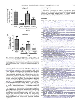
![S. Mukherjee et al. / The International Journal of Biochemistry & Cell Biology 45 (2013) 1516–1524 1517
Fig. 1. (A) Expression level of collagen A1 (Col1), fibronectin 1 (Fn1) and smooth muscle actin (SMA) genes in the cell culture we are working with. All results are shown
as mean ± SEM, *p < 0.05 vs. SMA gene expression; n = 6 in each group. (B) Human pulmonary fibroblasts at rest (without any treatment). Transmitted light image (i), and
confocal fluorimetric emitted images (ii). (C) Representative trace of fluorimetric activity in one naive fibroblast: no Ca2+
waves are evident.
and glomerular mesangial cells. PDGF plays a key role in several
critical biological functions and various types of tissue diseases
including tissue remodeling, scarring and fibrosis (Donovan et al.,
2013). PDGF receptors are up-regulated in fibroblasts and smooth
muscle cells during inflammation (Terracio et al., 1988) and in
patients with severe asthma (Lewis et al., 2005). PDGF stimulates
macrophages and several other cell types to trigger wound heal-
ing, and stimulates the production of several ECM molecules, such
as fibronectin (Blatti et al., 1988), collagen (Canalis, 1981), proteo-
glycans (Schönherr et al., 1991), and hyaluronic acid (Heldin et al.,
1989). In addition to these, several observations suggest that over-
activity of PDGF is associated with various fibrotic conditions in the
lung (Heldin and Westermark, 1999). Studies of severe asthmatic
patients show that PDGF significantly enhances fibroblast procol-
lagen I expression (Lewis et al., 2005). All these lines of evidence
suggest that PDGF plays a key role in fibrosis and lung disease: how-
ever, the molecular mechanisms by which it acts are still not totally
clear.
PDGF binds to specific receptors (PDGFR) on the cell membrane
and activates them. Ligand-induced PDGFR activation stimulates
autophosphorylation of tyrosine residues within the intracellu-
lar kinase domain and upregulates the catalytic activity of the
kinases. This autophosphorylation also leads to formation of dock-
ing sites for downstream signal transduction molecule containing
SH2 domains, such as Crk, Grb2, Grb7, and Nck. (Heldin et al.,
1998). PDGFRs can activate several major kinase-mediated path-
ways, including ERK, Jak/STAT, PI3-kinase/Akt, and NFB (Ball et al.,
2010). PDGF is also known to modulate cytosolic levels of cal-
cium ([Ca2+]i) (Bisaillon et al., 2010; Cuddon et al., 2008; Egan
et al., 2005; Espinosa-Tanguma et al., 2011), which may in turn
modulate other cellular events including the kinase pathways just
mentioned: however, many aspects of this action of PDGF are far
from clear.
We hypothesize that PDGF-induced gene expression in human
pulmonary fibroblasts can be regulated by Ca2+-wave frequency.
For this study, we cultured human pulmonary fibroblasts and
treated them with PDGF and/or a variety of blockers. Confocal
[Ca2+]i fluorimetry and quantitative RT-PCR were used to monitor
changes in Ca2+ waves and gene expression, respectively. Further-
more, the data presented herein suggest that PDGF acts through
IP3-gated channels to initiate Ca2+ wave activity, which in turn
modulates gene expression. This work builds on our previous study
of TGF-evoked responses (Mukherjee et al., 2012).
2. Materials and methods
2.1. Chemicals
PDGF-AB (PeproTech Inc., NJ, USA) was prepared in 0.25% BSA
in 10 mM acetic acid in PBS solution (pH 4.5). D-PBS was obtained
from Thermo Fisher Scientific (ON, Canada). Oregon Green calcium
dye, RPMI medium and Hank’s Balanced Salt Solution (HBSS) were
obtained from Invitrogen (CA, USA). Ryanodine and Xestospon-
gin C were obtained from Tocris Biosciences (MN, USA). All other
chemicals were obtained from Sigma-Aldrich Chemical Company,
(ON, Canada) unless otherwise specified, and prepared in absolute
EtOH (ryanodine, U73122), DMSO (CPA), or as aqueous solutions.
Aliquots were then diluted with HBSS to get the desired concentra-
tion.
2.2. Isolation and culture of fibroblasts
All experimental procedures were approved by the St. Joseph’s
Hospital Board of Ethics. Human primary fibroblasts were obtained
from the lungs of ten patients (5 male, 5 female; aged 60–80
years) undergoing lung surgery for pulmonary nodules following
informed consent. None of the patients had major respiratory co-
morbidities or lung function abnormalities. Normal lung tissues
were taken from macroscopically normal lung areas, as distant
from the nodules as possible, and cultivated in 35 mm tissue cul-
ture dishes in explant medium (DMEM +20% FBS + antibiotics) at
37 ◦C in 95% air, 5% CO2. After 2–3 weeks, cells were trypsinized by
trypsin–EDTA treatment and subcultured in 75 cm2 culture flask](https://image.slidesharecdn.com/92a9cc2d-f147-4824-811d-403b872592bf-160309045035/85/1-s2-0-S1357272513001234-main-2-320.jpg)
![1518 S. Mukherjee et al. / The International Journal of Biochemistry & Cell Biology 45 (2013) 1516–1524
Fig. 2. PDGF-evoked Ca2+
waves in human fibroblasts. (A) Confocal fluorimetric images of PDGF (10 ng/ml, O/N) treated fibroblasts at various time points (as indicated)
during a full-length recording; changes in [Ca2+
]i are evident in the fibroblasts indicated by white arrows. (B) [Ca2+
]i transients propagated throughout the cell as waves:
white arrows indicate the position of the [Ca2+
]i wave at different time points (as indicated). Background intensity was subtracted using Image J software. (C) Representative
fluorimetric traces from a cell treated with vehicle alone (0.25% BSA in 10 mM acetic acid in PBS; i) or PDGF (10 ng/ml; ii). (D) Mean (± SEM) Ca2+
frequency after vehicle and
PDGF treatments. *p < 0.05 vs. vehicle, n = 5 in each group.
for further growth. These cells, designated passage 1, were culti-
vated in standard growth medium (RPMI + 10% FBS + antibiotics) at
37 ◦C in 95% air, 5% CO2 and the culture medium was changed three
times a week. For confocal microscopy, cells were cultured in glass
bottom Petri dishes till they became confluent in the same growth
medium. All cells in the experiments were used between passages
5–10. For overnight (O/N) PDGF treatment and/or different blocker
treatment, confluent cells were incubated with medium containing
the required amount of reagent (∼18–20 h).
2.3. Ca2+-fluorimetry
A stock solution of Oregon Green (acetoxymethyl ester; Invitro-
gen, USA) was prepared in DMSO and 20% pluronic acid and stored
in small aliquots at −20 ◦C. Cells were incubated with Oregon Green
(5 M) and sulfobromophthalein (100 M) for 40 min at 37 ◦C, then
placed in a Plexiglass recording chamber and perfused with HBSS
solution for a period of 30 min prior to experimentation to allow for
complete dye hydrolysis. Confocal microscopy was then performed
at room temperature (21–23 ◦C) using a custom-built apparatus
described previously (Janssen et al., 2009); recording rate was
generally 1 frame/10 s. Blockers were delivered via the bathing
solution while PDGF for acute treatment was delivered via
a micropipette (PicospritzerTM II, General Valve, Fairfield, NJ)
brought into close proximity of the cell (∼100 m). Picture frames
were stored in TIF stacks of several hundred frames on a local hard
drive using image acquisition software (Video Savant 4.0; IO Indus-
tries, London, ON). Image files were then imported into Scion (Scion
Corporation; free download: www.scioncorp.com) for subsequent
analysis using a custom written macro designed to determine aver-
age fluorescence intensity over a user-defined region of interest
(10 × 10 pixels).
2.4. RNA isolation and RT-PCR
Total RNA isolation was done from cultured fibroblasts with
1 ml TRIzol reagent (Invitrogen, Carlsbad, CA), according to the
manufacturer’s instructions, and dissolved in DEPC-treated water.
Total RNA concentration and integrity were determined with a
microgel bioanalyzer (Agilent Bioanalyzer 2100; Agilent, Missis-
sauga, ON, Canada). 1 g of RNA was treated with DNAse and
reverse-transcribed according to the manufacturer’s instructions](https://image.slidesharecdn.com/92a9cc2d-f147-4824-811d-403b872592bf-160309045035/85/1-s2-0-S1357272513001234-main-3-320.jpg)
![S. Mukherjee et al. / The International Journal of Biochemistry & Cell Biology 45 (2013) 1516–1524 1519
Fig. 3. Effects of acute PDGF treatment on [Ca2+
]i. (A) Fluorescent images of fibroblasts at rest (i) and after application of PDGF into the vicinity of the cells (ii–vi). Changes in
[Ca2+
]i are evident in the fibroblasts indicated by white arrows. (B) Representative fluorimetric traces from cells treated with vehicle alone (0.25% BSA in 10 mM acetic acid
in PBS; top) or with PDGF (10 ng/ml; bottom). Black arrows indicate the time of vehicle or PDGF treatment. (C) Mean (± SEM) Ca2+
frequency after acute vehicle and PDGF
treatment. *p < 0.05 vs. vehicle, n = 5 in each group.
(Invitrogen). Quantitative real-time PCR was conducted by Taqman
method using the ABI Prism 7500 PCR system (Applied Biosystems,
Foster City, CA) according to manufacturer’s protocol. RT-PCR probe
and primer sets (gene expression assays) were purchased from
Applied Biosystems. Results were normalized to 2-microglobulin.
Relative gene expression was calculated using the CT method
(Applied Biosystems).
2.5. Data analysis
Growth factor-evoked changes in fluorescence were expressed
as a fraction of the baseline fluorescence at the beginning of
the experiment (F/Fo). Ca2+ wave frequencies were calculated by
counting the numbers of Ca2+ fluorimetric peaks (we defined a
Ca2+-spike as a transient (<50 s) elevation of F510 of 30% above
baseline,) in a fixed time period (e.g., 9–10 min). Data are reported
as mean ± SEM; n refers to the number of donors (more than 5).
Statistical comparisons were made using Student’s t-test; p < 0.05
was considered statistically significant.
3. Results
3.1. Baseline recording
We first confirmed that the cells we cultured out of the lung tissues are indeed
fibroblasts, as culture from lung explants may lead to a mixed population of many
cell types, including smooth muscle. We checked the expression of collagen A1,
fibronectin 1 (both marker genes for fibroblasts) and smooth muscle actin (marker
for smooth muscle cells). We found that expression of collagen A1 and fibronectin 1
genes were significantly higher than that of the smooth muscle actin gene (Fig. 1A).
This result confirmed that the cells we are working with are actually fibroblast
cells. Very few (<5%) of these cells exhibited any kind of spontaneous Ca2+
wave
activity (i.e., in the absence of any applied stimuli) (Fig. 1C). Normal fibroblasts
were shown to spread out into a monolayer, with extended segments in various
directions making contact with other cells (Fig. 1B).
3.2. Effect of PDGF on Ca2+
wave activity
Next, we examined the effect of externally applied PDGF on [Ca2+
]i. We divided
the cells in two groups. One group of cells was incubated O/N (∼18–20 h) with vehi-
cle, 0.25% BSA in 10 mM acetic acid in PBS solution (used as solvent for PDGF), and
another group of cells was incubated O/N with PDGF in BSA/acetic acid at a con-
centration of 10 ng/ml. We did not find any Ca2+
wave activity in vehicle-treated
cells (Fig. 2C(i)), but almost all of the cells treated with PDGF exhibited recurring
spike-like elevations in [Ca2+
]i (Fig. 2C(ii)). These PDGF-evoked [Ca2+
]i transients
propagated throughout the cell as waves (Fig. 2B, Video S1). Interestingly, we found
that some cells also exhibited mechanical responses (retraction, shifting of cyto-
solic contents, etc.), but this did not seem to depend on the Ca2+
waves: that is,
these cellular responses were often not concurrent or even present in the same cell.
Supplementary material related to this article found, in the online version, at
http://dx.doi.org/10.1016/j.biocel.2013.04.018.
3.3. Acute treatment of PDGF
After confirming the O/N or prolonged effect of PDGF, we examined its acute
effect on Ca2+
wave activity. PDGF or vehicle (0.25% BSA in 10 mM acetic acid in PBS
solution) was applied as a bolus from a micropipette into the vicinity of the cells. We
filled the micropipette with 25 ng/ml PDGF in order to ensure maximal activation,](https://image.slidesharecdn.com/92a9cc2d-f147-4824-811d-403b872592bf-160309045035/85/1-s2-0-S1357272513001234-main-4-320.jpg)
![1520 S. Mukherjee et al. / The International Journal of Biochemistry & Cell Biology 45 (2013) 1516–1524
Fig. 4. Concentration-dependence of PDGF-mediated Ca2+
wave activity. (A) Representative fluorimetric traces of fibroblasts pretreated with 1 ng/ml, 3 ng/ml, 10 ng/ml,
100 ng/ml, PDGF (O/N) (i–iv, respectively). Horizontal bar indicates the treatment with different concentrations of PDGF. (B) Sigmoidal relationship between PDGF concen-
tration and mean frequency (± SEM) of Ca2+
wave. *p < 0.05 vs. control, and †
p < 0.05 vs. 1 ng/ml PDGF. Each point represents repetitions of the experiments with cells derived
from 5 donors (n = 5).
because the concentration of PDGF at the leading edge of the bolus is expected to
decrease (due to diffusion and geometric expansion of the bolus). Cells puffed with
vehicle did not show any significant change in Ca2+
fluorescence. On the other hand,
acute PDGF treatment elicited recurring Ca2+
transients within seconds after onset
of application, which spread throughout the cell (Fig. 3; Video S2).
Supplementary material related to this article found, in the online version, at
http://dx.doi.org/10.1016/j.biocel.2013.04.018.
3.4. Concentration-dependence of PDGF-stimulation
To assess the concentration-dependence of PDGF-evoked Ca2+
wave activity,
we treated the fibroblasts with 1, 3, 10 or 100 ng/ml PDGF or vehicle and incubated
O/N. All sets of cells were then subjected to confocal fluorimetry. We found very
few or no Ca2+
transients in the vehicle-treated group or 1 ng/ml PDGF O/N treated
group. However, those cells incubated O/N with 3, 10 or 100 ng/ml PDGF exhibited
recurring spike-like elevations in [Ca2+
]i (Fig. 4). The mean frequency of Ca2+
waves
(see Section 2) showed a distinct sigmoidal relationship with PDGF concentration,
with an estimated half-maximally effective concentration of approximately 4 ng/ml
(Fig. 4B).
3.5. Ca2+
pool involved in mediating the PDGF-evoked responses
To assess the relative contributions of various Ca2+
pools to the above mentioned
PDGF-mediated Ca2+
wave activity, we treated the cells with cyclopiazonic acid
(CPA) (inhibitor of the internal Ca2+
-pump (Uyama et al., 1992)), ryanodine (blocker
of ryanodine receptors; RyR), U73122 (phospholipase C inhibitor) or nominally Ca2+
-
free HBSS medium via perfusing buffer. PDGF was not included in perfusing buffers
used (both normal HBSS and Ca2+
-free HBSS).
To check whether influx of external Ca2+
has any effect on PDGF-mediated Ca2+
activity, we perfused PDGF-treated (O/N ∼18–20 h) cells with Ca2+
free media for
10 min, finding this immediately reduced the baseline fluorescence and completely
occluded all Ca2+
wave activity (Fig. 5A). Re-introduction of external Ca2+
(by
reperfusion with normal HBSS solution) resulted in an immediate reversal of those
changes (Fig. 5A).
We used CPA (10−5
M) to determine the contribution of internally sequestered
Ca2+
in these responses. When the overnight PDGF-treated cells were perfused
with CPA for 10 min, we found that PDGF-mediated Ca2+
wave activity was totally
occluded in all cells tested and a sustained elevation in baseline [Ca2+
]i was noted
(Fig. 5B). Upon wash-out of CPA, Ca2+
waves seemed to re-appear, but had a much
lower frequency, although this generally required extensive periods of time beyond
the length of our recordings: we did not pursue this recovery in detail.
It is known that release of internally sequestered Ca2+
occurs through RyR
and/or IP3-gated channels (Janssen et al., 2009). In our PDGF-treated cells, ryanodine
(10−5
M) had no effect on PDGF-evoked Ca2+
wave activity (Fig. 6A). On the other
hand, treatment with 10−6
M U73122 inhibited the response of PDGF; this inhibitory
effect of U73122 was irreversible, at least over the course of 20 min (Fig. 6B). To fur-
ther clarify the role of IP3-gated channels in PDGF-mediated Ca2+
wave activity, we
treated the cells with 2 M Xestospongin C either 30 min prior to the treatment with
PDGF or after overnight pre-treatment with PDGF for 30 min prior to microscopy. In
both cases Xestospongin C reduced the Ca2+
wave activity evoked by PDGF treatment
(Fig. 6C).
3.6. Effect of Ca2+
on PDGF-mediated gene expression
To determine whether Ca2+
waves play any role in PDGF-mediated gene expres-
sion in human pulmonary fibroblasts, cells were pretreated with vehicle or PDGF
O/N (20 h), some of the latter also being treated with 10−5
M CPA or 10−5
M ryan-
odine or 10−6
M U73122 for 6 h before flash-freezing and quantifying expression
of two matrix genes (collagen A1 and fibronectin 1). PDGF rapidly and dramati-
cally increased the expression of both matrix genes: interestingly, however, CPA
and U73122 decreased the expression of those genes whereas ryanodine treatment
had no effect (Fig. 7).](https://image.slidesharecdn.com/92a9cc2d-f147-4824-811d-403b872592bf-160309045035/85/1-s2-0-S1357272513001234-main-5-320.jpg)
![S. Mukherjee et al. / The International Journal of Biochemistry & Cell Biology 45 (2013) 1516–1524 1521
Fig. 5. Role of extracellular Ca2+
influx and intracellular Ca2+
release in PDGF-mediated Ca2+
wave activity. Representative fluorimetric responses of PDGF (10 ng/ml) in
absence and presence of Ca2+
-deficient HBSS medium (A) or 10−5
M CPA (B) in bath medium. The horizontal filled bars indicate different treatments. Dashed lines and arrows
in B indicate the base line fluorescence value and the rise in baseline fluorescence after CPA treatment, respectively. A(ii) and B(ii) indicate mean (± SEM) Ca2+
frequency
(number of Ca2+
wave peaks) before and during treatment with Ca2+
-deficient HBSS medium and CPA, respectively. *p < 0.05 vs. normal HBSS medium. Bars represent
repetitions of the experiments with cells derived from 5 donors (n = 5).
4. Discussion
Almost all organ systems including the lung, heart, kidney,
liver, skin and bone can be affected by diseases related to fibrotic
disorder. Lung fibrosis in particular involves proliferation of myo-
fibroblasts, but fibrotic reactions are also involved in diseases such
as asthma, chronic bronchitis and chronic obstructive pulmonary
disease. Many different cytokines including TGF- and PDGF are
related to these abnormal healing processes. In a recent study, we
examined the effects of the prototypical fibrogenic cytokine TGF-
on human pulmonary fibroblasts (Mukherjee et al., 2012). Whilst
critically important in fibrogenesis, TGF- is by far not the only rel-
evant factor. PDGF is another prominent cytokine in fibrosis and it
is not yet totally clear how it acts on fibroblasts. In this study, we
examined novel effects of PDGF treatment on human pulmonary
fibroblasts.
There are several salient features in the present study. We show
for the first time that: (i) both acute and overnight PDGF treatment
dramatically evoked Ca2+ wave activity in cultured human pul-
monary fibroblasts; (ii) there is a distinct sigmoidal relationship
between PDGF concentration and mean frequency of Ca2+-waves;
(iii) removal of external Ca2+ or disruption of Ca2+ release using
CPA or U73122 occluded the PDGF-evoked Ca2+ waves; (iv) CPA
and U73122 reduced the PDGF-mediated over-expression of
fibronectin and collagen A1 gene; and (v) ryanodine had no effect
on PDGF-mediated Ca2+ waves nor expression of fibronectin and
collagen A1 gene.
It is well known that PDGF contributes to expansion of myofi-
broblast population and production of ECM proteins (Bonner,
2004); the common perception is that it exerts its function mainly
via phosphorylation and activating signaling pathways such as
ERK, Jak/STAT, PI3-kinase/Akt, and NFB (Ball et al., 2010). Sev-
eral recent studies indicated that PDGF also works by altering
[Ca2+]i (Bisaillon et al., 2010; Cuddon et al., 2008; Egan et al.,
2005; Espinosa-Tanguma et al., 2011; Estaciona and Mordan, 1997;
Ogawa et al., 2012). For example, PDGF-BB treatment caused mul-
tiple Ca2+ transients in human internal mammary artery SMCs
(Scherberich et al., 2000), and elevated Ca2+-wave activity in HITC6
smooth muscle cells (Espinosa-Tanguma et al., 2011). Several stud-
ies reported that PDGF activated store-operated calcium entry in
human neurosphere-derived cells (NDCs) (Cuddon et al., 2008)
and human pulmonary arterial smooth muscle cells (Ogawa et al.,
2012). PDGF is also known to stimulate intracellular Ca2+ signal in
preneoplastic clones derived from C3H 10T1/2 mouse fibroblasts
(Estaciona and Mordan, 1997).
Our study places human pulmonary fibroblasts within the list
of cells responding to PDGF by exhibiting recurring Ca2+ transients
that propagate throughout the cell as waves. We found a sigmoidal
relationship between growth factor concentration and Ca2+ wave
frequency, with moderate Ca2+ wave activity occurring at [PDGF]
of 3 ng/ml and maximal wave activity occurring at 10–100 ng/ml
PDGF. We therefore chose 10 ng/ml as the working concentration
of PDGF for this study, which is within the physiologically relevant
range of [PDGF].
Generally, cells maintain [Ca2+]i at a very low level, since it is
involved in various cell functions such as gene expression, secretion
of proteins, cytoskeletal rearrangement, metabolism, and apopto-
sis. In response to excitatory stimulation, [Ca2+]i rises via a complex
interaction between calcium entry and extrusion across the plas-
malemma and release and reuptake of Ca2+ from the internal store.](https://image.slidesharecdn.com/92a9cc2d-f147-4824-811d-403b872592bf-160309045035/85/1-s2-0-S1357272513001234-main-6-320.jpg)
![1522 S. Mukherjee et al. / The International Journal of Biochemistry & Cell Biology 45 (2013) 1516–1524
Fig. 6. Effects of ryanodine, U73122 and xestospongin C on PDGF-mediated Ca2+
wave activity. Representative fluorimetric traces of PDGF (10 ng/ml (O/N)) treated fibroblasts
in absence and presence of 10−5
M ryanodine (A), 10−6
M U73122 in bath medium (B), or xestospongin C (C). The horizontal filled bar indicates different treatments. A(ii) and
B(ii) indicate mean (± SEM) Ca2+
frequency (number of Ca2+
wave peak) before and during treatment with ryanodine and U73122, respectively. *p < 0.05 vs. Normal HBSS
medium. Bars represent repetitions of the experiments with cells derived from 5 donors (n = 5).
The sarcoplasmic/endoplasmic reticulum is the most important
intracellular store of Ca2+. Two types of calcium release channels,
ryanodine receptors (RyR) and inositol 1,4,5-trisphosphate recep-
tors (IP3R) are present on this organelle. After confirming that PDGF
evoked Ca2+ waves in human pulmonary fibroblasts, we asked
whether external and/or internal Ca2+ is responsible for Ca2+ wave
activity. Our results show that removal of external Ca2+ imme-
diately and completely occluded PDGF-evoked Ca2+ waves in a
fully reversible fashion. This observation confirms the involvement
of external Ca2+ influx across the plasmalemma in PDGF-evoked
Ca2+ wave activity. On the other hand, when we disrupted the
internal Ca2+ store using CPA, a marked and significant reduction
in Ca2+ wave frequency and amplitude were noticed; CPA treat-
ment also caused a sustained elevation of basal [Ca2+]i, which is
consistent with observations made by other investigators (Chen
et al., 1992; Ethier et al., 2001; Putney, 1986). This elevation of
basal [Ca2+]i is likely due to an unmasking of a persistent release
or “leak” of Ca2+from the internal store. Some of that released
Ca2+ would be ejected from the cell by the plasmalemmal Ca2+-
pump and/or Na+/Ca2+ exchange, and would therefore need to be
replaced by some form of Ca2+-influx in order to maintain a full
internal store. So altogether our results would suggest that both
extracellular Ca2+ influx and intracellular Ca2+ release are impor-
tant in PDGF-mediated Ca2+ wave activity. With respect to the
involvement of intracellular Ca2+ storage in the PDGF responses,
we went on to find that PDGF-mediated Ca2+ wave activity was
unchanged after the treatment with ryanodine but was totally
occluded by U73122. These findings indicate a role for phospho-
lipase C, possibly through activation of IP3-gated channels, rather
than RyR, in mediating the PDGF response. To confirm whether
it is regulating via IP3-gated channels or not, we blocked IP3
receptors using the selective and membrane-permeable inhibitor
Xextospongin C. We found that 2 M Xestospongin C signifi-
cantly reduced PDGF-mediated Ca2+ wave activity. This result
confirmed that IP3-gated channels are involved in PDGF-mediated
response. It is interesting to note that ryanodine had little effect
against the responses to PDGF (this study) or ATP (Janssen et al.,
2009), but strongly inhibited responses to TGF- (Mukherjee et al.,
2012).
We also considered the physiological response to which these
calcium waves were coupled in these cells. Although some cells
showed a mechanical response (retraction, shifting of cytosolic
contents, etc.), those mechanical responses were rarely coincident
with any type of Ca2+ transients, or vice versa: we therefore con-
clude that the contractions are not Ca2+ dependent. In a variety
of cell types, gene expression during cellular growth and differ-
entiation is known to be modulated by [Ca2+]i (Bridges et al.,
1981; De Smedt et al., 1991; Dolmetsch et al., 1998; Faletto and
Macara, 1985; Hensold et al., 1991; Li et al., 1998; Marks et al.,
1991; Poon et al., 1990; Rodland et al., 1990). In particular, dif-
ferent studies have shown that recurring Ca2+ waves can up- or
down-regulate gene expression in a manner dependent upon Ca2+
wave frequency (Dolmetsch et al., 1998; Li et al., 1998). Previ-
ous studies from our lab have shown this phenomenon in human](https://image.slidesharecdn.com/92a9cc2d-f147-4824-811d-403b872592bf-160309045035/85/1-s2-0-S1357272513001234-main-7-320.jpg)


![Pells et al [2015] PLoS ONE 10[7] e0131102](https://cdn.slidesharecdn.com/ss_thumbnails/f79bb09e-8eb1-41e7-8042-28c0aa4a48c6-150720142907-lva1-app6891-thumbnail.jpg?width=640&height=640&fit=bounds)


