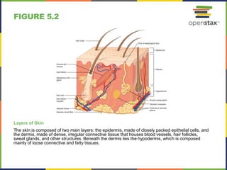
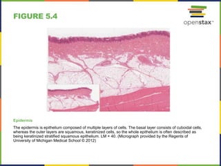
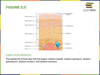
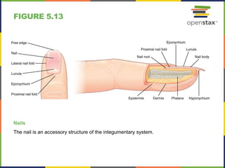
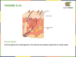
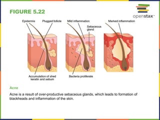
ANATOMY & PHYSIOLOGY
Chapter 5 THE INTEGUMENTARY SYSTEM
PowerPoint Image Slideshow
FIGURE 5.1
Your skin is a vital part of your life and appearance (a–d). Some people choose to
embellish it with tattoos (a), makeup (b), and even piercings (c). (credit a: Steve Teo;
credit b: “spaceodissey”/flickr; credit c: Mark/flickr; credit d: Lisa Schaffer)
FIGURE 5.2
Layers of Skin
The skin is composed of two main layers: the epidermis, made of closely packed epithelial cells, and
the dermis, made of dense, irregular connective tissue that houses blood vessels, hair follicles,
sweat glands, and other structures. Beneath the dermis lies the hypodermis, which is composed
mainly of loose connective and fatty tissues.
FIGURE 5.3
Thin Skin versus Thick Skin
These slides show cross-sections of the
epidermis and dermis of (a) thin and (b)
thick skin. Note the significant difference
in the thickness of the epithelial layer of
the thick skin. From top, LM × 40, LM ×
40. (Micrographs provided by the
Regents of University of Michigan
Medical School © 2012)
FIGURE 5.4
Epidermis
The epidermis is epithelium composed of multiple layers of cells. The basal layer consists of cuboidal cells,
whereas the outer layers are squamous, keratinized cells, so the whole epithelium is often described as
being keratinized stratified squamous epithelium. LM × 40. (Micrograph provided by the Regents of
University of Michigan Medical School © 2012)
FIGURE 5.5
Layers of the Epidermis
The epidermis of thick skin has five layers: stratum basale, stratum spinosum, stratum
granulosum, stratum lucidum, and stratum corneum.
FIGURE 5.6
Cells of the Epidermis
The cells in the different layers of the epidermis originate from basal cells located in the
stratum basale, yet the cells of each layer are distinctively different. EM ✕ 2700.
(Micrograph provided by the Regents of University of Michigan Medical School © 2012)
FIGURE 5.7
Layers of the Dermis
This stained slide shows the two
components of the dermis—the papillary
layer and the reticular layer. Both are
made of connective tissue with fibers of
collagen extending from one to the other,
making the border between the two
somewhat indistinct. The dermal papillae
extending into the epidermis belong to
the papillary layer, whereas the dense
collagen fiber bundles below belong to
the reticular layer. LM × 10. (credit:
modification of work by
“kilbad”/Wikimedia Commons)
FIGURE 5.8
Skin Pigmentation
The relative coloration of the skin depends of the amount of melanin produced by
melanocytes in the stratum basale and taken up by keratinocytes.
FIGURE 5.9
Moles
Moles range from benign accumulations of melanocytes to melanomas. These
structures populate the landscape of our skin. (credit: the National Cancer Institute)
FIGURE 5.10
Vitiligo
Individuals with vitiligo experience
depigmentation that results in lighter
colored patches of skin. The condition is
especially noticeable on darker skin.
(credit: Klaus D. Peter)
FIGURE 5.11
Hair
Hair follicles originate in the epidermis
and have many different parts.
FIGURE 5.12
Hair Follicle
The slide shows a cross-section of a hair follicle. Basal cells of the hair matrix in the center
differentiate into cells of the inner root sheath. Basal cells at the base of the hair root form the
outer root sheath. LM ✕ 4. (credit: modification of work by “kilbad”/Wikimedia Commons)
FIGURE 5.13
Nails
The nail is an accessory structure of the integumentary system.
FIGURE 5.14
Eccrine Gland
Eccrine glands are coiled glands in the dermis that release sweat that is mostly water.
FIGURE 5.15
Light Micrograph of a Meissneer Corpuscle
In this micrograph of a skin cross-section, you can see a Meissner corpuscle (arrow), a type
of touch receptor located in a dermal papilla adjacent to the basement membrane and
stratum basale of the overlying epidermis. LM × 100. (credit: “Wbensmith”/Wikimedia
Commons)
FIGURE 5.16
Thermoregulation
During strenuous physical activities, such as skiing (a) or running (c), the dermal blood vessels
dilate and sweat secretion increases (b). These mechanisms prevent the body from overheating. In
contrast, the dermal blood vessels constrict to minimize heat loss in response to low temperatures
(b). (credit a: “Trysil”/flickr; credit c: Ralph Daily)
FIGURE 5.17
Aging
Generally, skin, especially on the face and hands, starts to display the first noticeable
signs of aging, as it loses its elasticity over time. (credit: Janet Ramsden)
FIGURE 5.18
Basal Cell Carcinoma
Basal cell carcinoma can take several different forms. Similar to other forms of skin
cancer, it is readily cured if caught early and treated. (credit: John Hendrix, MD)
FIGURE 5.19
Squamous Cell Carcinoma
Squamous cell carcinoma presents here as a lesion on an individual’s nose. (credit: the
National Cancer Institute)
FIGURE 5.20
Squamous Cell Carcinoma
Squamous cell carcinoma presents here as a lesion on an individual’s nose. (credit: the
National Cancer Institute)
FIGURE 5.21
Eczema
Eczema is a common skin disorder that presents as a red, flaky rash. (credit:
“Jambula”/Wikimedia Commons)
FIGURE 5.22
Acne
Acne is a result of over-productive sebaceous glands, which leads to formation of
blackheads and inflammation of the skin.
FIGURE 5.23
Calculating the Size of a Burn
The size of a burn will guide decisions made about the need for specialized treatment.
Specific parts of the body are associated with a percentage of body area.
This OpenStax ancillary resource is © Rice University under a CC-BY 4.0
International license; it may be reproduced or modified but must be
attributed to OpenStax, Rice University and any changes must be noted.

Open stax anatomy& physiology ch05

  • 1.
    ANATOMY & PHYSIOLOGY Chapter5 THE INTEGUMENTARY SYSTEM PowerPoint Image Slideshow
  • 2.
    FIGURE 5.1 Your skinis a vital part of your life and appearance (a–d). Some people choose to embellish it with tattoos (a), makeup (b), and even piercings (c). (credit a: Steve Teo; credit b: “spaceodissey”/flickr; credit c: Mark/flickr; credit d: Lisa Schaffer)
  • 3.
    FIGURE 5.2 Layers ofSkin The skin is composed of two main layers: the epidermis, made of closely packed epithelial cells, and the dermis, made of dense, irregular connective tissue that houses blood vessels, hair follicles, sweat glands, and other structures. Beneath the dermis lies the hypodermis, which is composed mainly of loose connective and fatty tissues.
  • 4.
    FIGURE 5.3 Thin Skinversus Thick Skin These slides show cross-sections of the epidermis and dermis of (a) thin and (b) thick skin. Note the significant difference in the thickness of the epithelial layer of the thick skin. From top, LM × 40, LM × 40. (Micrographs provided by the Regents of University of Michigan Medical School © 2012)
  • 5.
    FIGURE 5.4 Epidermis The epidermisis epithelium composed of multiple layers of cells. The basal layer consists of cuboidal cells, whereas the outer layers are squamous, keratinized cells, so the whole epithelium is often described as being keratinized stratified squamous epithelium. LM × 40. (Micrograph provided by the Regents of University of Michigan Medical School © 2012)
  • 6.
    FIGURE 5.5 Layers ofthe Epidermis The epidermis of thick skin has five layers: stratum basale, stratum spinosum, stratum granulosum, stratum lucidum, and stratum corneum.
  • 7.
    FIGURE 5.6 Cells ofthe Epidermis The cells in the different layers of the epidermis originate from basal cells located in the stratum basale, yet the cells of each layer are distinctively different. EM ✕ 2700. (Micrograph provided by the Regents of University of Michigan Medical School © 2012)
  • 8.
    FIGURE 5.7 Layers ofthe Dermis This stained slide shows the two components of the dermis—the papillary layer and the reticular layer. Both are made of connective tissue with fibers of collagen extending from one to the other, making the border between the two somewhat indistinct. The dermal papillae extending into the epidermis belong to the papillary layer, whereas the dense collagen fiber bundles below belong to the reticular layer. LM × 10. (credit: modification of work by “kilbad”/Wikimedia Commons)
  • 9.
    FIGURE 5.8 Skin Pigmentation Therelative coloration of the skin depends of the amount of melanin produced by melanocytes in the stratum basale and taken up by keratinocytes.
  • 10.
    FIGURE 5.9 Moles Moles rangefrom benign accumulations of melanocytes to melanomas. These structures populate the landscape of our skin. (credit: the National Cancer Institute)
  • 11.
    FIGURE 5.10 Vitiligo Individuals withvitiligo experience depigmentation that results in lighter colored patches of skin. The condition is especially noticeable on darker skin. (credit: Klaus D. Peter)
  • 12.
    FIGURE 5.11 Hair Hair folliclesoriginate in the epidermis and have many different parts.
  • 13.
    FIGURE 5.12 Hair Follicle Theslide shows a cross-section of a hair follicle. Basal cells of the hair matrix in the center differentiate into cells of the inner root sheath. Basal cells at the base of the hair root form the outer root sheath. LM ✕ 4. (credit: modification of work by “kilbad”/Wikimedia Commons)
  • 14.
    FIGURE 5.13 Nails The nailis an accessory structure of the integumentary system.
  • 15.
    FIGURE 5.14 Eccrine Gland Eccrineglands are coiled glands in the dermis that release sweat that is mostly water.
  • 16.
    FIGURE 5.15 Light Micrographof a Meissneer Corpuscle In this micrograph of a skin cross-section, you can see a Meissner corpuscle (arrow), a type of touch receptor located in a dermal papilla adjacent to the basement membrane and stratum basale of the overlying epidermis. LM × 100. (credit: “Wbensmith”/Wikimedia Commons)
  • 17.
    FIGURE 5.16 Thermoregulation During strenuousphysical activities, such as skiing (a) or running (c), the dermal blood vessels dilate and sweat secretion increases (b). These mechanisms prevent the body from overheating. In contrast, the dermal blood vessels constrict to minimize heat loss in response to low temperatures (b). (credit a: “Trysil”/flickr; credit c: Ralph Daily)
  • 18.
    FIGURE 5.17 Aging Generally, skin,especially on the face and hands, starts to display the first noticeable signs of aging, as it loses its elasticity over time. (credit: Janet Ramsden)
  • 19.
    FIGURE 5.18 Basal CellCarcinoma Basal cell carcinoma can take several different forms. Similar to other forms of skin cancer, it is readily cured if caught early and treated. (credit: John Hendrix, MD)
  • 20.
    FIGURE 5.19 Squamous CellCarcinoma Squamous cell carcinoma presents here as a lesion on an individual’s nose. (credit: the National Cancer Institute)
  • 21.
    FIGURE 5.20 Squamous CellCarcinoma Squamous cell carcinoma presents here as a lesion on an individual’s nose. (credit: the National Cancer Institute)
  • 22.
    FIGURE 5.21 Eczema Eczema isa common skin disorder that presents as a red, flaky rash. (credit: “Jambula”/Wikimedia Commons)
  • 23.
    FIGURE 5.22 Acne Acne isa result of over-productive sebaceous glands, which leads to formation of blackheads and inflammation of the skin.
  • 24.
    FIGURE 5.23 Calculating theSize of a Burn The size of a burn will guide decisions made about the need for specialized treatment. Specific parts of the body are associated with a percentage of body area.
  • 25.
    This OpenStax ancillaryresource is © Rice University under a CC-BY 4.0 International license; it may be reproduced or modified but must be attributed to OpenStax, Rice University and any changes must be noted.