Download to read offline


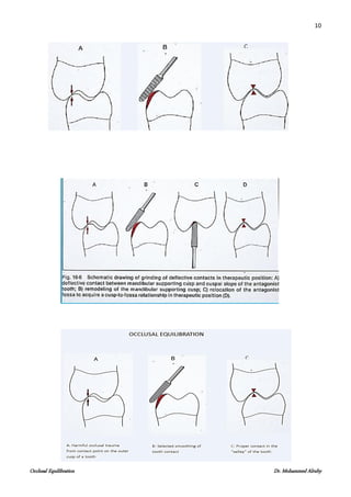

The document outlines the process and importance of occlusal equilibration, including causes of occlusal interference, recognition of stable and unstable occlusion, and treatment goals related to selective grinding. It details various technologies and techniques for diagnosing occlusal interferences and emphasizes the need for proper orthodontic knowledge in managing occlusal adjustments. The document also explores the significance of precise occlusal contact patterns and procedures for eliminating interferences during dental treatment.


