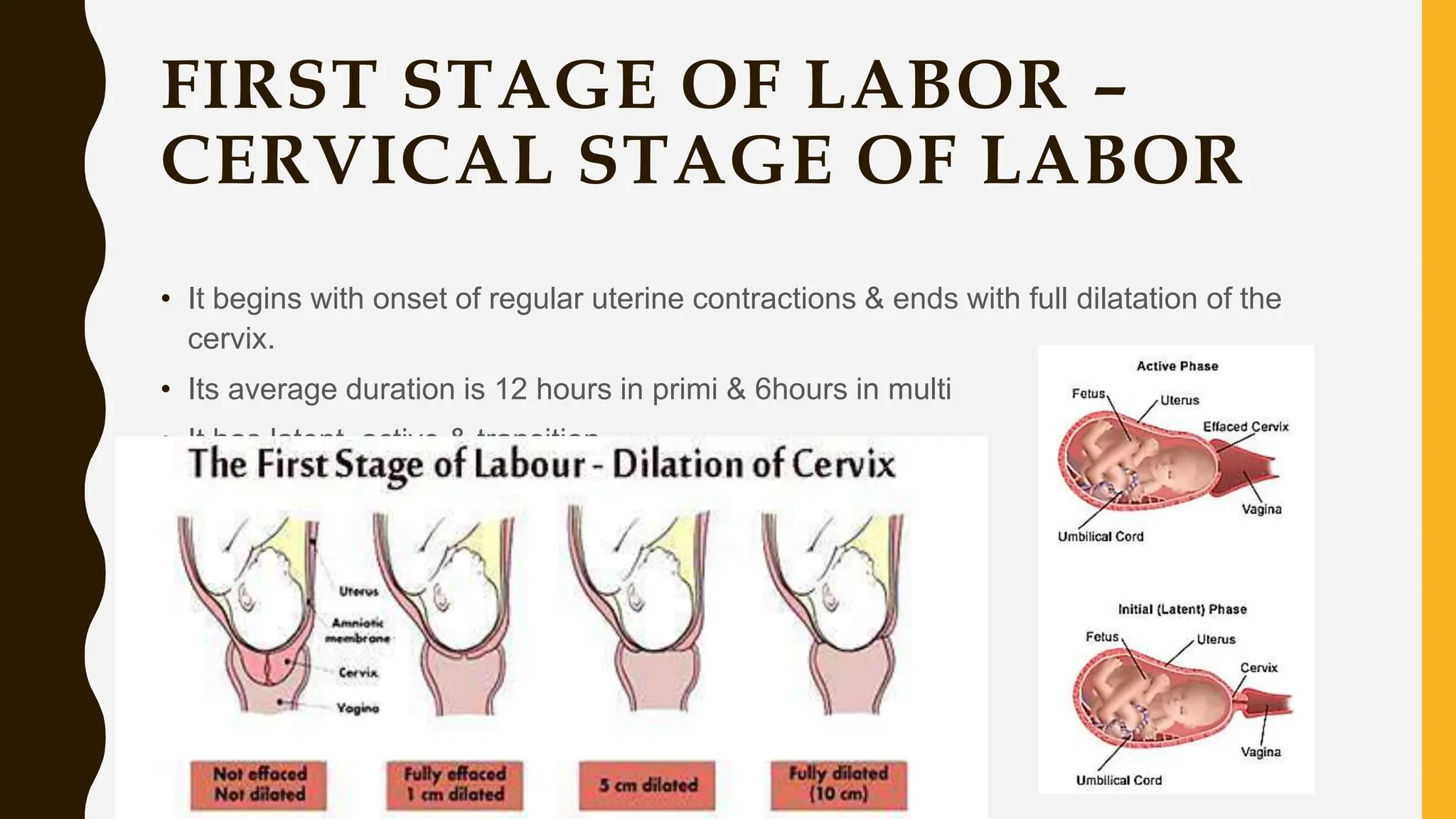
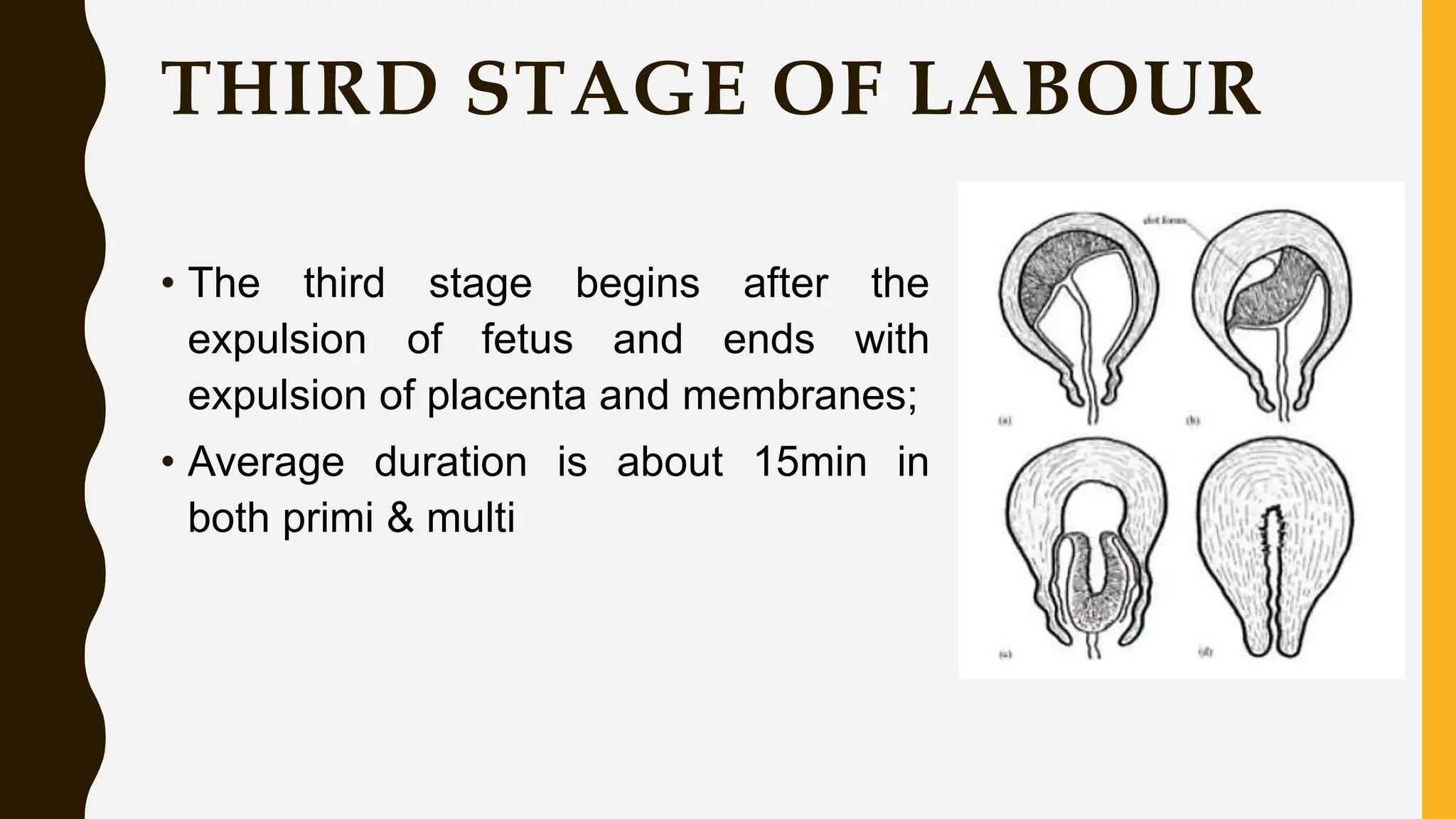
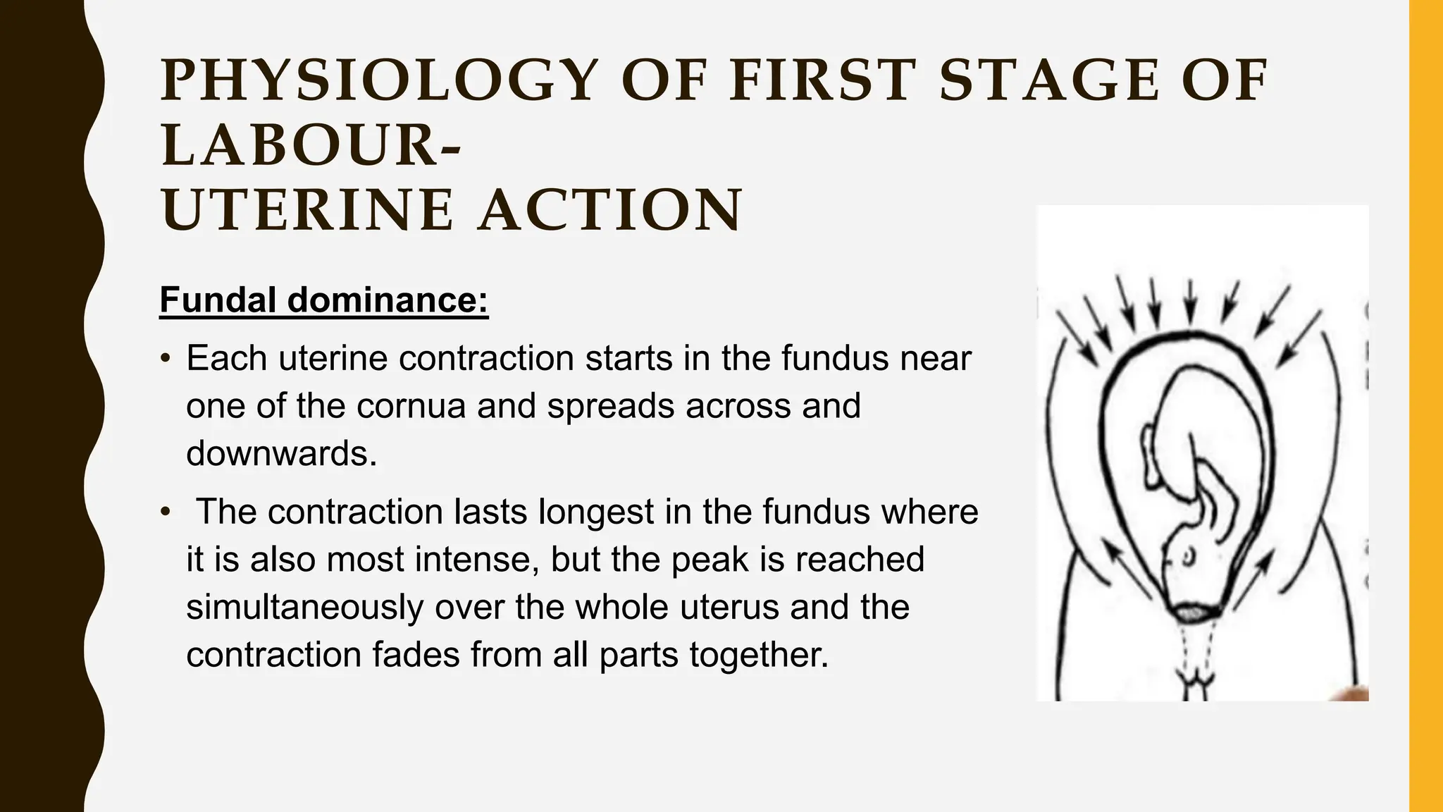
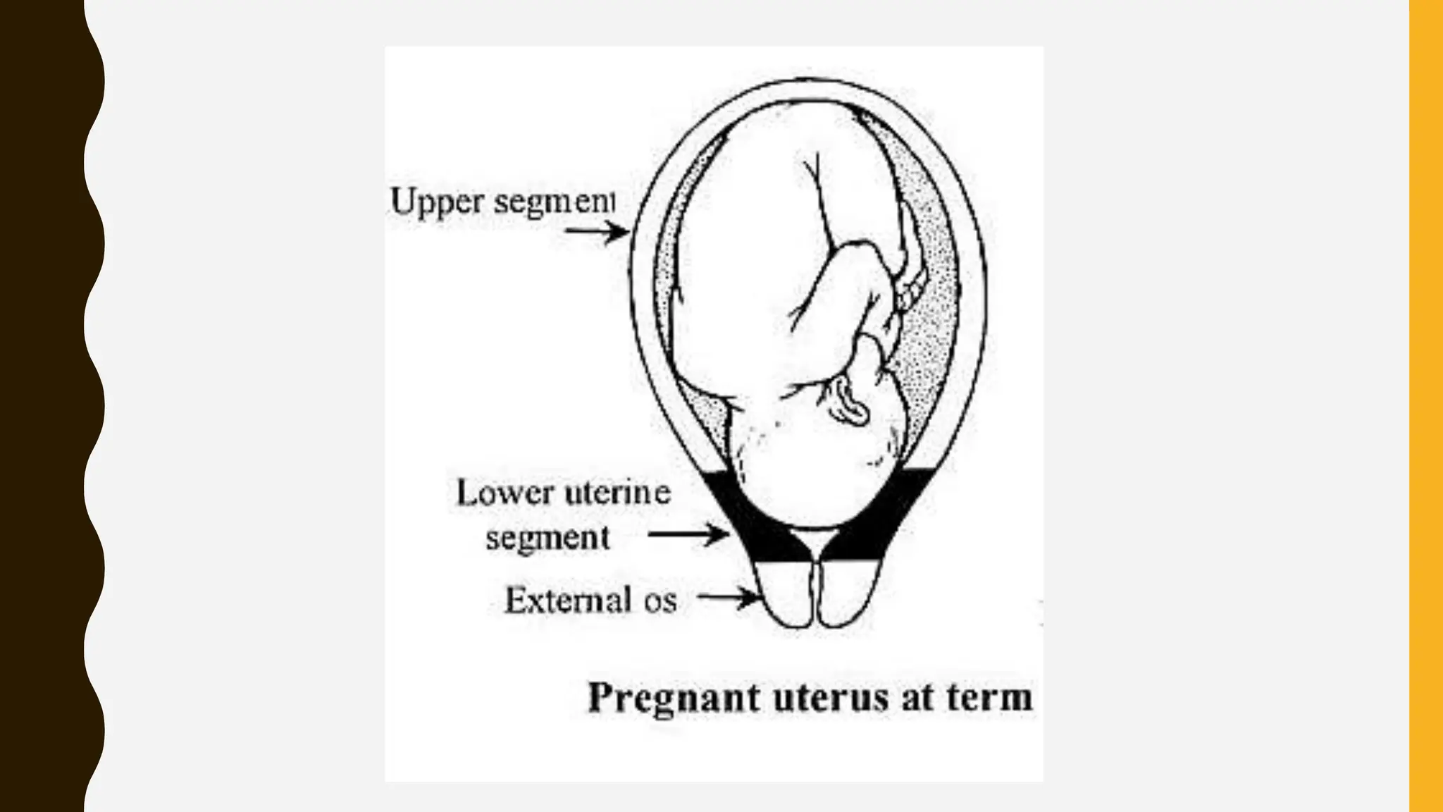
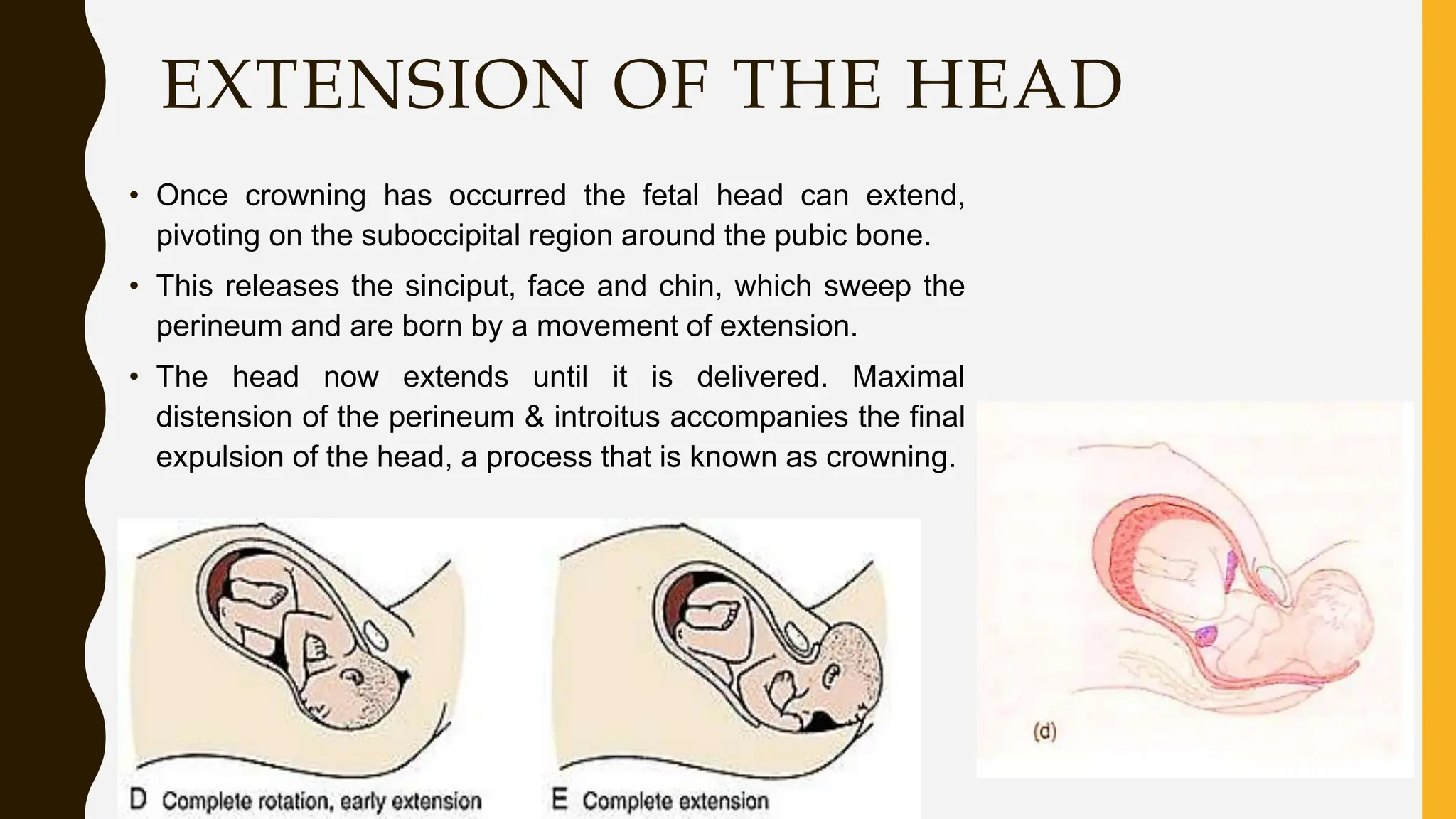
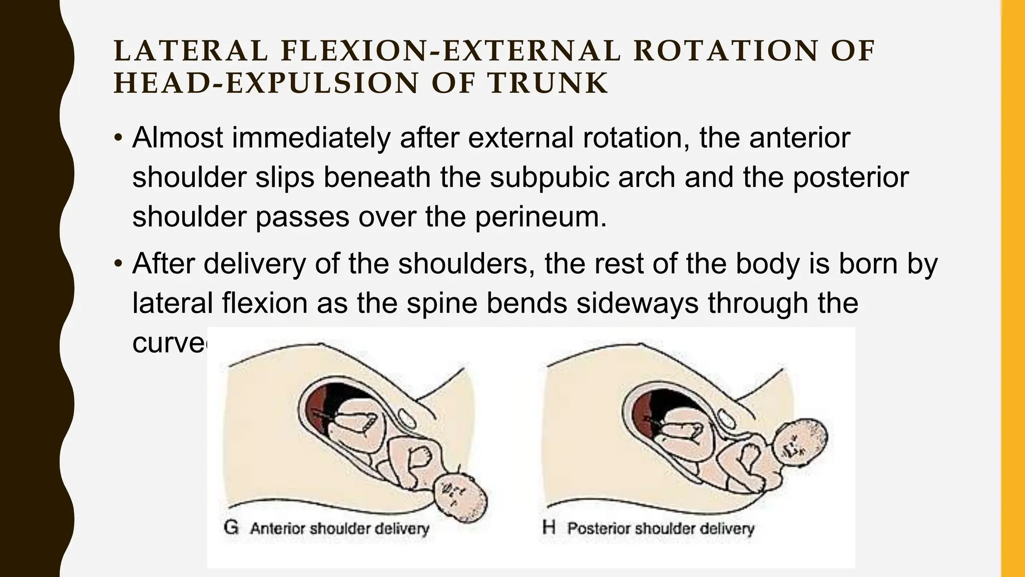
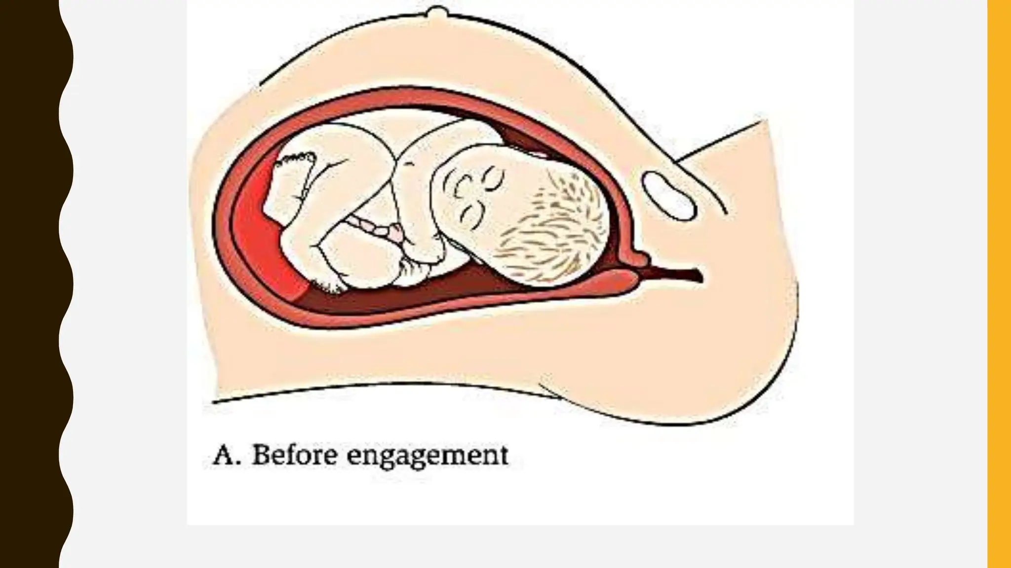
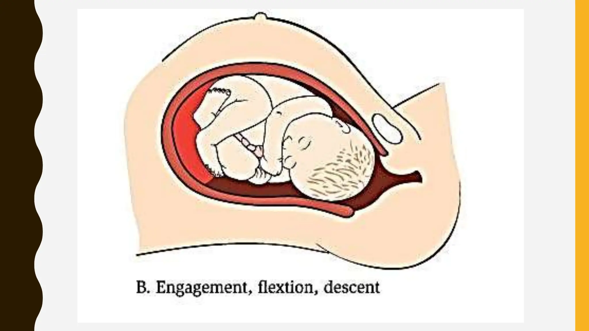
LABOUR, STAGES AND ITS
PHYSIOLOGY
INTRODUCTION
• Labor or parturition or childbirth is the physiologic process by which
regularly occurring uterine contractions results in progressive effacement
and dilatation of the cervix. These cervical changes permit passage of the
fetus & other products of conception from the uterus through the birth
canal, resulting in delivery.
• Pre term or premature labor – happens before 37 completed weeks
• Post term – happens after 42 weeks
• Parturient – women in labor / birth process
• Eutocia – Normal labor
• Dystocia – abnormal labor (mal presentation, mal positions etc)
CONTENTS
• Definitions
• Normal and abnormal labour
• Causes of onset of labour
• False labour pain and true labour pain
• Stages of labour
• Physiology of first stage of labour
• Physiology of second stage of labour
• Mechanism of normal labour
• Physiology of third stage of labour
DEFINITION
• Series of events that takes place in the genital organ in an effort to expel the
viable products of conception out of the womb through the vagina into the
outer world is called labour.
• Delivery is the expulsion or extraction of viable fetus out of the womb
• Normal labor is defined as spontaneous in onset, remains low risk through
out labor & delivery, infant is born spontaneously in vertex position between
37-41 completed weeks of gestation. Both mother & infant are in good
condition after birth
- WHO
Delivery is the spontaneous expulsion or aided extraction of a viable fetus
from the uterus vaginally (normal delivery) or through abdominal route
(Caesarean delivery)
NORMAL LABOUR
(EUTOCIA)
Labour is called normal if it fulfills the following criteria:
• Spontaneous in onset and at term.
• With vertex presentation
• Without undue prolongation
• Natural termination with minimal aids
• Without having any complications affecting the health of
mother and/or baby.
ABNORMAL LABOUR
(DYSTOCIA)
•Any deviation from the definition of normal
labour is called abnormal labour.
DEFINITION
• Premature Labour
–Labour occurring before the commencement of the 37th
week of gestation.
• Prolonged Labour
–Labour lasting in excess of:
• 24 hours in a primigravida
• 16 hours in a multigravida
DATE OF ONSET OF LABOUR
• The onset of Labor is defined as the time of onset of
regular, painful uterine contractions, which produce
progressive effacement and dilatation of the cervix.
• Calculation from Naegele‘s formula is only a rough
guide.
• Based on the formula, labour starts approx.
–on the expected date in 4%,
–one week on either side in 50%,
–2 weeks earlier and 1 week later in 80%,
ESSENTIAL FACTORS OF LABOR
The factors affecting Labour (5P’s)
• The power (uterine activity in labour)-contraction (frequency,
duration, strength) - Voluntary bearing down effort
• The passages (birth canal)and the pelvis – feto pelvic
diameter, cervical dilatation
• The passenger (fetus) – presentation of fetus and its position
and size of fetus
• Position: maternal postures & physical positions
• Psyche: the response of the mother – emotional factor
CAUSES OF ONSET OF
LABOUR
1. HORMONAL FACTORS
OESTROGEN
• Increase the release of oxytocin from maternal pituitary.
• Promotes the synthesis of receptors for oxytocin in the
myometrium and decidua.
• Increases the excitability of the myometrial cell membranes.-
last trimester
PROGESTERONE
• Increased fetal production of dehydroepiandrosterone
sulphate (DHEA-S) and cortisol inhibits the conversion of fetal
pregnenolone to progesterone. Progesterone levels therefore
fall before labour.
• It is the alteration in the oestrogen: progesterone ratio rather
than the fall in the absolute concentration of progesterone
which is linked with the prostaglandin synthesis.
PROSTAGLANDINS
• Prostaglandins are the important factor which initiate and
maintain labour.
• The major sites of synthesis of prostaglandins are --- amnion,
chorion, decidual cells and myometrium.
• Synthesis is triggered by –rise in oestrogen level,
glucocorticoids, mechanical stretching in the late pregnancy,
increase in cytokines, infection, vaginal examination,
separation or rupture of membranes
OXYTOCIN
• Oxytocin receptors are increased in the uterus with the onset
of labour.
• Oxytocin promotes the release of prostaglandins from the
decidua.
• Oxytocin synthesis is increased in the decidua and in the
placenta.
• Vaginal examination and amniotomy cause rise in maternal
plasma oxytocin level (Ferguson reflex).
• Presence of this hormone causes the initiation of contraction
of the smooth muscles of the body & the labor pain starts
NEUROLOGICAL FACTOR
• Both α and β adrenergic receptors are present in the
myometrium; oestrogen causing the α receptors and
progesterone the β receptoors to function predominantly.
• The contractile response is initiated through the α receptors
of the post ganglionic nerve fibres in and around the cervix
and the lower part of the uterus.
FETAL CORTISOL THEORY
• Increased cortisol production from the fetal adrenal gland
before labor may influence the onset of labor by increasing
Estrogen production from the placenta.
II. MECHANICAL FACTORS
UTERINE DISTENSION THEORY
• When the uterus is distended to a certain limit, it starts
contraction to evacuate its contents.
• Stretch of the lower uterine segment
• It is dine by the presenting part near term.
PREMONITORY STAGE-PRELABOR STAGE
Lightening
Shelfing
Vaginal secretions increase in amount
Loss of weight – excretion of body water
Sciatic nerve pressure
Cervical ripening
Appearance of false labor pain
Show
Dilatation of internal os
Greater frequency of urination
Spurt of energy
Occasional rupture of membranes
LIGHTENING
SHELFING
• It is falling forwards of the uterine fundus making the
upper abdomen looks like a shelf during standing
position.
CERVICAL RIPENING
FALSE LABOUR PAIN
• Features
1.Dull in nature
2.Confined to the lower abdomen and groin.
3.Continuous and unrelated with hardening of the uterus
4.Without any effect on dilatation of the cervix.
5.Usually relieved by medications.
TRUE LABOUR PAIN
Features of true labour pain:
• Painful uterine contractions (labour pain) at regular intervals
• Contraction with increasing frequency, intensity and duration
• Show
• Progressive effacement and dilatation of the cervix
• Formation of the ―bag of waters.
• Not relieved by enema or sedatives
TRUE VS. FALSE LABOR
True False
Contraction
Regular, become closer &
stronger
Irregular
Timing Last 30-60 sec May last 1-2 min
Contraction position Lower abdomen & back Upper abdomen
Position
Get stronger with changing
position
Go away with changing
position, walking, hot bath
Cervix Dilation & effacement No changes
Fetus Drops into pelvis No significant changes
LABOR PAIN
• Throughout pregnancy, painless Braxton hick
contractions with simultaneous hardening of the
uterus occur.
SHOW
DILATATION OF INTERNAL OS
PRE LABOUR (PREMONITORY
STAGE) BEGINS:
• Primigravida: 2 or 3 weeks before the onset of true labour.
• Multigravida: few days prior.
STAGES OF LABOUR
First stage of labour
Second stage of labour
Third stage of labour
Fourth stage of labour
FIRST STAGE OF LABOR –
CERVICAL STAGE OF LABOR
• It begins with onset of regular uterine contractions & ends with full dilatation of the
cervix.
• Its average duration is 12 hours in primi & 6hours in multi
• It has latent, active & transition
SECOND STAGE OF LABOUR
• Starts from full dilatation of cervix to
expulsion of the fetus from the birth
canal.
• It has two phases
– Propulsive phase – starts from full
dilatation to descent of presenting
part to pelvic floor
– Expulsive phase – distinguished
by maternal bearing down efforts &
ends with the delivery.
The average duration is 2hours in
primi gravida and 30 min in multi
THIRD STAGE OF LABOUR
• The third stage begins after the
expulsion of fetus and ends with
expulsion of placenta and membranes;
• Average duration is about 15min in
both primi & multi
FOURTH STAGE OF LABOUR
• It is the stage of observation for atleast one
hour after expulsion of the after birth
• Begins with delivery of placenta and
extends to the first 1-4hours post partum.
• The maternal vitals, uterine retraction &
vaginal bleeding are monitored, baby is
examined during this period.
PHYSIOLOGY OF FIRST STAGE OF
LABOUR-
UTERINE ACTION
Fundal dominance:
• Each uterine contraction starts in the fundus near
one of the cornua and spreads across and
downwards.
• The contraction lasts longest in the fundus where
it is also most intense, but the peak is reached
simultaneously over the whole uterus and the
contraction fades from all parts together.
POLARITY
• Polarity is the term used to describe the neuromuscular harmony that
prevails between the two poles or segments of the uterus throughout
labour. During each uterine contraction, these two poles act
harmoniously.
• The upper pole contracts strongly and retracts to expel the fetus; the
lower pole contracts slightly and dilates to allow expulsion to take place.
If polarity is disorganized then the progress of labour is inhibited.
CONTRACTION AND RETRACTION
CHARACTERISTICS OF UTERINE
CONTRACTIONS
• Frequency: contractions occurs intermittently throughout
labor, they begin at 20-30 m apart & become closer together
until 2-3min
• Regularity – contractions occur more regularly as labor
becomes more established
• Duration – contraction may last from 30sec to between 60-90
sec near full dilatation of the cervix
• Intensity – the strength of the contraction increases as labor
progresses, from weak contractions noted early in labor,
strong expulsive contraction.
FORMATION OF UPPER AND
LOWER UTERINE SEGMENTS
• The upper uterine segment, having been formed from the
body of the fundus, is mainly concerned with contraction and
retraction; it is thick and muscular.
• The lower uterine segment is formed of the isthmus and the
cervix, and is about 8-10 cm in length. The lower segment is
prepared for distention and dilatation.
• The muscle content reduces from the fundus to the cervix,
where it is thinner.
FORMATION OF UPPER AND
LOWER UTERINE SEGMENTS
CONT…
• When the labour begins, the retracted longitudinal fibres in the upper
segment pull on the lower segment causing it to stretch; this is aided by
the descending presenting part.
THE RETRACTION RING
• The ridge forms between the upper
and lower uterine segments; this is
known as the retraction ring.
• The physiological ring gradually
rises as the upper uterine segment
contracts and retracts and the lower
uterine segment thins out to
accommodate the descending fetus.
Once the cervix is fully dilated and
the fetus can leave the uterus, the
retraction ring rises no further.
CERVICAL EFFACEMENT
• Effacement refers to the inclusion of the cervical canal into
the lower uterine segment.
• It takes place from above downward; that is, the muscle fibres
surrounding the internal os are drawn upwards by the
retracted upper segment and the cervix merges into the lower
uterine segment.
• The cervical canal widens at the level of the internal os,
where the condition of the external os remains unchanged.
CERVICAL EFFACEMENT
CONT…
CERVICAL DILATATION
• Dilatation of cervix is the process of enlargement of the os uteri from a
tightly closed aperture to an opening large enough to permit the
passage of the fetal head. Dilatation is measured in centimeters and full
dilatation at term equates to about 10 cm.
CERVICAL DILATATION
SHOW
• As a result of the dilatation of the cervix, the operculum,
which formed the cervical plug during pregnancy, is lost. The
woman may see a blood stained mucoid discharge a few
hours before, or within a few hours after, labour starts.
• The blood comes from the ruptured capillaries in the parietal
decidua where the chorion has become detached from the
dilating cervix.
FORMATION OF FORE WATER
• As the lower uterine segment forms and stretches, the
chorion becomes detached from it and the increased
intrauterine pressure causes its loosened part of the sac of
fluid to bulge downwards into the internal os, to the depth of
6-12 mm.
• The well flexes head fits snugly into the cervix and cuts off the
fluid in front of the head from that which surrounds the body.
• The former is known as ‗forewaters‘ and the latter the
‗hindwaters‘.
FORMATION OF FORE WATER
GENERAL FLUID PRESSURE
• While the membranes remain
intact, the pressure of the uterine
contractions is exerted on the
fluid and, as fluid is not
compressible, the pressure is
equalized throughout the uterus
and the fetal body; it is known as
general fluid pressure‘.
RUPTURE OF MEMBRANE
• The optimal physiological time for
the membranes to rupture
spontaneously is at the end of the
first stage of labour after the cervix
becomes fully dilated and no
longer supports the bag of
forewaters.
FETAL AXIS PRESSURE
• During each contraction the
uterus rises forward and the
force of the fundal contraction is
transmitted to the upper pole of
the fetus down the long axis of
the fetus and applied by the
presenting part to the cervix.
This is known as fetal axis
pressure.
PHYSIOLOGY OF SECOND
STAGE OF LABOUR
INTRODUCTION TO 2ND STAGE OF LABOR
• The 2nd stage begins to complete dilataion of cervix (10cm) &
ends with the expulsion of the fetus. This stage is concerned
with the descent and delivery of the fetus through the birth
canal – Expulsion
• The expulsive force of uterine contractions is added by
voluntary contraction of the abdominal muscles called
‘Bearing down Efforts’.
• The force at work inn this stage are Uterine Contractions,
which occur every 2-3mins & lasts 50-60 sec.
PHASES OF 2ND STAGE
Latent, descent & transition are characterised by maternal &
verbal behaviours, uterine activity, the urge to bear down &
fetal descent.
• The latent phase
This is a period of rest & relative calm. The urge to bear down is not
well established.
• The Descent phase
Characterized by strong urge to bear down as the nerve reflex is
activated when the presenting part presses on the vagina & there is
release of oxytocin from the posterior pituitary gland, which promotes
stronger expulsive uterine contractions
• The transition phase
The presenting part is on the perineum & bearing down efforts are
most effective for promoting birth.
SIGNS OF 2ND STAGE OF LABOR
• Imminent Signs
– Increased bloody show
– Desire to bear down or have bowel movement (result of descent of
presenting part)
– Bulging of the perineum
– Dilatation of the anal orifice
• Impending Signs
– Nausea & retching
– Irritability & uncooperativeness
– c/o severe discomfort
EVENTS IN THE 2ND STAGE
OF LABOR
UTERINE ACTION
• Contractions become stronger and longer but may be less
frequent, allowing both mother and fetus regular recovery
periods.
• The membrane often rupture spontaneously towards the end
of the first stage or during transition to the second stage.
• The consequent drainage of liquor allows the hard, round
fetal head to be directly applied to the vaginal tissues. This
pressure aids distension.
• Fetal axis pressure increases flexion of the head, which
results in smaller presenting diameters, more rapid progress
and less trauma to both mother and fetus
UTERINE ACTION CONTD…
• The contraction becomes expulsive as the fetus descends
further into the vagina.
• Pressure from the presenting part stimulates nerve receptors
in the pelvic floor - this is termed the ‘Ferguson reflex’ and the
woman experiences the need to push.
• The mother‘s response is to employ her secondary powers of
expulsion by contracting her abdominal muscles and
diaphragm.
SOFT TISSUE DISPLACEMENT
• As the hard fetal head descends, the soft tissues of the pelvis
becomes displaced.
– Anteriorly-Bladder
– Posteriorly- Rectum
– The levator ani muscles
– Perineal body
SOFT TISSUE DISPLACEMENT CONTD…
• The fetal head becomes visible at the vulva, advancing each
contraction and receding between contractions until crowning
takes place.
• The head is then born.
• The shoulders and body follow with next contraction,
accompanied by gush of amniotic fluid and sometimes of
blood.
• The second stage culminates in the birth of the baby.
MATERNAL PHYSIOLOGICAL CHANGES IN
2ND STAGE OF LABOR
BP
• Rise by 15-20mmHg with contractions in 2nd stage
Metabolism
• Maternal pushing efforts adds further skeletal muscle activity that contributes to the
increase in metabolism
Pulse rate
• Increases during each pushing effort
Temperture
• Highest elevation is at the time of delivery, an increase of 0.5 to 16c is considered
normal
GI changes
• There is reduction in gastric motility & absorption continues through 2nd stage
Renal & hematologic changes
• There is increased filtration & reabsorption bcoz of increased co2, decrease renal
vascular resistance
MECHANISM OF NORMAL
LABOUR
LANDMARKS OF PELVIS
DIAMETER OF PELVIS
FETAL SKULL
LIE
• It refers to the relationship of the long axis of the fetus to the long axis of
the centralized uterus or maternal spine.
PRESENTATION
PRESENTING PART
• Is defined as the part of the
presentation which overlies the
internal os and is felt by the
examining finger through the
cervical opening.
ATTITUDE
• The relation of the different parts of the fetus to one another is
called attitude of the fetus. The universal attitude is that of
flexion.
DENOMINATOR
• It is an arbitrary bony fixed point on the presenting part which
comes in relation with the various quadrants of the maternal
pelvis. The following are denominators of the different
presentations- occiput in vertex, mentum in face, frontal
eminence in brow, sacrum in breech and acromion in
shoulder
POSITION
MECHANISM OF LABOUR
•As the fetus descends, soft tissue and bony
structures exert pressures which lead to
descent through the birth canal by a series of
movements. Collectively, these movements are
called the mechanism of labour.
DEFINITION
•The series of movements that occur on the
head in the process if adaptation, during its
journey through the pelvis
PRINCIPLES COMMON TO ALL
MECHANISM
• Descent takes place
• Whichever part leads and first meets the resistance of the
pelvic floor will rotate forwards until it comes under the
symphysis pubis.
• Whatever emerges from the pelvis will pivot around the pubic
bone.
SIX CONSIDERATIONS FOR
NORMAL LABOUR
• The lie is longitudinal
• The presentation is cephalic
• The position is right or left occipitoanterior
• The attitude is one of the good flexion
• The denominator is the occiput
• The presenting part is the posterior part of the anterior
parietal bone.
CARDINAL MOVEMENT
• Engagement
• Descent
• Flexion
• Internal rotation of the head
• Extension of the head
• External Rotation/Restitution
• Internal rotation of the shoulders
• Lateral flexion
ENGAGEMENT
• The mechanism by which the
biparietal diameter—the greatest
transverse diameter in an occiput
presentation—passes through the
pelvic inlet is designated
engagement.
DESCENT
• This movement is the first requisite for
birth of the newborn.
• Different in nulliparous and multigravida
women.
• Throughout the first stage of labour the
contraction and retraction of the uterine
muscles allow less room in the uterus,
exerting pressure on the fetus to
descend.
• Following rupture of the fore waters and
the exertion of maternal effort, progress
FACTORS FACILITATING
DESCENT
•Uterine contraction & retraction
•Pressure of amniotic fluid
•Bearing down efforts
•Extension & straightening of fetal body
FLEXION
• As soon as the descending head meets
resistance, whether from the cervix,
walls of the pelvis, or pelvic floor, then
flexion of the head normally results.
• Suboccipitobregmatic diameter (9.5 cm)
is substituted for the longer
occipitofrontal diameter (10 cm). The
occiput becomes the leading part.
INTERNAL ROTATION OF THE HEAD
• During contraction, the leading part is
pushed downwards onto the pelvic floor. The
resistance of this muscular diaphragm brings
about rotation.
• Occiput gradually moves toward the
symphysis pubis anteriorly.
• Whichever part of the fetus meets the lateral
half of this slope will be directed forwards
and towards the center in a well flexed
vertex presentation the occiput leads, and
rotates anteriorly through 1/8th of a circle
when it meets the pelvic floor. This causes a
slight twist in the neck as the head is no
INTERNAL ROTATION CONTD…
• The anteroposterior diameter of the head now lies in the
widest (anteroposterior) diameter of the pelvic outlet.
• The occiput slips beneath the sub-pubic arch and crowning
occurs when the head no longer recedes between contraction
and the widest transverse diameter is born.
• Of flexion is maintained, the suboccipito bregmatic diameter,
usually distends the vaginal orifice.
EXTENSION OF THE HEAD
• Once crowning has occurred the fetal head can extend,
pivoting on the suboccipital region around the pubic bone.
• This releases the sinciput, face and chin, which sweep the
perineum and are born by a movement of extension.
• The head now extends until it is delivered. Maximal
distension of the perineum & introitus accompanies the final
expulsion of the head, a process that is known as crowning.
RESTITUTION
• The twist in the neck of the fetus that resulted from internal
rotation is now corrected by a slight untwisting movement.
• The occiput moves one-eight of a circle towards the side from
which it started
INTERNAL ROTATION OF THE
SHOULDERS
• The shoulders undergo a similar
rotation to that of the head to lie in the
widest diameter of the pelvic outlet,
namely anteroposterior.
• The anterior shoulder is first to reach
the levator ani muscle and is therefore
rotates anteriorly to lie under the
symhysis pubis.
• It occurs in the same direction as
restitution, and the occiput of the fetal
head now lies laterally.
LATERAL FLEXION-EXTERNAL ROTATION OF
HEAD-EXPULSION OF TRUNK
• Almost immediately after external rotation, the anterior
shoulder slips beneath the subpubic arch and the posterior
shoulder passes over the perineum.
• After delivery of the shoulders, the rest of the body is born by
lateral flexion as the spine bends sideways through the
curved birth canal.
SECOND STAGE ENDS WITH DELIVERY
OF BABY.
PHYSIOLOGY OF THIRD STAGE OF LABOUR
THIRD STAGE OF LABOUR
• This stage begins immediately after delivery of the
fetus and involves the separation and expulsion of the
placenta and membranes, involving the separation,
descent and expulsion of placenta and membranes
and control of hemorrhage from the placenta site.
• The third stage usually lasts between 5 and 15
minutes, but any period upto 30 minutes is considered
to be within normal limits.
MECHANICAL FACTORS
• As the neonate is born, the uterus spontaneously contracts
around its diminishing contents.
• The uterine fundus now lies just below the level of the
umbilicus.
• Thus, by the beginning of the third stage, the placental site
has already diminished in area by about 75%.
• As this occurs the placenta becomes compressed and the
blood in the intervillous spaces is forced back into the spongy
layer of the decidua basalis.
SCHULTZE METHOD
• Separation usually begins centrally so that retroplacental clot
is formed.
• Increased weight helps to strip the adherent lateral borders
and peel the membranes off the uterine wall so that the clot
thus formed becomes enclosed in a membranous bag as the
placenta descends, fetal surface first.
• This process of separation is associated with more shearing
of both placenta and membranes and less fluid blood loss.
SCHULTZ METHOD
MATTHEWS DUNCAN
METHOD
• The placenta may begin to separate unevenly at one of its
lateral borders.
• The blood escapes so that separation is unaided by the
formation of a retroplacental clot.
• The placenta descends, slipping sideways, maternal surface
first.
• This process takes longer and is associated with ragged,
incomplete expulsion of the membranes and a higher fluid
blood loss.
SEPARATION OF FETAL
MEMBRANES
• The great decrease in uterine cavity surface area
simultaneously throws the fetal membranes—the amnion,
chorion and the parietal decidua—into innumerable folds.
• Membranes usually remain in situ until placental separation is
nearly completed.
• These are then peeled off the uterine wall, partly by further
contraction of the myometrium and partly by traction that is
exerted by the separated placenta, which lies in the lower
segment or upper vagina.
HOMEOSTASIS
• Retraction of the oblique uterine muscle fibres in the upper
uterine segment through which the tortuous blood vessels
interwine- the resultant thickening of the muscles exert
pressure on the torn vessels, acting as clamps, so securing a
ligature action.
HOMEOSTASIS CONT…
• Vigorous uterine contraction following separation-this brings
the walls into apposition so that further pressure is exerted on
the placental site.
• There is transitory activation of the coagulation and
fibrinolytic systems during, and immediately following
placental separation
SIGNS OF SEPARATION OF PLACENTA
Uterus becomes smaller, harder, more globular & is
below umbilicus on abdominal examination
Uterine height increases & the separated placenta
passes into the lower segment – shroedkar sign
Sudden gush of vaginal bleeding
There is no receding of the umbilical cord on pushing the
uterus upwards with an abdominal hand (kustner sign)
placenta followed by membranes is either
delivered spontaneously or by controlled cord
traction method followed by 200-300ml blood
loss.
• Modified Brandt-Andrew’s
Technique (Controlled cord
Traction method)
Crede’s Method
MANUAL REMOVAL OF PLACENTA
FOURTH STAGE OF LABOUR
• The first 4hours postpartum, sometimes referred to as 4th
stage of labor; (stage of observation for atleast 1hour after the
delivery of the baby, placenta & the membranes to ensure
that voth the mother & the baby are well). It is the time that
physiologic stability is restored.
• During this period myometrial contraction & retraction,
accompanied by vessel thrombosis, operate effectively to
control bleeding from the placental site.

Labour, Stages and its Physiology in obg

  • 1.
    LABOUR, STAGES ANDITS PHYSIOLOGY
  • 2.
    INTRODUCTION • Labor orparturition or childbirth is the physiologic process by which regularly occurring uterine contractions results in progressive effacement and dilatation of the cervix. These cervical changes permit passage of the fetus & other products of conception from the uterus through the birth canal, resulting in delivery. • Pre term or premature labor – happens before 37 completed weeks • Post term – happens after 42 weeks • Parturient – women in labor / birth process • Eutocia – Normal labor • Dystocia – abnormal labor (mal presentation, mal positions etc)
  • 3.
    CONTENTS • Definitions • Normaland abnormal labour • Causes of onset of labour • False labour pain and true labour pain • Stages of labour • Physiology of first stage of labour • Physiology of second stage of labour • Mechanism of normal labour • Physiology of third stage of labour
  • 4.
    DEFINITION • Series ofevents that takes place in the genital organ in an effort to expel the viable products of conception out of the womb through the vagina into the outer world is called labour. • Delivery is the expulsion or extraction of viable fetus out of the womb • Normal labor is defined as spontaneous in onset, remains low risk through out labor & delivery, infant is born spontaneously in vertex position between 37-41 completed weeks of gestation. Both mother & infant are in good condition after birth - WHO Delivery is the spontaneous expulsion or aided extraction of a viable fetus from the uterus vaginally (normal delivery) or through abdominal route (Caesarean delivery)
  • 5.
    NORMAL LABOUR (EUTOCIA) Labour iscalled normal if it fulfills the following criteria: • Spontaneous in onset and at term. • With vertex presentation • Without undue prolongation • Natural termination with minimal aids • Without having any complications affecting the health of mother and/or baby.
  • 6.
    ABNORMAL LABOUR (DYSTOCIA) •Any deviationfrom the definition of normal labour is called abnormal labour.
  • 7.
    DEFINITION • Premature Labour –Labouroccurring before the commencement of the 37th week of gestation. • Prolonged Labour –Labour lasting in excess of: • 24 hours in a primigravida • 16 hours in a multigravida
  • 8.
    DATE OF ONSETOF LABOUR • The onset of Labor is defined as the time of onset of regular, painful uterine contractions, which produce progressive effacement and dilatation of the cervix. • Calculation from Naegele‘s formula is only a rough guide. • Based on the formula, labour starts approx. –on the expected date in 4%, –one week on either side in 50%, –2 weeks earlier and 1 week later in 80%,
  • 9.
    ESSENTIAL FACTORS OFLABOR The factors affecting Labour (5P’s) • The power (uterine activity in labour)-contraction (frequency, duration, strength) - Voluntary bearing down effort • The passages (birth canal)and the pelvis – feto pelvic diameter, cervical dilatation • The passenger (fetus) – presentation of fetus and its position and size of fetus • Position: maternal postures & physical positions • Psyche: the response of the mother – emotional factor
  • 10.
    CAUSES OF ONSETOF LABOUR
  • 11.
  • 12.
    OESTROGEN • Increase therelease of oxytocin from maternal pituitary. • Promotes the synthesis of receptors for oxytocin in the myometrium and decidua. • Increases the excitability of the myometrial cell membranes.- last trimester
  • 13.
    PROGESTERONE • Increased fetalproduction of dehydroepiandrosterone sulphate (DHEA-S) and cortisol inhibits the conversion of fetal pregnenolone to progesterone. Progesterone levels therefore fall before labour. • It is the alteration in the oestrogen: progesterone ratio rather than the fall in the absolute concentration of progesterone which is linked with the prostaglandin synthesis.
  • 14.
    PROSTAGLANDINS • Prostaglandins arethe important factor which initiate and maintain labour. • The major sites of synthesis of prostaglandins are --- amnion, chorion, decidual cells and myometrium. • Synthesis is triggered by –rise in oestrogen level, glucocorticoids, mechanical stretching in the late pregnancy, increase in cytokines, infection, vaginal examination, separation or rupture of membranes
  • 15.
    OXYTOCIN • Oxytocin receptorsare increased in the uterus with the onset of labour. • Oxytocin promotes the release of prostaglandins from the decidua. • Oxytocin synthesis is increased in the decidua and in the placenta. • Vaginal examination and amniotomy cause rise in maternal plasma oxytocin level (Ferguson reflex). • Presence of this hormone causes the initiation of contraction of the smooth muscles of the body & the labor pain starts
  • 16.
    NEUROLOGICAL FACTOR • Bothα and β adrenergic receptors are present in the myometrium; oestrogen causing the α receptors and progesterone the β receptoors to function predominantly. • The contractile response is initiated through the α receptors of the post ganglionic nerve fibres in and around the cervix and the lower part of the uterus.
  • 17.
    FETAL CORTISOL THEORY •Increased cortisol production from the fetal adrenal gland before labor may influence the onset of labor by increasing Estrogen production from the placenta.
  • 18.
  • 19.
    UTERINE DISTENSION THEORY •When the uterus is distended to a certain limit, it starts contraction to evacuate its contents. • Stretch of the lower uterine segment • It is dine by the presenting part near term.
  • 21.
    PREMONITORY STAGE-PRELABOR STAGE Lightening Shelfing Vaginalsecretions increase in amount Loss of weight – excretion of body water Sciatic nerve pressure Cervical ripening Appearance of false labor pain Show Dilatation of internal os Greater frequency of urination Spurt of energy Occasional rupture of membranes
  • 22.
  • 23.
    SHELFING • It isfalling forwards of the uterine fundus making the upper abdomen looks like a shelf during standing position.
  • 24.
  • 26.
    FALSE LABOUR PAIN •Features 1.Dull in nature 2.Confined to the lower abdomen and groin. 3.Continuous and unrelated with hardening of the uterus 4.Without any effect on dilatation of the cervix. 5.Usually relieved by medications.
  • 27.
    TRUE LABOUR PAIN Featuresof true labour pain: • Painful uterine contractions (labour pain) at regular intervals • Contraction with increasing frequency, intensity and duration • Show • Progressive effacement and dilatation of the cervix • Formation of the ―bag of waters. • Not relieved by enema or sedatives
  • 28.
    TRUE VS. FALSELABOR True False Contraction Regular, become closer & stronger Irregular Timing Last 30-60 sec May last 1-2 min Contraction position Lower abdomen & back Upper abdomen Position Get stronger with changing position Go away with changing position, walking, hot bath Cervix Dilation & effacement No changes Fetus Drops into pelvis No significant changes
  • 29.
    LABOR PAIN • Throughoutpregnancy, painless Braxton hick contractions with simultaneous hardening of the uterus occur.
  • 30.
  • 31.
  • 33.
    PRE LABOUR (PREMONITORY STAGE)BEGINS: • Primigravida: 2 or 3 weeks before the onset of true labour. • Multigravida: few days prior.
  • 34.
    STAGES OF LABOUR Firststage of labour Second stage of labour Third stage of labour Fourth stage of labour
  • 35.
    FIRST STAGE OFLABOR – CERVICAL STAGE OF LABOR • It begins with onset of regular uterine contractions & ends with full dilatation of the cervix. • Its average duration is 12 hours in primi & 6hours in multi • It has latent, active & transition
  • 37.
    SECOND STAGE OFLABOUR • Starts from full dilatation of cervix to expulsion of the fetus from the birth canal. • It has two phases – Propulsive phase – starts from full dilatation to descent of presenting part to pelvic floor – Expulsive phase – distinguished by maternal bearing down efforts & ends with the delivery. The average duration is 2hours in primi gravida and 30 min in multi
  • 38.
    THIRD STAGE OFLABOUR • The third stage begins after the expulsion of fetus and ends with expulsion of placenta and membranes; • Average duration is about 15min in both primi & multi
  • 39.
    FOURTH STAGE OFLABOUR • It is the stage of observation for atleast one hour after expulsion of the after birth • Begins with delivery of placenta and extends to the first 1-4hours post partum. • The maternal vitals, uterine retraction & vaginal bleeding are monitored, baby is examined during this period.
  • 40.
    PHYSIOLOGY OF FIRSTSTAGE OF LABOUR- UTERINE ACTION Fundal dominance: • Each uterine contraction starts in the fundus near one of the cornua and spreads across and downwards. • The contraction lasts longest in the fundus where it is also most intense, but the peak is reached simultaneously over the whole uterus and the contraction fades from all parts together.
  • 41.
    POLARITY • Polarity isthe term used to describe the neuromuscular harmony that prevails between the two poles or segments of the uterus throughout labour. During each uterine contraction, these two poles act harmoniously. • The upper pole contracts strongly and retracts to expel the fetus; the lower pole contracts slightly and dilates to allow expulsion to take place. If polarity is disorganized then the progress of labour is inhibited.
  • 42.
  • 43.
    CHARACTERISTICS OF UTERINE CONTRACTIONS •Frequency: contractions occurs intermittently throughout labor, they begin at 20-30 m apart & become closer together until 2-3min • Regularity – contractions occur more regularly as labor becomes more established • Duration – contraction may last from 30sec to between 60-90 sec near full dilatation of the cervix • Intensity – the strength of the contraction increases as labor progresses, from weak contractions noted early in labor, strong expulsive contraction.
  • 44.
    FORMATION OF UPPERAND LOWER UTERINE SEGMENTS • The upper uterine segment, having been formed from the body of the fundus, is mainly concerned with contraction and retraction; it is thick and muscular. • The lower uterine segment is formed of the isthmus and the cervix, and is about 8-10 cm in length. The lower segment is prepared for distention and dilatation. • The muscle content reduces from the fundus to the cervix, where it is thinner.
  • 45.
    FORMATION OF UPPERAND LOWER UTERINE SEGMENTS CONT… • When the labour begins, the retracted longitudinal fibres in the upper segment pull on the lower segment causing it to stretch; this is aided by the descending presenting part.
  • 47.
    THE RETRACTION RING •The ridge forms between the upper and lower uterine segments; this is known as the retraction ring. • The physiological ring gradually rises as the upper uterine segment contracts and retracts and the lower uterine segment thins out to accommodate the descending fetus. Once the cervix is fully dilated and the fetus can leave the uterus, the retraction ring rises no further.
  • 48.
    CERVICAL EFFACEMENT • Effacementrefers to the inclusion of the cervical canal into the lower uterine segment. • It takes place from above downward; that is, the muscle fibres surrounding the internal os are drawn upwards by the retracted upper segment and the cervix merges into the lower uterine segment. • The cervical canal widens at the level of the internal os, where the condition of the external os remains unchanged.
  • 49.
  • 50.
    CERVICAL DILATATION • Dilatationof cervix is the process of enlargement of the os uteri from a tightly closed aperture to an opening large enough to permit the passage of the fetal head. Dilatation is measured in centimeters and full dilatation at term equates to about 10 cm.
  • 51.
  • 53.
    SHOW • As aresult of the dilatation of the cervix, the operculum, which formed the cervical plug during pregnancy, is lost. The woman may see a blood stained mucoid discharge a few hours before, or within a few hours after, labour starts. • The blood comes from the ruptured capillaries in the parietal decidua where the chorion has become detached from the dilating cervix.
  • 54.
    FORMATION OF FOREWATER • As the lower uterine segment forms and stretches, the chorion becomes detached from it and the increased intrauterine pressure causes its loosened part of the sac of fluid to bulge downwards into the internal os, to the depth of 6-12 mm. • The well flexes head fits snugly into the cervix and cuts off the fluid in front of the head from that which surrounds the body. • The former is known as ‗forewaters‘ and the latter the ‗hindwaters‘.
  • 55.
  • 56.
    GENERAL FLUID PRESSURE •While the membranes remain intact, the pressure of the uterine contractions is exerted on the fluid and, as fluid is not compressible, the pressure is equalized throughout the uterus and the fetal body; it is known as general fluid pressure‘.
  • 57.
    RUPTURE OF MEMBRANE •The optimal physiological time for the membranes to rupture spontaneously is at the end of the first stage of labour after the cervix becomes fully dilated and no longer supports the bag of forewaters.
  • 58.
    FETAL AXIS PRESSURE •During each contraction the uterus rises forward and the force of the fundal contraction is transmitted to the upper pole of the fetus down the long axis of the fetus and applied by the presenting part to the cervix. This is known as fetal axis pressure.
  • 59.
  • 60.
    INTRODUCTION TO 2NDSTAGE OF LABOR • The 2nd stage begins to complete dilataion of cervix (10cm) & ends with the expulsion of the fetus. This stage is concerned with the descent and delivery of the fetus through the birth canal – Expulsion • The expulsive force of uterine contractions is added by voluntary contraction of the abdominal muscles called ‘Bearing down Efforts’. • The force at work inn this stage are Uterine Contractions, which occur every 2-3mins & lasts 50-60 sec.
  • 61.
    PHASES OF 2NDSTAGE Latent, descent & transition are characterised by maternal & verbal behaviours, uterine activity, the urge to bear down & fetal descent. • The latent phase This is a period of rest & relative calm. The urge to bear down is not well established. • The Descent phase Characterized by strong urge to bear down as the nerve reflex is activated when the presenting part presses on the vagina & there is release of oxytocin from the posterior pituitary gland, which promotes stronger expulsive uterine contractions • The transition phase The presenting part is on the perineum & bearing down efforts are most effective for promoting birth.
  • 62.
    SIGNS OF 2NDSTAGE OF LABOR • Imminent Signs – Increased bloody show – Desire to bear down or have bowel movement (result of descent of presenting part) – Bulging of the perineum – Dilatation of the anal orifice • Impending Signs – Nausea & retching – Irritability & uncooperativeness – c/o severe discomfort
  • 63.
    EVENTS IN THE2ND STAGE OF LABOR
  • 64.
    UTERINE ACTION • Contractionsbecome stronger and longer but may be less frequent, allowing both mother and fetus regular recovery periods. • The membrane often rupture spontaneously towards the end of the first stage or during transition to the second stage. • The consequent drainage of liquor allows the hard, round fetal head to be directly applied to the vaginal tissues. This pressure aids distension. • Fetal axis pressure increases flexion of the head, which results in smaller presenting diameters, more rapid progress and less trauma to both mother and fetus
  • 65.
    UTERINE ACTION CONTD… •The contraction becomes expulsive as the fetus descends further into the vagina. • Pressure from the presenting part stimulates nerve receptors in the pelvic floor - this is termed the ‘Ferguson reflex’ and the woman experiences the need to push. • The mother‘s response is to employ her secondary powers of expulsion by contracting her abdominal muscles and diaphragm.
  • 66.
    SOFT TISSUE DISPLACEMENT •As the hard fetal head descends, the soft tissues of the pelvis becomes displaced. – Anteriorly-Bladder – Posteriorly- Rectum – The levator ani muscles – Perineal body
  • 67.
    SOFT TISSUE DISPLACEMENTCONTD… • The fetal head becomes visible at the vulva, advancing each contraction and receding between contractions until crowning takes place. • The head is then born. • The shoulders and body follow with next contraction, accompanied by gush of amniotic fluid and sometimes of blood. • The second stage culminates in the birth of the baby.
  • 68.
    MATERNAL PHYSIOLOGICAL CHANGESIN 2ND STAGE OF LABOR BP • Rise by 15-20mmHg with contractions in 2nd stage Metabolism • Maternal pushing efforts adds further skeletal muscle activity that contributes to the increase in metabolism Pulse rate • Increases during each pushing effort Temperture • Highest elevation is at the time of delivery, an increase of 0.5 to 16c is considered normal GI changes • There is reduction in gastric motility & absorption continues through 2nd stage Renal & hematologic changes • There is increased filtration & reabsorption bcoz of increased co2, decrease renal vascular resistance
  • 69.
  • 70.
  • 71.
  • 72.
  • 73.
    LIE • It refersto the relationship of the long axis of the fetus to the long axis of the centralized uterus or maternal spine.
  • 74.
  • 75.
    PRESENTING PART • Isdefined as the part of the presentation which overlies the internal os and is felt by the examining finger through the cervical opening.
  • 76.
    ATTITUDE • The relationof the different parts of the fetus to one another is called attitude of the fetus. The universal attitude is that of flexion.
  • 77.
    DENOMINATOR • It isan arbitrary bony fixed point on the presenting part which comes in relation with the various quadrants of the maternal pelvis. The following are denominators of the different presentations- occiput in vertex, mentum in face, frontal eminence in brow, sacrum in breech and acromion in shoulder
  • 78.
  • 79.
    MECHANISM OF LABOUR •Asthe fetus descends, soft tissue and bony structures exert pressures which lead to descent through the birth canal by a series of movements. Collectively, these movements are called the mechanism of labour.
  • 80.
    DEFINITION •The series ofmovements that occur on the head in the process if adaptation, during its journey through the pelvis
  • 81.
    PRINCIPLES COMMON TOALL MECHANISM • Descent takes place • Whichever part leads and first meets the resistance of the pelvic floor will rotate forwards until it comes under the symphysis pubis. • Whatever emerges from the pelvis will pivot around the pubic bone.
  • 82.
    SIX CONSIDERATIONS FOR NORMALLABOUR • The lie is longitudinal • The presentation is cephalic • The position is right or left occipitoanterior • The attitude is one of the good flexion • The denominator is the occiput • The presenting part is the posterior part of the anterior parietal bone.
  • 83.
    CARDINAL MOVEMENT • Engagement •Descent • Flexion • Internal rotation of the head • Extension of the head • External Rotation/Restitution • Internal rotation of the shoulders • Lateral flexion
  • 84.
    ENGAGEMENT • The mechanismby which the biparietal diameter—the greatest transverse diameter in an occiput presentation—passes through the pelvic inlet is designated engagement.
  • 86.
    DESCENT • This movementis the first requisite for birth of the newborn. • Different in nulliparous and multigravida women. • Throughout the first stage of labour the contraction and retraction of the uterine muscles allow less room in the uterus, exerting pressure on the fetus to descend. • Following rupture of the fore waters and the exertion of maternal effort, progress
  • 87.
    FACTORS FACILITATING DESCENT •Uterine contraction& retraction •Pressure of amniotic fluid •Bearing down efforts •Extension & straightening of fetal body
  • 88.
    FLEXION • As soonas the descending head meets resistance, whether from the cervix, walls of the pelvis, or pelvic floor, then flexion of the head normally results. • Suboccipitobregmatic diameter (9.5 cm) is substituted for the longer occipitofrontal diameter (10 cm). The occiput becomes the leading part.
  • 89.
    INTERNAL ROTATION OFTHE HEAD • During contraction, the leading part is pushed downwards onto the pelvic floor. The resistance of this muscular diaphragm brings about rotation. • Occiput gradually moves toward the symphysis pubis anteriorly. • Whichever part of the fetus meets the lateral half of this slope will be directed forwards and towards the center in a well flexed vertex presentation the occiput leads, and rotates anteriorly through 1/8th of a circle when it meets the pelvic floor. This causes a slight twist in the neck as the head is no
  • 90.
    INTERNAL ROTATION CONTD… •The anteroposterior diameter of the head now lies in the widest (anteroposterior) diameter of the pelvic outlet. • The occiput slips beneath the sub-pubic arch and crowning occurs when the head no longer recedes between contraction and the widest transverse diameter is born. • Of flexion is maintained, the suboccipito bregmatic diameter, usually distends the vaginal orifice.
  • 92.
    EXTENSION OF THEHEAD • Once crowning has occurred the fetal head can extend, pivoting on the suboccipital region around the pubic bone. • This releases the sinciput, face and chin, which sweep the perineum and are born by a movement of extension. • The head now extends until it is delivered. Maximal distension of the perineum & introitus accompanies the final expulsion of the head, a process that is known as crowning.
  • 93.
    RESTITUTION • The twistin the neck of the fetus that resulted from internal rotation is now corrected by a slight untwisting movement. • The occiput moves one-eight of a circle towards the side from which it started
  • 94.
    INTERNAL ROTATION OFTHE SHOULDERS • The shoulders undergo a similar rotation to that of the head to lie in the widest diameter of the pelvic outlet, namely anteroposterior. • The anterior shoulder is first to reach the levator ani muscle and is therefore rotates anteriorly to lie under the symhysis pubis. • It occurs in the same direction as restitution, and the occiput of the fetal head now lies laterally.
  • 95.
    LATERAL FLEXION-EXTERNAL ROTATIONOF HEAD-EXPULSION OF TRUNK • Almost immediately after external rotation, the anterior shoulder slips beneath the subpubic arch and the posterior shoulder passes over the perineum. • After delivery of the shoulders, the rest of the body is born by lateral flexion as the spine bends sideways through the curved birth canal.
  • 96.
    SECOND STAGE ENDSWITH DELIVERY OF BABY.
  • 106.
    PHYSIOLOGY OF THIRDSTAGE OF LABOUR
  • 107.
    THIRD STAGE OFLABOUR • This stage begins immediately after delivery of the fetus and involves the separation and expulsion of the placenta and membranes, involving the separation, descent and expulsion of placenta and membranes and control of hemorrhage from the placenta site. • The third stage usually lasts between 5 and 15 minutes, but any period upto 30 minutes is considered to be within normal limits.
  • 108.
    MECHANICAL FACTORS • Asthe neonate is born, the uterus spontaneously contracts around its diminishing contents. • The uterine fundus now lies just below the level of the umbilicus. • Thus, by the beginning of the third stage, the placental site has already diminished in area by about 75%. • As this occurs the placenta becomes compressed and the blood in the intervillous spaces is forced back into the spongy layer of the decidua basalis.
  • 110.
    SCHULTZE METHOD • Separationusually begins centrally so that retroplacental clot is formed. • Increased weight helps to strip the adherent lateral borders and peel the membranes off the uterine wall so that the clot thus formed becomes enclosed in a membranous bag as the placenta descends, fetal surface first. • This process of separation is associated with more shearing of both placenta and membranes and less fluid blood loss.
  • 111.
  • 112.
    MATTHEWS DUNCAN METHOD • Theplacenta may begin to separate unevenly at one of its lateral borders. • The blood escapes so that separation is unaided by the formation of a retroplacental clot. • The placenta descends, slipping sideways, maternal surface first. • This process takes longer and is associated with ragged, incomplete expulsion of the membranes and a higher fluid blood loss.
  • 114.
    SEPARATION OF FETAL MEMBRANES •The great decrease in uterine cavity surface area simultaneously throws the fetal membranes—the amnion, chorion and the parietal decidua—into innumerable folds. • Membranes usually remain in situ until placental separation is nearly completed. • These are then peeled off the uterine wall, partly by further contraction of the myometrium and partly by traction that is exerted by the separated placenta, which lies in the lower segment or upper vagina.
  • 115.
    HOMEOSTASIS • Retraction ofthe oblique uterine muscle fibres in the upper uterine segment through which the tortuous blood vessels interwine- the resultant thickening of the muscles exert pressure on the torn vessels, acting as clamps, so securing a ligature action.
  • 116.
    HOMEOSTASIS CONT… • Vigorousuterine contraction following separation-this brings the walls into apposition so that further pressure is exerted on the placental site. • There is transitory activation of the coagulation and fibrinolytic systems during, and immediately following placental separation
  • 117.
    SIGNS OF SEPARATIONOF PLACENTA Uterus becomes smaller, harder, more globular & is below umbilicus on abdominal examination Uterine height increases & the separated placenta passes into the lower segment – shroedkar sign Sudden gush of vaginal bleeding There is no receding of the umbilical cord on pushing the uterus upwards with an abdominal hand (kustner sign)
  • 118.
    placenta followed bymembranes is either delivered spontaneously or by controlled cord traction method followed by 200-300ml blood loss. • Modified Brandt-Andrew’s Technique (Controlled cord Traction method) Crede’s Method
  • 119.
  • 120.
  • 121.
    • The first4hours postpartum, sometimes referred to as 4th stage of labor; (stage of observation for atleast 1hour after the delivery of the baby, placenta & the membranes to ensure that voth the mother & the baby are well). It is the time that physiologic stability is restored. • During this period myometrial contraction & retraction, accompanied by vessel thrombosis, operate effectively to control bleeding from the placental site.