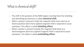
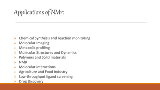
TOPIC:
Nuclear Magnetic Resonance(NMR)
Spectroscopy
Hamna Shahzad
Syed Jihan
8TH Semester
Bs Biochemistry
WhatisSpectroscopy
• Spectroscopy involves the study of interaction of radiation with matter.
Introduction
NMR spectroscopy stands for nuclear magnetic resonance
spectroscopy.
 NMR spectroscopy is a analytical technique which is used in
characterizing organic molecules by identifying carbon hydrogen
frameworks.
This technique utilizes the magnetic properties of some nmr active
substances.
 Nmr spectroscopy works on the spin of the nucleus as well as the
standard absobtion phenomenon of spectroscopy.
Historyof NMR
The Purcell group at Harvard University and the Bloch group at Stanford
University independently developed NMR spectroscopy in the late 1940s and
early 1950s.
Edward Mills Purcell and Felix Bloch shared the 1952 Nobel Prize in Physics for
their discoveries.
Prof. R.R. Ernst received the Nobel Prize in Chemistry, in 1991, for the
development of the NMR techniques.
Types ofNMR
 There are various types of NMR SPECTROSCOPY but the
most common are of two types which are charachterized
on the basis of atoms used.
 Hi NMR -: it is used to study the structure of compound
with refrence of hydrogen atoms.
 C13 NMR-: it is used to study the structure of compound
with refrence to carbon atoms.
● Sample
● Permanent magnet
● CRT display
● Magnetic coils
● Radio frequency generator
● Sample holder
● Detector and Amplifier
Instrumentation
Sourcesof NMR
 The source of nmr is radio waves which have a wavelength having more
than 107 nm and thus low frequency and energy.
 When these low energy waves interact with a molecule they can change the
nuclear spins of the some elements including 'H and C.
 The sample is dissolved in a solvent and placed in a magnetic field.
 A radiofrequency generator then irradiates the sample with a short pulse
of radiation, causing resonance of the proton.
 When the nuclie fall back to their lowest energy state ,the detector
measures the energy released and a spectrum is recorded in the form of
graph.
WorkingofNMR
NMR Active and Inactive Nucleus:
 The principle behind NMR comes from
 The spin of the nucleus and it generates a magnetic field without an external
applied magnetic field, the nuclear spins are random in directions.
 But when an external field is applied the nuclei align themselves in the
direction of the magnetic field or opposite this is called spin flip.
 If an external field is applied, an energy transfer is possible between ground
state to excited state. When the spin returns to the ground state the absorbed
radiofrequency energy is emitted at the same frequency level.
 This emitted radio frequency gives the nmr spectrum of the concerned
nucleus
PrinciplesofNMR
 The shift in the position of the NMR region resulting from the shielding
and deshelling by electrons is called chemical shift.
 When a proton is present inside the magnetic field more close to an
electro positive atom more applied magnetic field is required to cause
excitation. This effect is called shielding effect.
 When a proton is present outside the magnetic field close to a
electronegative atom less applied magnetic field is required to cause
excitation. This effect is called deshielding effect.
What ischemicalshift?
NMRspectrium
 Chemical Synthesis and reaction monitoring
 Molecular Imaging
 Metabolic profiling
 Molecular Structures and Dynamics
 Polymers and Solid materials
 NMR
 Molecular interactions
 Agriculture and Food Industry
 Low-throughput ligand screening
 Drug Discovery
Applicationsof NMr:
The two major areas where NMR has proven to be of critical importance is in fields of
medicine and chemistry, with new applications being developed daily.
Nuclear magnetic resonance imaging, better known as magnetic resonance imaging (MRI) is
an important medical diagnostic tool used to study the function and structure of the human
body. It provides detailed images of any part of the body, especially soft tissue, in all possible
planes and has been used in the areas of cardiovascular, neurological, musculoskeletal and
oncological imaging.
Applications:
In many laboratories today, chemists use nuclear magnetic resonance to determine
structures of important chemical and biological compounds. In NMR spectra, different peaks
give information about different atoms in a molecule according specific chemical
environments and bonding between atoms. The most common isotopes used to detect
NMR signals are 1H and 13C but there are many others, such as 2H, 3He, 15N, 19F, etc., that are
also in use.
NMR has also proven to be very useful in other area such as environmental testing,
petroleum industry, process control, earth’s field NMR and magnetometers. Non-destructive
testing saves a lot of money for expensive biological samples and can be used again if more
trials need to be run. The petroleum industry uses NMR equipment to measure porosity of
different rocks and permeability of different underground fluids. Magnetometers are used
to measure the various magnetic fields that are relevant to one’s study.
Applications:
NMR Technology in Tumor Metabolism
Cancer is one of the most serious diseases that cause an enormous number of deaths all over
the world. Tumor metabolism has great discrimination from that of normal tissues. Exploring the
tumor metabolism may be one of the best ways to find biomarkers for cancer detection,
diagnosis and to provide novel insights into internal physiological state where subtle changes
may happen in metabolite concentrations. Nuclear Magnetic Resonance (NMR) technique
nowadays is a popular tool to analyze cell extracts, tissues and biological fluids, etc, since it is a
relatively fast and an accurate technique to supply abundant biochemical information at
molecular levels for tumor research.
ARTICLE LINK:
https://doi.org/10.1016/j.gendis.2016.12.001
Steps:
1) Sample preparation
2) NMR spectrum
3) Statistical Analysis
4) Pathway Validation
In case of Colorectal Cancer:
Colorectal cancer is a disease in which cells in the colon or rectum grow out of
control. Sometimes it is called colon cancer, for short. The colon is the large
intestine or large bowel. The rectum is the passageway that connects the colon
to the anus
PCA (software use to do statistical analysis) results showed that advanced
colorectal tumor demonstrated increased fecal concentrations of four short-
chain fatty acids (valerate, acetate, propionate and butyrate) and decreased
signals relating to β-glucose, Gln, and glutamate.
Cancer type sample Metabolic Changes in cancer
tumor vs control
Colorectal serum (+) acetate, acetoacetate, 3-
hydroxybutyrate,
lactate,pyruvate
(−) glucose, myo-
inositol,taurine, dimethylglycine
Fecal extracts (+)acetate,valerate, propionate,
butyrate
(−)β-glucose, Gln, glutamat
biopsies (+)taurine, glutamate,
aspartate, lactate
(−)myo-inositol, β-glucose
NMR PRESENTATION.pptx

NMR PRESENTATION.pptx

  • 1.
    TOPIC: Nuclear Magnetic Resonance(NMR) Spectroscopy HamnaShahzad Syed Jihan 8TH Semester Bs Biochemistry
  • 2.
    WhatisSpectroscopy • Spectroscopy involvesthe study of interaction of radiation with matter.
  • 3.
    Introduction NMR spectroscopy standsfor nuclear magnetic resonance spectroscopy.  NMR spectroscopy is a analytical technique which is used in characterizing organic molecules by identifying carbon hydrogen frameworks. This technique utilizes the magnetic properties of some nmr active substances.  Nmr spectroscopy works on the spin of the nucleus as well as the standard absobtion phenomenon of spectroscopy.
  • 4.
    Historyof NMR The Purcellgroup at Harvard University and the Bloch group at Stanford University independently developed NMR spectroscopy in the late 1940s and early 1950s. Edward Mills Purcell and Felix Bloch shared the 1952 Nobel Prize in Physics for their discoveries. Prof. R.R. Ernst received the Nobel Prize in Chemistry, in 1991, for the development of the NMR techniques.
  • 5.
    Types ofNMR  Thereare various types of NMR SPECTROSCOPY but the most common are of two types which are charachterized on the basis of atoms used.  Hi NMR -: it is used to study the structure of compound with refrence of hydrogen atoms.  C13 NMR-: it is used to study the structure of compound with refrence to carbon atoms.
  • 6.
    ● Sample ● Permanentmagnet ● CRT display ● Magnetic coils ● Radio frequency generator ● Sample holder ● Detector and Amplifier Instrumentation
  • 8.
    Sourcesof NMR  Thesource of nmr is radio waves which have a wavelength having more than 107 nm and thus low frequency and energy.  When these low energy waves interact with a molecule they can change the nuclear spins of the some elements including 'H and C.
  • 9.
     The sampleis dissolved in a solvent and placed in a magnetic field.  A radiofrequency generator then irradiates the sample with a short pulse of radiation, causing resonance of the proton.  When the nuclie fall back to their lowest energy state ,the detector measures the energy released and a spectrum is recorded in the form of graph. WorkingofNMR
  • 10.
    NMR Active andInactive Nucleus:
  • 12.
     The principlebehind NMR comes from  The spin of the nucleus and it generates a magnetic field without an external applied magnetic field, the nuclear spins are random in directions.  But when an external field is applied the nuclei align themselves in the direction of the magnetic field or opposite this is called spin flip.  If an external field is applied, an energy transfer is possible between ground state to excited state. When the spin returns to the ground state the absorbed radiofrequency energy is emitted at the same frequency level.  This emitted radio frequency gives the nmr spectrum of the concerned nucleus PrinciplesofNMR
  • 14.
     The shiftin the position of the NMR region resulting from the shielding and deshelling by electrons is called chemical shift.  When a proton is present inside the magnetic field more close to an electro positive atom more applied magnetic field is required to cause excitation. This effect is called shielding effect.  When a proton is present outside the magnetic field close to a electronegative atom less applied magnetic field is required to cause excitation. This effect is called deshielding effect. What ischemicalshift?
  • 16.
  • 17.
     Chemical Synthesisand reaction monitoring  Molecular Imaging  Metabolic profiling  Molecular Structures and Dynamics  Polymers and Solid materials  NMR  Molecular interactions  Agriculture and Food Industry  Low-throughput ligand screening  Drug Discovery Applicationsof NMr:
  • 18.
    The two majorareas where NMR has proven to be of critical importance is in fields of medicine and chemistry, with new applications being developed daily. Nuclear magnetic resonance imaging, better known as magnetic resonance imaging (MRI) is an important medical diagnostic tool used to study the function and structure of the human body. It provides detailed images of any part of the body, especially soft tissue, in all possible planes and has been used in the areas of cardiovascular, neurological, musculoskeletal and oncological imaging. Applications:
  • 19.
    In many laboratoriestoday, chemists use nuclear magnetic resonance to determine structures of important chemical and biological compounds. In NMR spectra, different peaks give information about different atoms in a molecule according specific chemical environments and bonding between atoms. The most common isotopes used to detect NMR signals are 1H and 13C but there are many others, such as 2H, 3He, 15N, 19F, etc., that are also in use. NMR has also proven to be very useful in other area such as environmental testing, petroleum industry, process control, earth’s field NMR and magnetometers. Non-destructive testing saves a lot of money for expensive biological samples and can be used again if more trials need to be run. The petroleum industry uses NMR equipment to measure porosity of different rocks and permeability of different underground fluids. Magnetometers are used to measure the various magnetic fields that are relevant to one’s study. Applications:
  • 20.
    NMR Technology inTumor Metabolism Cancer is one of the most serious diseases that cause an enormous number of deaths all over the world. Tumor metabolism has great discrimination from that of normal tissues. Exploring the tumor metabolism may be one of the best ways to find biomarkers for cancer detection, diagnosis and to provide novel insights into internal physiological state where subtle changes may happen in metabolite concentrations. Nuclear Magnetic Resonance (NMR) technique nowadays is a popular tool to analyze cell extracts, tissues and biological fluids, etc, since it is a relatively fast and an accurate technique to supply abundant biochemical information at molecular levels for tumor research. ARTICLE LINK: https://doi.org/10.1016/j.gendis.2016.12.001
  • 21.
    Steps: 1) Sample preparation 2)NMR spectrum 3) Statistical Analysis 4) Pathway Validation
  • 23.
    In case ofColorectal Cancer: Colorectal cancer is a disease in which cells in the colon or rectum grow out of control. Sometimes it is called colon cancer, for short. The colon is the large intestine or large bowel. The rectum is the passageway that connects the colon to the anus PCA (software use to do statistical analysis) results showed that advanced colorectal tumor demonstrated increased fecal concentrations of four short- chain fatty acids (valerate, acetate, propionate and butyrate) and decreased signals relating to β-glucose, Gln, and glutamate.
  • 24.
    Cancer type sampleMetabolic Changes in cancer tumor vs control Colorectal serum (+) acetate, acetoacetate, 3- hydroxybutyrate, lactate,pyruvate (−) glucose, myo- inositol,taurine, dimethylglycine Fecal extracts (+)acetate,valerate, propionate, butyrate (−)β-glucose, Gln, glutamat biopsies (+)taurine, glutamate, aspartate, lactate (−)myo-inositol, β-glucose