
Recommended
Recommended
More Related Content
Similar to ANATOMY AND PHYSIOLOGY OF NMJ Prabhat (3).pptx
Similar to ANATOMY AND PHYSIOLOGY OF NMJ Prabhat (3).pptx (20)
Recently uploaded
Recently uploaded (20)
ANATOMY AND PHYSIOLOGY OF NMJ Prabhat (3).pptx
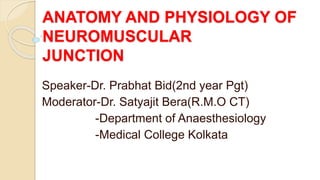
- 1. ANATOMY AND PHYSIOLOGY OF NEUROMUSCULAR JUNCTION Speaker-Dr. Prabhat Bid(2nd year Pgt) Moderator-Dr. Satyajit Bera(R.M.O CT) -Department of Anaesthesiology -Medical College Kolkata
- 2. What is the NMJ The NMJ is a synapses at which electrical impulses travelling down the motor nerve, releases the chemical messengers/transmitter which causes the muscle to contract. The NMJ is the chemical synapse between an alpha motor neuron and a muscle cell. The transmission of motor action potentials or indeed their prevention is an obvious importance to anaesthesists. The α-motor neuron originates in the ventral horn of the spinal cord. Its axon is myelinated, as the conduction of motor action potentials needs to be rapid. Before the axon reaches the NMJ, it branches to innervate several muscle cells. A motor unit consists of an α-motor neuron and the muscle cells that it
- 3. For neuromuscular transmission there are 3 essential components: 1.PRESYNAPTIC PART(MOTOR NERVE TERMINAL) 2.SYNAPTIC CLEFT 3 POSTSYNAPTIC MEMBRANE (MUSCLE END PLATE)
- 4. ANATOMY
- 6. MOTOR UNITS Motor unit consist of an alpha- motor neuron and the muscle fibre/fibres it innervate. Each motor neuron has its origin in the ventral horn of the spinal cord or medulla and runs an uninterrupted course as a large, myelinated axon to the neuromuscular junction. Each neuron branches and supplies several muscle cells, which together form the motor unit.
- 7. The NMJ junction or the endplate is a highly specialized synapse at which presynaptic motor nerve endings meet the post synaptic membranes of skeletal muscles. Each motor neuron approaches its target muscle fiber, its loses its myelin sheath and makes a contact with a single muscle fiber to form a NMJ. The more delicate the movements, the fewer muscle fibres per motor neuron. More intense contraction = more motor unit & long muscle fibres.
- 8. PRESYNAPTIC NERVE ENDING Presynaptic part consists of distal demyelinated part of the motor nerve axon and is separated from the extra cellular fluid by extension of the terminal Schwann cells and insulates the entire structure. Contents of the presynatic end: - Calcium channel (P type- fast) - Acetylcholine vesicles(3 types) - Mitochondria - Active zones - Proteins: Synaptotagmin,Synaptobrevin, Syntaxin, Synaptosome associated protein 25(SNAP 25) - Presynaptic nicotinic AchRs.
- 9. ACETYLCHOLINE VESICLES 3 lacs vesicles in an end plate 45 nm : bound by lipid bilayer membrane. Active zones 5000-10,000 molecules of Ach in 1 vesicle, loaded by Mg++ dependent proton pumping ATPase 1% - Releasable store(VP2) 80% - Reserve pool(VP1) Rest - Stationary pool
- 11. The SNARE (soluble N-ethylmaleimide-sensitive attachment protein receptors) proteins are involved in fusion, docking, and release of acetylcholine at the active zone Synaptophysin is a glycoprotein component of the vesicle membrane. Phosphorylation of another membrane protein, synapsin, facilitates vesicular trafficking to the release site. Synaptotagmin is the protein on the vesicular membrane acts as a calcium sensor and localize the synaptic vesicles to synaptic zones rich in calcium
- 12. Synaptobrevin is a vesicle associated membrane protein (VAMP). During depolarisation & entry of Ca it unfolds & forms a ternary complex with syntaxin/SNAP-25. Assembly of this complex guides the vesicle to the active zone.(Docking and Fusion) Then ca influx sensed by Synaptotagmine leads to burst release of Ach.
- 13. MODEL OF PROTEIN MEDIATED MEMBRANE FUSION AND EXOCYTOSIS
- 15. Acetylcholine Storage Once synthesised Ach is packaged into vesicles. Each vesicle contains around 5000 Ach molecules known as QUANTUM. THERE ARE FUNCTIONALLY 3 TYPES OF VESICLES : 1.Vesicles in the active zones VP2(1%)-this vesicle are docked at presynaptic membrane ready for immediate realease. 2.Vesicles in the reserve pools VP1(80%)-this vesicles move forwards to replace the vessicles in the active zone as they are used. 3.Vesicles in the stationary store(19%)-this vesicles cannot released their Ach
- 16. Vesicle Types at presynaptic nerve terminal
- 17. SYNAPTIC CLEFT The nerve is separated from the muscle by a gap of 20 – 50 nm, called the synaptic cleft. Contents: 1. Extracellular Fluid 2.Acetylcholinestera se
- 18. MUSCLE END PLATE The motor end plate is a specialized region of the sarcolemma (muscle membrane). It is oval, spanning an area of 3000 micrometre square. It is heavily corrugated, with deep invaginations, called the primary and the secondary clefts, to increase the surface area. The nicotinic Ach receptors are densely populated in the shoulders of the clefts (5 million AChR per junction), while the sodium channels are located in the depths of the folds.
- 19. MUSCLE END PLATE 1.nAchRs 2.Na Channels -Voltage dependent gate (VDG) -Timed dependent gate (TDG) VDG opens till depolarisation persists but TDG closes and cuts off the flow of sodium. TDG does not open again until VDG closes and reopens with a fresh depolarisation.
- 20. Synthesised in muscle cells Cylindrical receptor – central pore as ion channel MW-250000 Dalton. Each subunit consist 400-500 amino acids. One molecule of Ach binds with each α subunits at amino acid sequence 192-193 ACh RECEPTORS
- 21. Acetylcholine Receptor 2 TYPES :– MUSCARINIC ( M1 –M5) NICOTINIC- Nm – skeletal muscle end plate. Nn – adrenal medulla, ganglionic cells etc.
- 22. nAchRs 1.Adults nAchRs is a pentameric complex consist of 2α, 1β, 1, 1ϵ. 2.Fetal/ Extrajunctional- 2α, 1β, 1, 1. These subunits form a transmembrane pore and a pocket for acetylcholine binding.
- 23. Adult versus Fetal nAchR
- 24. Depolarisation of Nr terminal ↓ Opening of voltage gated Ca channel ↓ Entry of Ca in nerve terminal ↓ Mobiliation of synaptic vesicles ↓ Binding to docking protein ↓ Fusion of vesicles ↓ Release of ACh into
- 25. Action Potential
- 26. At the presynaptic zone…. Nerve action potential causes membrane depolarization. Activation of voltage dependent p-type calcium channel occurs. Entry of calcium into the axoplasm. Release of vesicles (VP2) from active zone . Docking and pore formation occurs via SNARE protein. Release of Ach in the synaptic cleft. Neuro muscular transmission
- 27. Post-junctional zone…….. Two molecules of Ach binds with the 2 alpha subunits of Ach receptors . Conformational change of the receptors leads to opening of the channel. Inward flow of Na+ and Ca2+ and outward flow of K+ starts. Change of membrane potential occurs. Activation of voltage sensitive Na+ channels. Neuro-Muscular transmission cont’d…
- 28. Post-junctional zone…….. Na entry across the muscle membrane via Na channel causes initiation and propagation of action potential to the T- tubules(upstokes of AP) DHPR(L-VGCC) types of ca channels get activated in the T- tubules.(voltage sensor) Activates RyR1 receptor in sarcoplasmic reticulum. Muscle contraction occurs. Neuro-Muscular transmission cont’d…
- 29. Termination of Neurotransmission Ach rapidly removed from the synaptic cleft mainly by degradation. 1.Ach is rapidly hydrolysed by the enzyme AchE to choline and acetic acid, this product are actively transported into presynaptic membrane for resynthesis of Ach. 2.AchE is mainly found in the junctional fold of the synaptic cleft.
- 31. Structures of skeletal muscle Two type of contractile filaments- a)thick – myosin b)thin - 3 types actin. tropomyosin. troponin.
- 32. Thick filaments Myosin – 2 heavy chains + 4 light chains. Heavy chain twist over each other and form a double helix . Each heavy chain has 2 light chain heads. Light chain – ATP ase action.
- 33. Thin filaments Actin – two types – G actin and F actin. Tropomysoin – covers active site of actin molecules to which myosin heads can attach. Troponin – 3 types :- I, T, C.
- 34. Clinical relevance Neurotransmission at the NMJ can be blocked by following mechanism 1. inhibision of Ach synthesis- Hemicholinium blocks the uptake of choline in the nerve action. 2.inhibition of vesical exocytosis – a-Mg and amynoglycosides-block the presynapic voltage gated ca channel. b- Botulinum toxin-It degrades a protein known as SNAP-25 required for vesicle docking.
- 35. Clinical relevance continue 3.blockage of Ach receptor- a.Depolarising MR b.Non depolarising MR
- 36. Depolarization of membrane opens the voltage dependent gate of peri junctional Na+ channels. Influx of Na+ occurs. After some time, Time dependent gate of Na+ channel closes. But the voltage dependent gate remains open as there is presence of Sch in the environment and Na+ channels remains in that state and propagation of depolarization stops. This is also called Phase I block. Characterized by a) decrease in twitch tension, (b) no fade during repetitive stimulation (tetanic or TOF), and (c) no post tetanic potentiation. Action of depolarizing MR
- 37. ILLUSTRATION OF SODIUM CHANNEL 1.At rest lower gate is opened and upper gate is closed.(resting state) 2.When muscle get depolarised the upper gate opens and sodiun enter.(active state) 3.shortly after that the lower gate closed (inactive state)
- 38. It is a complex phenomenon associated with typical fade in muscle during continuous exposure to depolarizing drug. Causes due to depolarizing action of Succinyl choline on pre junctional neuronal Ach receptors. Higher concentration of succinyl choline is needed. During repetitive nerve stimulation post junctional receptors also get involved. Repeated opening of channels causes Na+ inflow and K+ outflow results in abnormal membrane potential. Inflow of Ca2+ causes disruption of receptors and sub end plate structures. The activity of Na+-K+-ATPase is increased to restore membrane potentials. Phase II Block
- 39. Normally, the two ACh molecules have to combine with the two alpha 1 subunits of the nAChR to cause conformational change resulting in opening of the channel and flow of Na and Ca into the muscle and potassium ions flow outside. The channel excludes anions like chloride. The influx of ions depolarizes the adjacent membrane and causes the muscle to contract. When one or both ACh molecules detach from the receptor, the channel closes by a reversed mechanical conformation. This stops the depolarization. Actions of acetylcholine on end plate receptor
- 40. Classic Action of NDMR Both α subunits of nAchR, must be occupied by agonist for cationic flow across the membrane NDMRs inhibits depolarization by competitive inhibition of Ach from its binding site. Relative concentration and affinity determines the effectiveness of a particular NDMR. When it binds to one or both of the binding site conformational change of Ach receptor no cationic flow across membrane occurs.
- 41. Continued.... If choline esterase inhibitor is added - neostigmine Ach concentration remains high increase the chance of binding of 2 Ach even though NDMR is present. After large doses of NDMR / absence of clinical signs of reversal (high concentration of NDMR) neostigmine ineffective until the concentration of the relaxant lowers in perijunctional area by elimination / redistribution. Anticholinesterase is not indicated at deep block.
- 42. Actions of acetylcholine or Non depolarising muscle relaxant(NDMR) on end plate receptor
- 44. Receptor desensitization AchRs Bind with agonist (also with antagonist) but remains inactivated. Some Lipid soluble drugs also causes desensitization of receptors. Desensitized receptors potentiate action of muscle relaxants. The aberrant attachment of drugs to the recognition site probably responsible for this type of block.
- 45. Few drugs causes receptor desensitization
- 46. NEURO MUSCULAR DISORDER 1- MYASTHENIA GRAVIS 2-LAMBERT EATON MYASTHENIC SYNDROME 3-PERIODIC PARALYSIS
- 47. Is a chronic autoimmune disorder . Autoantibody forms against the α subunit of muscle type of nicotinic Ach receptors causes its destruction or inactivation. As many as 80% of functional receptors may be lost. Incidence varies between geographical regions ◦ Young age female > male ◦ >60 yrs male > female 70% thymic hyperplasia; 10% thymomas May occur as a part of paraneoplastic syndromes Myasthenia gravis
- 48. Ocular, pharyngeal and laryngeal muscles are involved causing ptosis, diplopia and dysphagia. Patchy distribution of muscle weakness ◦ Symptoms vary from day to day ◦ Remission of varying duration These patients are at the risk of pulmonary aspiration. Myocarditis may cause heart block, cardiomyopathy. Diagnosis :- ◦ Neurological examination ◦ Weakness, easy exhaustion of skeletal muscle and partial recovery with rest is the hallmark. Confirmation :- Tensilon test ◦ Administration of an anti-cholinesterase e.g. edrophonium ◦ Improvement in 5 minutes and lasts for 10 minutes Electrophysiologic evaluation a classic decrement in the compound muscle action potential after repetitive nerve stimulation Myasthenia gravis
- 49. Treatment :- ◦ Oral pyridostigmine is the drug of choice, maximum dose is 120mg every 3 hourly. ◦ Immunosuppressive therapy with corticosteroid, azathioprine, cyclosporin, mycophenolate. ◦ Plasmapheresis is done for Myasthenic crisis. ◦ Thymectomy is one of the treatment option. Anaesthetic considerations :- ◦ Pulmonary function tests ◦ Risk for post-operative mechanical ventilation ◦ Succinylcholine for tracheal intubation Increased dose requirement Prolonged neuromuscular block ◦ Potent volatile anaesthetics :- decreased margin of safety decreased requirements of muscle relaxants ◦ Epidural/ Spinal anaesthesia :- need for post-operative monitoring of muscle function and ventilation Myasthenia gravis
- 50. Frequently a part of Auto-antibody against P/Q type voltage sensitive calcium channel at prejunctional membrane and ANS. paraneoplastic syndromes – usually Small cell lung cancer. Decrease in number of channels. Restricted calcium entry during AP. Muscle weakness and fatigability ◦ Proximal > Distal ◦ Lower extremities > Extraocular/Bulbar muscle groups Unlike Myasthenia gravis ◦ Worse in morning Gradual improvement throughout the day ◦ Improvement of muscle function with exercise Eaton-Lambert Myasthenic syndrome
- 51. Treatment Plasmapheresis, Immunoglobulins, 3,4- diaminopyridine Temporary relief Anaesthetic implications :- ◦ Anticholinesterase drugs are ineffective as reversal of neuromuscular blockade ◦ Risk of postoperative respiratory failure need for prolonged respiratory monitoring in the postoperative period Eaton-Lambert Myasthenic syndrome
- 52. QUANTAL THEORY Spontaneous depolarizing potentials at neuromuscular junctions can be seen. This potentials have only one hundredth the amplitude of the evoked end-plate potentia produced when the motor nerve is stimulated. These small-amplitude potentials are called miniature end-plate potentials (MEPPs)
- 53. Because MEPPs are too big to be produced by a single molecule of acetylcholine, it was deduced that they are produced by uniformly sized packages, or quanta, of transmitter released from the nerve (in the absence of stimulation). The stimulus-evoked end-plate potential is the additive depolarization produced by the synchronous discharge of quanta from several hundred vesicles. The amount of Ach released by each nerve impulse is large, atleast 200 quanta of about 50,000 molecules each & the number of AchRs activated by transmitter released is about 500,000
- 54. THANK YOU