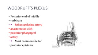
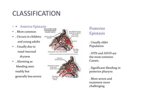
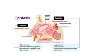
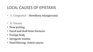
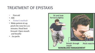
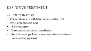
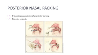
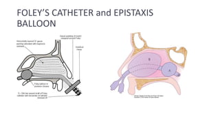
CAUSES AND MANAGEMENT OF EPISTAXIS
•INTRODUCTION
•  Bleeding from nostril, nasal cavity or nasopharynx
•  Most often self limited, but can often be serious and
• life threatening
•  5-10% of the population experience an episode of
• epistaxis each year, 10% of those will seek a physician
• and 1% of those will need a specialist
•  Can occur in all age groups
•REASON FOR EXCESSIVE BLEEDING
•  Rich vascularity
•  Supplied by both internal and external carotid system
•  Various anastomoses between arteries and veins
•  Blood vessels run under the mucosa unprotected
•  Larger vessels on the turbinate run in bony canals –
• cannot contract
Branches of internal
carotid system :
. Anterior Ethmoidal
artery
. Posterior ethmoidal
artery
 Branches of external
carotid system :
. Sphenopalatine
artery- major branch
. Greater palatine
artery
. Superior labial
branch of facial artery
. Infraorbital branch
of maxillary artery
KIESSELBACH’S PLEXUS (Little’s area)
•  In anterior inferior part of
• nasal septum
•  Most common site for
• epistaxis
•  Mainly anterior epistaxis
• 1. septal br. Of
• sphenopalatine
• 2. Anterior ethmoidal
• 3. Septal br. Of superior
• labial
• 4. greater palatine arteries
• anastomose here
WOODRUFF’S PLEXUS
• Posterior end of middle
• turbinate
•  Sphenopalatine artery
• anastomoses with
• posterior pharyngeal
• artery
•  Most common site for
• posterior epistaxis
CLASSIFICATION
•  Anterior Epistaxis
• . More common
• . Occurs in children
and young adults
• . Usually due to
nasal mucosal
dryness
• . Alarming as
bleeding seen
readily but
generally less severe
Posterior
Epistaxis
. Usually older
Population
. HTN and ASVD are
the most common
Causes.
. Significant bleeding in
posterior pharynx
. More severe and
treatment more
challenging
LOCAL CAUSES OF EPISTAXIS
• A. Congenital – Hereditary telangiectasia
• B. Trauma
• Nose picking
• Facial and skull bone fractures
• Foreign body
• Iatrogenic trauma
• Hard blowing, violent sneeze
• C. Inflammatory
Infective rhinitis
• D Specific
• Chronic granulomatous- TB, Leprosy, Syphilis,
• Rhinosporiodiasis
• E. Non Specific
• . Viral – Common cold, Influenza
• . Bacterial – Secondary bacterial rhinitis sinusitis
• . Fungal rhinosinusitis
• . Atrophic rhinitis
• F. Physiological
• . High altitude
• . Extreme cold or hot climate
• G. Neoplastic
• . Benign – Juvenile angiofibroma, angioma of
septum, capillary and cavernous hemangioma
• Malignant – SCC, Olfactory neuroblastoma,
Nasopharyngeal carcinoma
• H. Miscellaneous
• . Deviated septum & spur
• . Rhinitis sicca
• . Spontaneous rupture of vessels
• . Rhinolith
SYSTEMIC CAUSES
• Hypertension- commonest
•  Cardiac –CCF, Mitral stenosis
• Coagulopathies –
• Clotting disorders
• bleeding disorders
• Agranulocytosis
• Leukemia
• Vitamin K deficiency
• Exanthematous fevers
•  Hormonal – Vicarious
• Menstruation, endometriosis,
• granuloma gravidarum
•  Pulmonary –COPD
PATIENT HISTORY
 Previous bleeding episodes
 Onset, duration, frequency, amount of blood loss
 h/o trauma
 Family history of bleeding
 Hypertension
 Hepatic diseases
 Drug history
 Any other medical ailment
MANAGEMENT
• Locate the bleeding site
• Anterior and Posterior rhinoscopy
• Diagnostic Nasal Endoscopy
•
• INVESTIGATIONS :
• Hematological investigations – Hb%, TLC, DLC, BT, CT,
Platelet count, prothrombin time
• Blood urea, liver function tests
• Radiology – x-ray and CT scan of nose, PNS and
nasopharynx
• Other investigations depending upon the possible cause
TREATMENT OF EPISTAXIS
• First aid
• ABC
• Trotter’s method-
• Make patient sit up,
pinch the nose for 5-10
minutes. Head bent
forward. Open mouth
and breathe
• . Ice packs
DEFINITIVE TREATMENT
•  CAUTERIZATION
• Chemical cautery with Silver nitrate sticks, TCA
(3%), Chromic acid bead
• Electrocautery
• Vasoconstrictor sprays / anesthetics
• Anterior nasal packing or anterior epistaxis balloons
for refractory epistaxis
ANTERIOR NASAL PACKING
METHODS OF INSERTING
ANTERIOR NASAL PACK
POSTERIOR NASAL PACKING
•  If bleeding does not stop after anterior packing
•  Posterior epistaxis
FOLEY’S CATHETER and EPISTAXIS
BALLOON
COMPLICATIONS OF NASAL
PACKING
• SEPTAL HAEMATOMA / ABSCESS
• SINUSITIS
• PRESSURE NECROSIS
TOXIC SHOCK SYNDROME
NECROSIS OF ALA
PATIENTS ON NASAL PACK
•  Best to place patient on antibiotics to decrease risk of
sinusitis and toxic shock syndrome
•  Advise patient to avoid straining, bending forward or
removing pack early
•  If other nostril is unpacked advise patient topical
saline spray or saline gel to moisturize nasal mucosa
•  Admitted and monitored in severe cases
OTHER TREATMENTS FOR
REFRACTORY EPISTAXIS
•  Greater palatine foramen block
•  Septoplasty
•  Endoscopic cauterization
•  Internal maxillary artery ligation
•  Transantral sphenopalatine artery ligation
•  Intraoral ligation of maxillary artery
•  Anterior and posterior ethmoid artery ligation
•  Selective embolisation
•  External carotid artery ligation
NASAL BLEED.pptx
NASAL BLEED.pptx

NASAL BLEED.pptx

  • 1.
  • 2.
    •INTRODUCTION •  Bleedingfrom nostril, nasal cavity or nasopharynx •  Most often self limited, but can often be serious and • life threatening •  5-10% of the population experience an episode of • epistaxis each year, 10% of those will seek a physician • and 1% of those will need a specialist •  Can occur in all age groups
  • 3.
    •REASON FOR EXCESSIVEBLEEDING •  Rich vascularity •  Supplied by both internal and external carotid system •  Various anastomoses between arteries and veins •  Blood vessels run under the mucosa unprotected •  Larger vessels on the turbinate run in bony canals – • cannot contract
  • 4.
    Branches of internal carotidsystem : . Anterior Ethmoidal artery . Posterior ethmoidal artery  Branches of external carotid system : . Sphenopalatine artery- major branch . Greater palatine artery . Superior labial branch of facial artery . Infraorbital branch of maxillary artery
  • 5.
    KIESSELBACH’S PLEXUS (Little’sarea) •  In anterior inferior part of • nasal septum •  Most common site for • epistaxis •  Mainly anterior epistaxis • 1. septal br. Of • sphenopalatine • 2. Anterior ethmoidal • 3. Septal br. Of superior • labial • 4. greater palatine arteries • anastomose here
  • 6.
    WOODRUFF’S PLEXUS • Posteriorend of middle • turbinate •  Sphenopalatine artery • anastomoses with • posterior pharyngeal • artery •  Most common site for • posterior epistaxis
  • 7.
    CLASSIFICATION •  AnteriorEpistaxis • . More common • . Occurs in children and young adults • . Usually due to nasal mucosal dryness • . Alarming as bleeding seen readily but generally less severe Posterior Epistaxis . Usually older Population . HTN and ASVD are the most common Causes. . Significant bleeding in posterior pharynx . More severe and treatment more challenging
  • 9.
    LOCAL CAUSES OFEPISTAXIS • A. Congenital – Hereditary telangiectasia • B. Trauma • Nose picking • Facial and skull bone fractures • Foreign body • Iatrogenic trauma • Hard blowing, violent sneeze
  • 10.
    • C. Inflammatory Infectiverhinitis • D Specific • Chronic granulomatous- TB, Leprosy, Syphilis, • Rhinosporiodiasis
  • 11.
    • E. NonSpecific • . Viral – Common cold, Influenza • . Bacterial – Secondary bacterial rhinitis sinusitis • . Fungal rhinosinusitis • . Atrophic rhinitis • F. Physiological • . High altitude • . Extreme cold or hot climate
  • 12.
    • G. Neoplastic •. Benign – Juvenile angiofibroma, angioma of septum, capillary and cavernous hemangioma • Malignant – SCC, Olfactory neuroblastoma, Nasopharyngeal carcinoma • H. Miscellaneous • . Deviated septum & spur • . Rhinitis sicca • . Spontaneous rupture of vessels • . Rhinolith
  • 13.
    SYSTEMIC CAUSES • Hypertension-commonest •  Cardiac –CCF, Mitral stenosis • Coagulopathies – • Clotting disorders • bleeding disorders • Agranulocytosis • Leukemia • Vitamin K deficiency • Exanthematous fevers •  Hormonal – Vicarious • Menstruation, endometriosis, • granuloma gravidarum •  Pulmonary –COPD
  • 14.
    PATIENT HISTORY  Previousbleeding episodes  Onset, duration, frequency, amount of blood loss  h/o trauma  Family history of bleeding  Hypertension  Hepatic diseases  Drug history  Any other medical ailment
  • 15.
    MANAGEMENT • Locate thebleeding site • Anterior and Posterior rhinoscopy • Diagnostic Nasal Endoscopy • • INVESTIGATIONS : • Hematological investigations – Hb%, TLC, DLC, BT, CT, Platelet count, prothrombin time • Blood urea, liver function tests • Radiology – x-ray and CT scan of nose, PNS and nasopharynx • Other investigations depending upon the possible cause
  • 16.
    TREATMENT OF EPISTAXIS •First aid • ABC • Trotter’s method- • Make patient sit up, pinch the nose for 5-10 minutes. Head bent forward. Open mouth and breathe • . Ice packs
  • 17.
    DEFINITIVE TREATMENT • CAUTERIZATION • Chemical cautery with Silver nitrate sticks, TCA (3%), Chromic acid bead • Electrocautery • Vasoconstrictor sprays / anesthetics • Anterior nasal packing or anterior epistaxis balloons for refractory epistaxis
  • 18.
  • 19.
  • 20.
    POSTERIOR NASAL PACKING • If bleeding does not stop after anterior packing •  Posterior epistaxis
  • 21.
    FOLEY’S CATHETER andEPISTAXIS BALLOON
  • 22.
    COMPLICATIONS OF NASAL PACKING •SEPTAL HAEMATOMA / ABSCESS • SINUSITIS • PRESSURE NECROSIS TOXIC SHOCK SYNDROME NECROSIS OF ALA
  • 23.
    PATIENTS ON NASALPACK •  Best to place patient on antibiotics to decrease risk of sinusitis and toxic shock syndrome •  Advise patient to avoid straining, bending forward or removing pack early •  If other nostril is unpacked advise patient topical saline spray or saline gel to moisturize nasal mucosa •  Admitted and monitored in severe cases
  • 24.
    OTHER TREATMENTS FOR REFRACTORYEPISTAXIS •  Greater palatine foramen block •  Septoplasty •  Endoscopic cauterization •  Internal maxillary artery ligation •  Transantral sphenopalatine artery ligation •  Intraoral ligation of maxillary artery •  Anterior and posterior ethmoid artery ligation •  Selective embolisation •  External carotid artery ligation