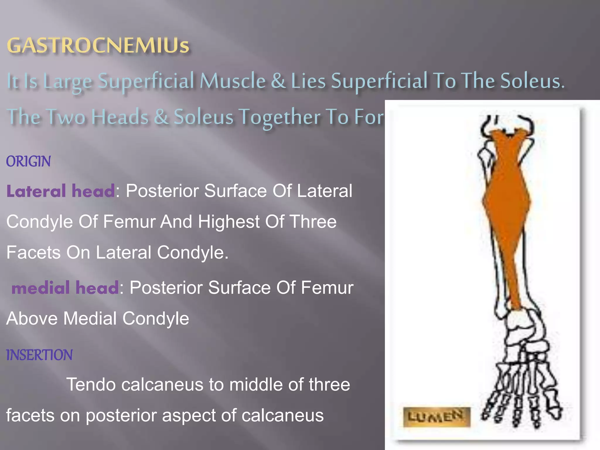
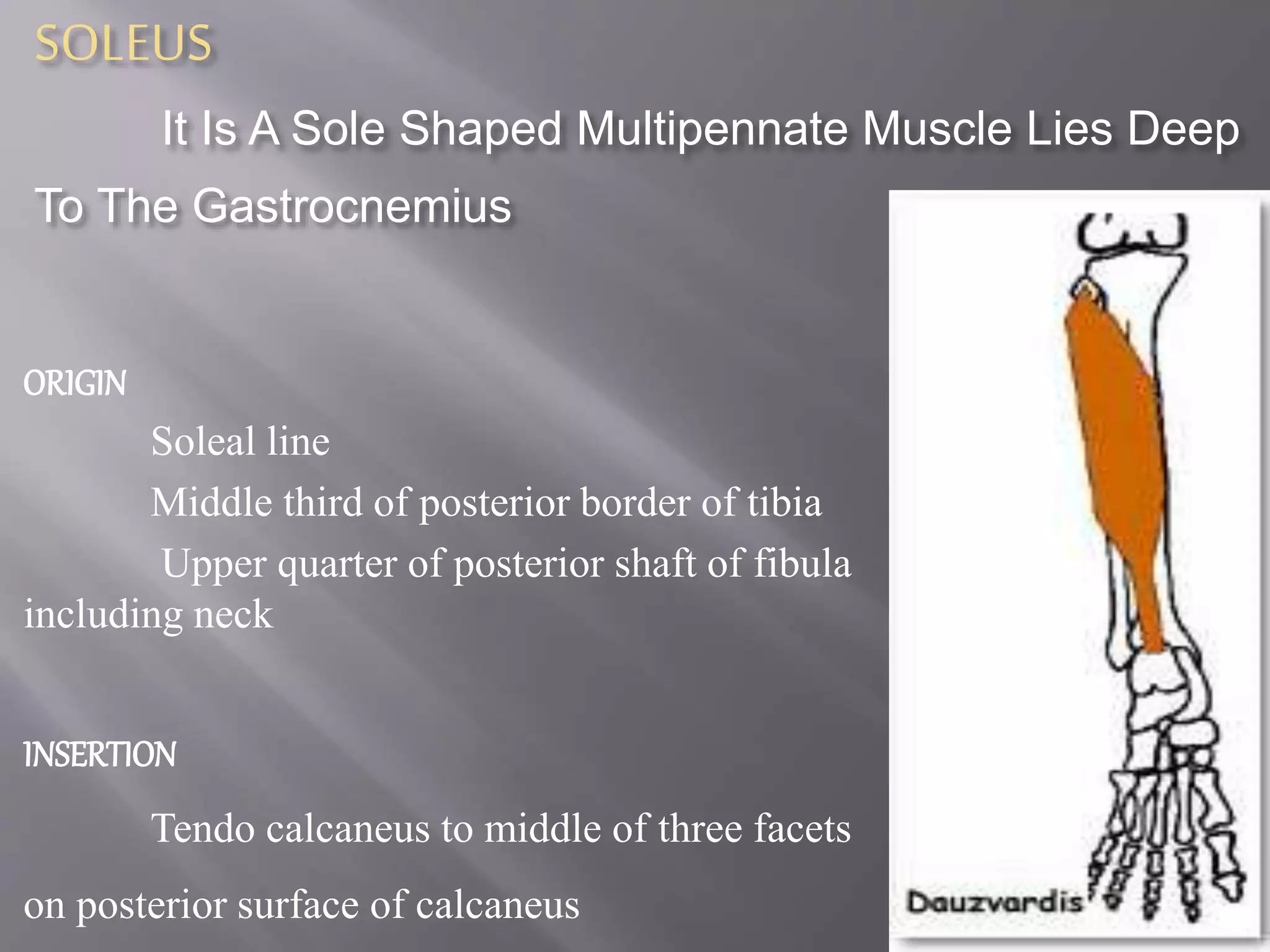
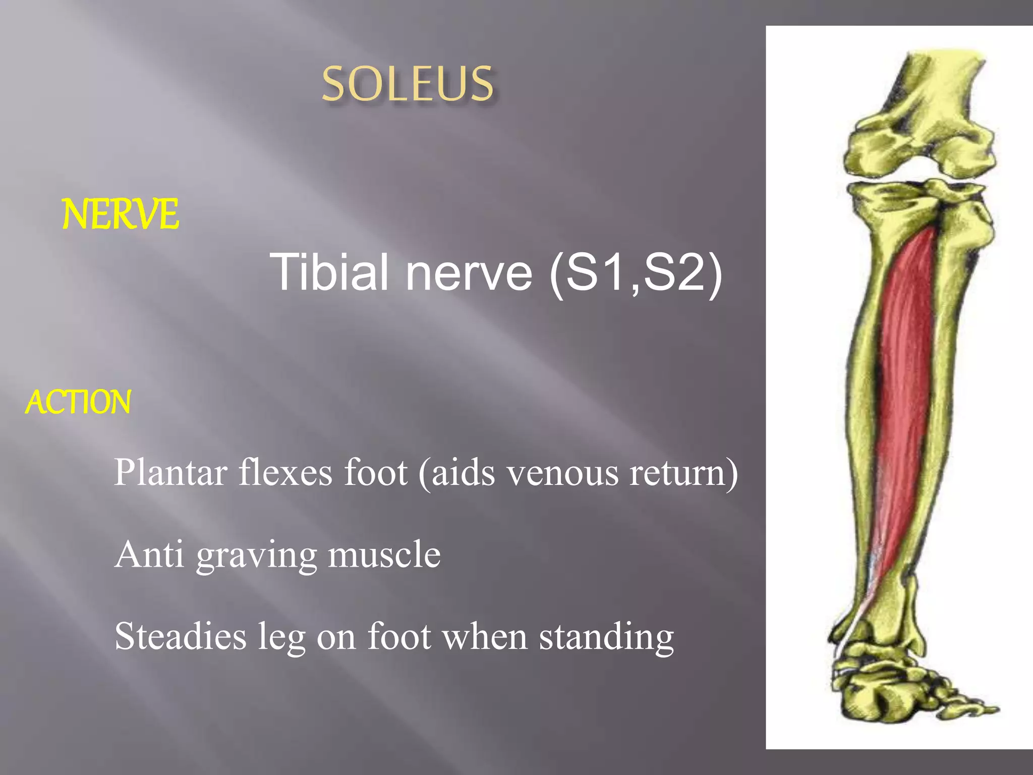
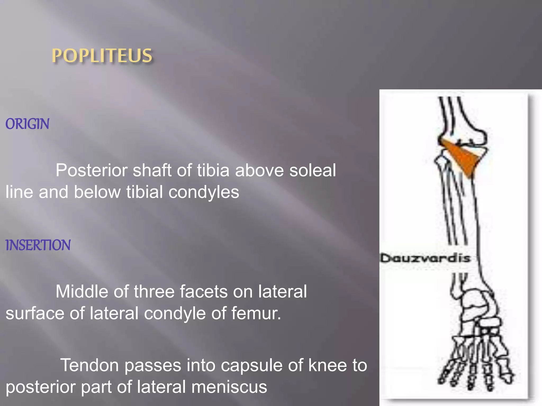
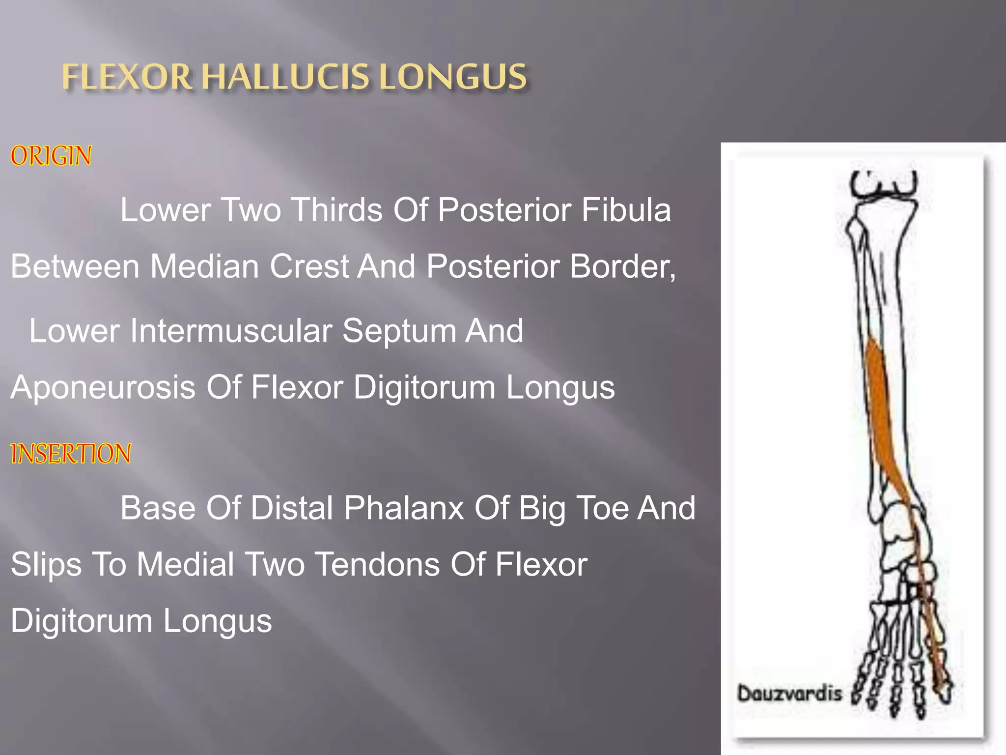
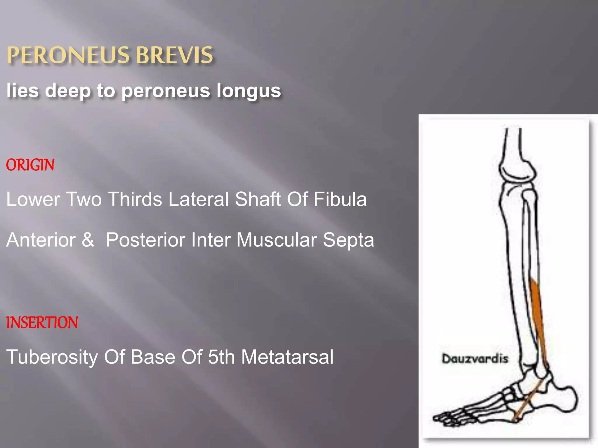
POSTERIOR & LATERAL COMPARTMENT
OF CALF
Presented By,
Posterior compartment of calf
muscles
Superficial set muscles
Gastrocnemius
Soleus
Plantaris
POPLITEUS
FLEXOR HALLUCIS LONGUS
FLEXOR DIGITORUM LONGUS
TIBIALIS POSTERIOR
GASTROCNEMIUs
It Is Large Superficial Muscle& Lies Superficial ToThe Soleus.
The Two Heads & SoleusTogether To Form Triceps Surae
ORIGIN
Lateral head: Posterior Surface Of Lateral
Condyle Of Femur And Highest Of Three
Facets On Lateral Condyle.
medial head: Posterior Surface Of Femur
Above Medial Condyle
INSERTION
Tendo calcaneus to middle of three
facets on posterior aspect of calcaneus
GASTROCNEMIUs
NERVE
Tibial nerve (S1, S2)
Action
Plantar flexes foot.
Flexes knee
SOLEUS
It Is A Sole Shaped Multipennate Muscle Lies Deep
To The Gastrocnemius
ORIGIN
Soleal line
Middle third of posterior border of tibia
Upper quarter of posterior shaft of fibula
including neck
INSERTION
Tendo calcaneus to middle of three facets
on posterior surface of calcaneus
SOLEUS
NERVE
Tibial nerve (S1,S2)
ACTION
Plantar flexes foot (aids venous return)
Anti graving muscle
Steadies leg on foot when standing
plantaris
it vestigeal in human beings. It has a short belly & a long tendon
ORIGIN
Lateral supracondylar ridge of femur
above lateral head of gastrocnemius and
oblique popliteal ligament of knee joint
INSERTION
Tendo calcaneus (medial side, deep
to gastrocnemius tendon)
PLANTARIS
NERVE
Tibial nerve (S1, 2)
ACTION
Plantar flexes foot and flexes knee
Assist gastrocnemius in action
POPLITEUS
ORIGIN
Posterior shaft of tibia above soleal
line and below tibial condyles
INSERTION
Middle of three facets on lateral
surface of lateral condyle of femur.
Tendon passes into capsule of knee to
posterior part of lateral meniscus
POPLITEUS
NERVE
Tibial nerve (L5,S1)
ACTION
Unlocks extended knee by lateral rotation of
femur on tibia.
Pulls back lateral meniscus
FLEXOR HALLUCIS LONGUS
Lower Two Thirds Of Posterior Fibula
Between Median Crest And Posterior Border,
Lower Intermuscular Septum And
Aponeurosis Of Flexor Digitorum Longus
Base Of Distal Phalanx Of Big Toe And
Slips To Medial Two Tendons Of Flexor
Digitorum Longus
FLEXORHALLUCIS LONGUS
NERVE
Tibial Nerve
ACTION
Flexes Distal Phalanx Of Big Toe
Flexes Foot At Ankle,
Supports Medial Longitudinal Arch
FLEXOR DIGITORUM LONGUS
Posterior Shaft Of Tibia Below Sole
Al Line And By Broad Aponeurosis From
Fibula
Base Of Distal Phalanges Of Lateral Four
Toes
FLEXORDIGITORUMLONGUS
NERVE
Tibial nerve (S1, S2)
ACTION
Flexes distal phalanges of lateral four toes
and foot at ankle.
Supports lateral longitudinal arch
TIBIALIS POSTERIOR
Origin
Upper Half Of Posterior Shaft Of Tibia
Upper Half Of Fibula Between Medial
Nerve Crest Interosseous Border, And
Interosseous Membrane
INSERTION
Tuberosity Of Navicular Bone And All
Tarsal Bones (Except Talus) And Spring
Ligament
NERVE
Tibial nerve (L4, L5)
ACTION
Plantar flexes and inverts foot
Supports medial longitudinal arch of foot
 Peroneus longus
 Peroneus brevis
Peroneus Longus
it is superficial to peroneus brevis
Origin
Head Of Fibula, Upper 1/2 - 2/3 Of
Lateral Fibular Shaft Surface; Also
Anterior And Posterior Intermuscular
Septa Of Leg
Insertion
Plantar Posterolateral Aspect Of Medial
Cuneiform And Lateral Side Of 1st
Innervation
Superficial Peroneal Nerve (L5, S1, S2); May Also
Receive Additional Innervation From Common Or Deep Peroneal
Nerves (L5, S1, S2)
ArterialSupply
Anterior Tibial And Peroneal Arteries
Action
Everts foot and plantar flexes ankle;
also helps to support the transverse arch of
the foot
PERONEUS BREVIS
lies deep to peroneus longus
ORIGIN
Lower Two Thirds Lateral Shaft Of Fibula
Anterior & Posterior Inter Muscular Septa
INSERTION
Tuberosity Of Base Of 5th Metatarsal
ACTION
Weak plantar flexor of foot
evertor of foot .
Supports lateral longitudinal arch
NERVE
Superficial peroneal nerve (L5, S1)
muscles of lower limb

muscles of lower limb

  • 1.
    POSTERIOR & LATERALCOMPARTMENT OF CALF Presented By,
  • 2.
    Posterior compartment ofcalf muscles Superficial set muscles Gastrocnemius Soleus Plantaris
  • 3.
    POPLITEUS FLEXOR HALLUCIS LONGUS FLEXORDIGITORUM LONGUS TIBIALIS POSTERIOR
  • 5.
    GASTROCNEMIUs It Is LargeSuperficial Muscle& Lies Superficial ToThe Soleus. The Two Heads & SoleusTogether To Form Triceps Surae ORIGIN Lateral head: Posterior Surface Of Lateral Condyle Of Femur And Highest Of Three Facets On Lateral Condyle. medial head: Posterior Surface Of Femur Above Medial Condyle INSERTION Tendo calcaneus to middle of three facets on posterior aspect of calcaneus
  • 6.
    GASTROCNEMIUs NERVE Tibial nerve (S1,S2) Action Plantar flexes foot. Flexes knee
  • 7.
    SOLEUS It Is ASole Shaped Multipennate Muscle Lies Deep To The Gastrocnemius ORIGIN Soleal line Middle third of posterior border of tibia Upper quarter of posterior shaft of fibula including neck INSERTION Tendo calcaneus to middle of three facets on posterior surface of calcaneus
  • 8.
    SOLEUS NERVE Tibial nerve (S1,S2) ACTION Plantarflexes foot (aids venous return) Anti graving muscle Steadies leg on foot when standing
  • 9.
    plantaris it vestigeal inhuman beings. It has a short belly & a long tendon ORIGIN Lateral supracondylar ridge of femur above lateral head of gastrocnemius and oblique popliteal ligament of knee joint INSERTION Tendo calcaneus (medial side, deep to gastrocnemius tendon)
  • 10.
    PLANTARIS NERVE Tibial nerve (S1,2) ACTION Plantar flexes foot and flexes knee Assist gastrocnemius in action
  • 12.
    POPLITEUS ORIGIN Posterior shaft oftibia above soleal line and below tibial condyles INSERTION Middle of three facets on lateral surface of lateral condyle of femur. Tendon passes into capsule of knee to posterior part of lateral meniscus
  • 13.
    POPLITEUS NERVE Tibial nerve (L5,S1) ACTION Unlocksextended knee by lateral rotation of femur on tibia. Pulls back lateral meniscus
  • 14.
    FLEXOR HALLUCIS LONGUS LowerTwo Thirds Of Posterior Fibula Between Median Crest And Posterior Border, Lower Intermuscular Septum And Aponeurosis Of Flexor Digitorum Longus Base Of Distal Phalanx Of Big Toe And Slips To Medial Two Tendons Of Flexor Digitorum Longus
  • 15.
    FLEXORHALLUCIS LONGUS NERVE Tibial Nerve ACTION FlexesDistal Phalanx Of Big Toe Flexes Foot At Ankle, Supports Medial Longitudinal Arch
  • 16.
    FLEXOR DIGITORUM LONGUS PosteriorShaft Of Tibia Below Sole Al Line And By Broad Aponeurosis From Fibula Base Of Distal Phalanges Of Lateral Four Toes
  • 17.
    FLEXORDIGITORUMLONGUS NERVE Tibial nerve (S1,S2) ACTION Flexes distal phalanges of lateral four toes and foot at ankle. Supports lateral longitudinal arch
  • 18.
    TIBIALIS POSTERIOR Origin Upper HalfOf Posterior Shaft Of Tibia Upper Half Of Fibula Between Medial Nerve Crest Interosseous Border, And Interosseous Membrane INSERTION Tuberosity Of Navicular Bone And All Tarsal Bones (Except Talus) And Spring Ligament
  • 19.
    NERVE Tibial nerve (L4,L5) ACTION Plantar flexes and inverts foot Supports medial longitudinal arch of foot
  • 20.
     Peroneus longus Peroneus brevis
  • 21.
    Peroneus Longus it issuperficial to peroneus brevis Origin Head Of Fibula, Upper 1/2 - 2/3 Of Lateral Fibular Shaft Surface; Also Anterior And Posterior Intermuscular Septa Of Leg Insertion Plantar Posterolateral Aspect Of Medial Cuneiform And Lateral Side Of 1st
  • 22.
    Innervation Superficial Peroneal Nerve(L5, S1, S2); May Also Receive Additional Innervation From Common Or Deep Peroneal Nerves (L5, S1, S2) ArterialSupply Anterior Tibial And Peroneal Arteries Action Everts foot and plantar flexes ankle; also helps to support the transverse arch of the foot
  • 23.
    PERONEUS BREVIS lies deepto peroneus longus ORIGIN Lower Two Thirds Lateral Shaft Of Fibula Anterior & Posterior Inter Muscular Septa INSERTION Tuberosity Of Base Of 5th Metatarsal
  • 24.
    ACTION Weak plantar flexorof foot evertor of foot . Supports lateral longitudinal arch NERVE Superficial peroneal nerve (L5, S1)