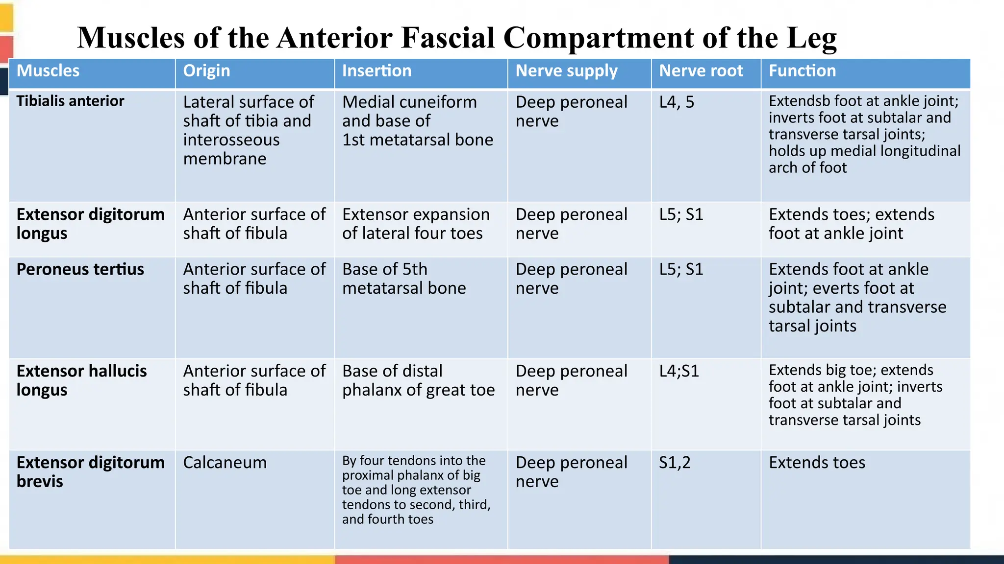
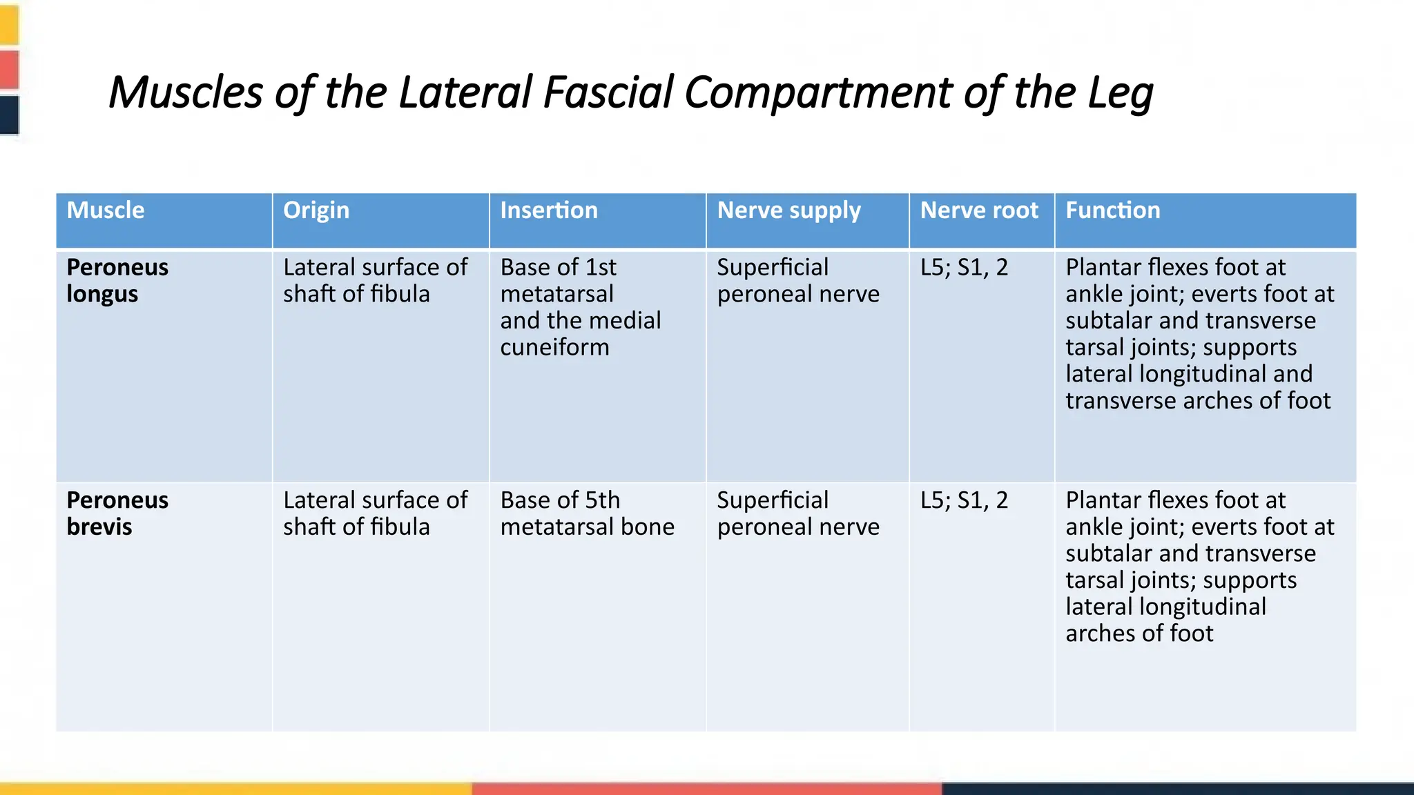
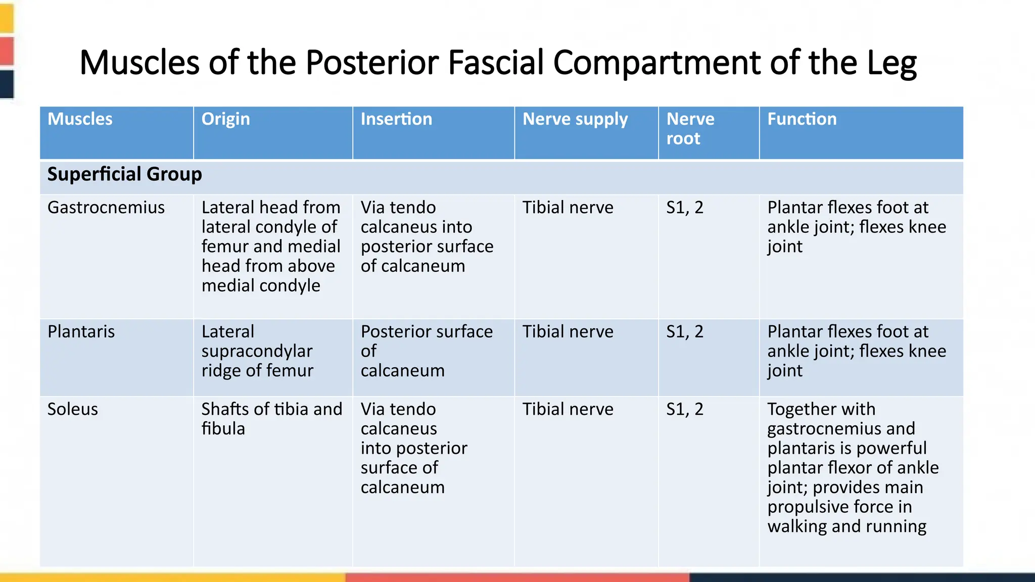
Fascial Compartments
of the Leg
Presented to:- Dr. Sahibzada Saifullah Khalid,
Presented by:- • Zainab Tariq
• Zainab Batool
• Umme Aimen
• Wijh-ur-Rehman
• Hamza Ahmad
OUTLINES
 Fascia of Leg
 The front of Leg
• Skin
• Anterior facial compartment ( Muscles,Nerve supply ,etc)
 Lateral side of leg
• Lateral facial compartment
 The Back of Leg
• Skin
• Posterior compartment of leg
Fascia of the leg
The deep fascia surrounds the leg and is
continuous above with the deep fascia of the
thigh. Below the tibial condyles, it is attached
to the periosteum on the anterior and medial
borders of the tibia.
Interosseous Membrane:
The interosseous membrane binds the tibia and
fibula together and provides attachment for
neighbouring muscles
Fascia of leg ( Cont....)
• Retinacula of the Ankle:
The retinacula are thickenings of the deep fascia that keep the long tendons around the ankle
joint in position and act as pulleys.
• Superior Extensor Retinaculum:
Superior extensor retinaculum is attached to the distal ends of the anterior borders of the fibula
and the tibia.
• Inferior Extensor Retinaculum:
The inferior extensor retinaculum is a Y-shaped band located in front of the ankle joint
• Flexor Retinaculum:
The flexor retinaculum extends from the medial malleolus downward and backward to be
attached to the medial surface of the calcaneum.
cont...
• Superior Peroneal Retinaculum:
The superior peroneal retinaculum connects the lateral malleolus to the lateral surface of the
calcaneum.
• Inferior Peroneal Retinaculum:
The inferior peroneal retinaculum binds the tendons of the peroneus longus and brevis muscles to
the lateral side of the calcaneum.
The Front of Leg
Skin Of Leg:
o Cutaneous Nerves
The lateral cutaneous nerve of the calf, a branch of the common peroneal nerve,
supplies the skin on the upper part of the lateral surface of the leg.
The superficial peroneal nerve, a branch of the common peroneal nerve, supplies
the skin of the lower part of the anterolateral surface of the leg.
The saphenous nerve, a branch of the femoral nerve, supplies the skin on the
anteromedial surface of the leg.
Cont....
• Superficial Veins:
Numerous small veins curve
around the medial aspect of the
leg and ultimately drain into the
great saphenous vein
Cont....
• Lymph vessels
• The greater part of the lymph from the skin and superficial fascia
on the front of the leg drains upward and medially in vessels that
follow the great saphenous vein, to end in the vertical group of
superficial inguinal lymph nodes . A small amount of lymph from
the upper lateral part of the front of the leg may pass via vessels
that accompany the small saphenous vein and drain into the
popliteal nodes
Contents of the Anterior Fascial Compartment
of the Leg
Muscles
• Tibialis anterior, Extensor digitorium longus
• Perioneus tertius, Extensor hallucis longus
• Extensor digitorium brevis
Nerve supply • Deep Peroneal Nerve
Blood Supply • Anterior Tibial artery
Muscles of the Anterior Fascial Compartment of the Leg
Muscles Origin Insertion Nerve supply Nerve root Function
Tibialis anterior Lateral surface of
shaft of tibia and
interosseous
membrane
Medial cuneiform
and base of
1st metatarsal bone
Deep peroneal
nerve
L4, 5 Extendsb foot at ankle joint;
inverts foot at subtalar and
transverse tarsal joints;
holds up medial longitudinal
arch of foot
Extensor digitorum
longus
Anterior surface of
shaft of fibula
Extensor expansion
of lateral four toes
Deep peroneal
nerve
L5; S1 Extends toes; extends
foot at ankle joint
Peroneus tertius Anterior surface of
shaft of fibula
Base of 5th
metatarsal bone
Deep peroneal
nerve
L5; S1 Extends foot at ankle
joint; everts foot at
subtalar and transverse
tarsal joints
Extensor hallucis
longus
Anterior surface of
shaft of fibula
Base of distal
phalanx of great toe
Deep peroneal
nerve
L4;S1 Extends big toe; extends
foot at ankle joint; inverts
foot at subtalar and
transverse tarsal joints
Extensor digitorum
brevis
Calcaneum By four tendons into the
proximal phalanx of big
toe and long extensor
tendons to second, third,
and fourth toes
Deep peroneal
nerve
S1,2 Extends toes
Contents of the Lateral Fascial Compartment
of the Leg
Muscles
Nerve supply
• Peroneus longus and peroneus brevis
• Superficial peroneal nerve
Blood supply • Branches from the peroneal artery
Muscles of the Lateral Fascial Compartment of the Leg
Muscle Origin Insertion Nerve supply Nerve root Function
Peroneus
longus
Lateral surface of
shaft of fibula
Base of 1st
metatarsal
and the medial
cuneiform
Superficial
peroneal nerve
L5; S1, 2 Plantar flexes foot at
ankle joint; everts foot at
subtalar and transverse
tarsal joints; supports
lateral longitudinal and
transverse arches of foot
Peroneus
brevis
Lateral surface of
shaft of fibula
Base of 5th
metatarsal bone
Superficial
peroneal nerve
L5; S1, 2 Plantar flexes foot at
ankle joint; everts foot at
subtalar and transverse
tarsal joints; supports
lateral longitudinal
arches of foot
The Back of the Leg
Skin
Cutaneous Nerves
The posterior cutaneous nerve of the thigh descends on the back of the thigh. In the popliteal fossa, it supplies
the skin over the popliteal fossa and the upper part of the back of the leg.
The lateral cutaneous nerve of the calf, a branch of the common peroneal nerve , supplies the skin on the upper
part of the posterolateral surface of the leg.
The sural nerve, a branch of the tibial nerve, supplies the skin on the lower part of the posterolat_x0002_eral
surface of the leg.
The saphenous nerve, a branch of the femoral nerve, gives off branches that supply the skin on the posteromedial
surface of the leg .
Contents of the Posterior Fascial Compartment of the Leg
Muscles Nerve Supply Blood Supply
Superficial group
Tibial Nerve Posterior Tibial artery
Deep group
Muscles of the Posterior Fascial Compartment of the Leg
Muscles Origin Insertion Nerve supply Nerve
root
Function
Superficial Group
Gastrocnemius Lateral head from
lateral condyle of
femur and medial
head from above
medial condyle
Via tendo
calcaneus into
posterior surface
of calcaneum
Tibial nerve S1, 2 Plantar flexes foot at
ankle joint; flexes knee
joint
Plantaris Lateral
supracondylar
ridge of femur
Posterior surface
of
calcaneum
Tibial nerve S1, 2 Plantar flexes foot at
ankle joint; flexes knee
joint
Soleus Shafts of tibia and
fibula
Via tendo
calcaneus
into posterior
surface of
calcaneum
Tibial nerve S1, 2 Together with
gastrocnemius and
plantaris is powerful
plantar flexor of ankle
joint; provides main
propulsive force in
walking and running
Cont....
Muscles Origin Insertion Nerve
supply
Nerve
root
Action
Deep group
Popliteus Lateral surface of
lateral condyle of
femur
Posterior surface
of shaft of tibia
above soleal line
Tibial nerve L4, 5; S1 Flexes leg at knee joint; unlocks
knee joint by lateral rotation of
femur on tibia and slackens
ligaments of joint
Flexor digitorum
longus
Posterior surface of
shaft of tibia
Bases of distal
phalanges of
lateral four toes
Tibial nerve S2, 3 Flexes distal phalanges of lateral
four toes; plantar flexes foot at ankle
joint; supports medial and lateral
longitudinal arches of foot
Flexor hallucis
longus
Posterior surface of
shaft of fibula
Base of distal
phalanx of big toe
Tibial nerve S2, 3 Flexes distal phalanx of big toe;
plantar flexes foot at ankle joint;
supports medial longitudinal arch of
foot
Tibialis
posterior
Posterior surface
of shafts of tibia
and fibula and
interosseous
membrane
Tuberosity of
navicular bone
and other
neighboring bones
Tibial nerve L4, 5 Plantar flexes foot at ankle joint;
inverts foot at subtalar and
transverse tarsal joints; supports
medial longitudinal arch of foot
Reference :
 Snells Clinical Anatomy by region, Edition 09,
Richard S Snells, MD PHD
Thats all from our side
anatomy of lower limb with nerve and blood supply.pptx

anatomy of lower limb with nerve and blood supply.pptx

  • 2.
    Fascial Compartments of theLeg Presented to:- Dr. Sahibzada Saifullah Khalid, Presented by:- • Zainab Tariq • Zainab Batool • Umme Aimen • Wijh-ur-Rehman • Hamza Ahmad
  • 3.
    OUTLINES  Fascia ofLeg  The front of Leg • Skin • Anterior facial compartment ( Muscles,Nerve supply ,etc)  Lateral side of leg • Lateral facial compartment  The Back of Leg • Skin • Posterior compartment of leg
  • 4.
    Fascia of theleg The deep fascia surrounds the leg and is continuous above with the deep fascia of the thigh. Below the tibial condyles, it is attached to the periosteum on the anterior and medial borders of the tibia. Interosseous Membrane: The interosseous membrane binds the tibia and fibula together and provides attachment for neighbouring muscles
  • 5.
    Fascia of leg( Cont....) • Retinacula of the Ankle: The retinacula are thickenings of the deep fascia that keep the long tendons around the ankle joint in position and act as pulleys. • Superior Extensor Retinaculum: Superior extensor retinaculum is attached to the distal ends of the anterior borders of the fibula and the tibia. • Inferior Extensor Retinaculum: The inferior extensor retinaculum is a Y-shaped band located in front of the ankle joint • Flexor Retinaculum: The flexor retinaculum extends from the medial malleolus downward and backward to be attached to the medial surface of the calcaneum.
  • 7.
    cont... • Superior PeronealRetinaculum: The superior peroneal retinaculum connects the lateral malleolus to the lateral surface of the calcaneum. • Inferior Peroneal Retinaculum: The inferior peroneal retinaculum binds the tendons of the peroneus longus and brevis muscles to the lateral side of the calcaneum.
  • 8.
    The Front ofLeg Skin Of Leg: o Cutaneous Nerves The lateral cutaneous nerve of the calf, a branch of the common peroneal nerve, supplies the skin on the upper part of the lateral surface of the leg. The superficial peroneal nerve, a branch of the common peroneal nerve, supplies the skin of the lower part of the anterolateral surface of the leg. The saphenous nerve, a branch of the femoral nerve, supplies the skin on the anteromedial surface of the leg.
  • 9.
    Cont.... • Superficial Veins: Numeroussmall veins curve around the medial aspect of the leg and ultimately drain into the great saphenous vein
  • 10.
    Cont.... • Lymph vessels •The greater part of the lymph from the skin and superficial fascia on the front of the leg drains upward and medially in vessels that follow the great saphenous vein, to end in the vertical group of superficial inguinal lymph nodes . A small amount of lymph from the upper lateral part of the front of the leg may pass via vessels that accompany the small saphenous vein and drain into the popliteal nodes
  • 11.
    Contents of theAnterior Fascial Compartment of the Leg Muscles • Tibialis anterior, Extensor digitorium longus • Perioneus tertius, Extensor hallucis longus • Extensor digitorium brevis Nerve supply • Deep Peroneal Nerve Blood Supply • Anterior Tibial artery
  • 12.
    Muscles of theAnterior Fascial Compartment of the Leg Muscles Origin Insertion Nerve supply Nerve root Function Tibialis anterior Lateral surface of shaft of tibia and interosseous membrane Medial cuneiform and base of 1st metatarsal bone Deep peroneal nerve L4, 5 Extendsb foot at ankle joint; inverts foot at subtalar and transverse tarsal joints; holds up medial longitudinal arch of foot Extensor digitorum longus Anterior surface of shaft of fibula Extensor expansion of lateral four toes Deep peroneal nerve L5; S1 Extends toes; extends foot at ankle joint Peroneus tertius Anterior surface of shaft of fibula Base of 5th metatarsal bone Deep peroneal nerve L5; S1 Extends foot at ankle joint; everts foot at subtalar and transverse tarsal joints Extensor hallucis longus Anterior surface of shaft of fibula Base of distal phalanx of great toe Deep peroneal nerve L4;S1 Extends big toe; extends foot at ankle joint; inverts foot at subtalar and transverse tarsal joints Extensor digitorum brevis Calcaneum By four tendons into the proximal phalanx of big toe and long extensor tendons to second, third, and fourth toes Deep peroneal nerve S1,2 Extends toes
  • 13.
    Contents of theLateral Fascial Compartment of the Leg Muscles Nerve supply • Peroneus longus and peroneus brevis • Superficial peroneal nerve Blood supply • Branches from the peroneal artery
  • 14.
    Muscles of theLateral Fascial Compartment of the Leg Muscle Origin Insertion Nerve supply Nerve root Function Peroneus longus Lateral surface of shaft of fibula Base of 1st metatarsal and the medial cuneiform Superficial peroneal nerve L5; S1, 2 Plantar flexes foot at ankle joint; everts foot at subtalar and transverse tarsal joints; supports lateral longitudinal and transverse arches of foot Peroneus brevis Lateral surface of shaft of fibula Base of 5th metatarsal bone Superficial peroneal nerve L5; S1, 2 Plantar flexes foot at ankle joint; everts foot at subtalar and transverse tarsal joints; supports lateral longitudinal arches of foot
  • 15.
    The Back ofthe Leg Skin Cutaneous Nerves The posterior cutaneous nerve of the thigh descends on the back of the thigh. In the popliteal fossa, it supplies the skin over the popliteal fossa and the upper part of the back of the leg. The lateral cutaneous nerve of the calf, a branch of the common peroneal nerve , supplies the skin on the upper part of the posterolateral surface of the leg. The sural nerve, a branch of the tibial nerve, supplies the skin on the lower part of the posterolat_x0002_eral surface of the leg. The saphenous nerve, a branch of the femoral nerve, gives off branches that supply the skin on the posteromedial surface of the leg .
  • 16.
    Contents of thePosterior Fascial Compartment of the Leg Muscles Nerve Supply Blood Supply Superficial group Tibial Nerve Posterior Tibial artery Deep group
  • 17.
    Muscles of thePosterior Fascial Compartment of the Leg Muscles Origin Insertion Nerve supply Nerve root Function Superficial Group Gastrocnemius Lateral head from lateral condyle of femur and medial head from above medial condyle Via tendo calcaneus into posterior surface of calcaneum Tibial nerve S1, 2 Plantar flexes foot at ankle joint; flexes knee joint Plantaris Lateral supracondylar ridge of femur Posterior surface of calcaneum Tibial nerve S1, 2 Plantar flexes foot at ankle joint; flexes knee joint Soleus Shafts of tibia and fibula Via tendo calcaneus into posterior surface of calcaneum Tibial nerve S1, 2 Together with gastrocnemius and plantaris is powerful plantar flexor of ankle joint; provides main propulsive force in walking and running
  • 18.
    Cont.... Muscles Origin InsertionNerve supply Nerve root Action Deep group Popliteus Lateral surface of lateral condyle of femur Posterior surface of shaft of tibia above soleal line Tibial nerve L4, 5; S1 Flexes leg at knee joint; unlocks knee joint by lateral rotation of femur on tibia and slackens ligaments of joint Flexor digitorum longus Posterior surface of shaft of tibia Bases of distal phalanges of lateral four toes Tibial nerve S2, 3 Flexes distal phalanges of lateral four toes; plantar flexes foot at ankle joint; supports medial and lateral longitudinal arches of foot Flexor hallucis longus Posterior surface of shaft of fibula Base of distal phalanx of big toe Tibial nerve S2, 3 Flexes distal phalanx of big toe; plantar flexes foot at ankle joint; supports medial longitudinal arch of foot Tibialis posterior Posterior surface of shafts of tibia and fibula and interosseous membrane Tuberosity of navicular bone and other neighboring bones Tibial nerve L4, 5 Plantar flexes foot at ankle joint; inverts foot at subtalar and transverse tarsal joints; supports medial longitudinal arch of foot
  • 19.
    Reference :  SnellsClinical Anatomy by region, Edition 09, Richard S Snells, MD PHD
  • 20.