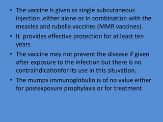
MUMPS VIRUS
• Mumps is an acute infectious disease
commonly affecting children and
characterised by non suppurative enlargement
of the parotid glands
• As epidermic parotitis it had been described
by hippocrates in the fifth century BC
• The viral origins of mumps was demonstrated
by johnson and goodpasture (1934) by its
experimental transmissionto monkeys
• 1945 - Habel cultivated the virus in
embryonated eggs
• 1955 - Henle and Deinhardt grew it in tissue
culture
PROPERTIES
• The mumps virus is a typical paramyxovirus
possessing both HN and F proteins
• It agglutinates the erythrocytes of fowls,
guinea pigs , humans , and many other species
• Hemagglutination is followed by hemolysis
and elution at 37⁰C
• The virus can be grown in chick embryos- in
the amniotic cavity for primary isolation and
allantonic cavity after adaption .
• The mumps virus is labile , being rapidly
inactivated at room temperature or by the
exposure to formaldehyde ether or ultraviolet
light
• It can be preserved at -70⁰C or by
lyophilisation
• The mumps virus is antigentically stable and
only one serotype exists.
• Two complement fixing antigens can be
recognised as in influenza viruses – the soluble
(S) antigen and the viral (V)antigen
EPIDERMIOLOGY
• Mumps is endemic world wide but has
become less common in the advanced nations
due to immunisation
• It often occurs as epidemic in children 5-15
years of age and also in young people living in
groups such as in army camps, house hold
spread is common
• Humans are the only natural host
• Eggs are inoculated at 6-8 days and incubated
at 35⁰C for five days before harvesting.
• Cell cultures are better suited for isolation –
primary monkey kidney being the preffered
cell.
• The cytopathic effect is slow and consists of
syncytium formation and the presence of
acidophilic cytoplasmic inclusions.
• Growth is best identified by hemadsorption
• The source of infection is a patients in the late
incubation or early clinical stage of illness
• No humans carrier or animal reservoirs exist
• Infection is transmitted by direct contact
,airborne droplets or fomites contaminated
with saliva and also possibly urine
• The virus is detectable in the saliva for about a
week before and a week or two after onset of
parotitis.
• Peak infectivity is about a day or two before
parotitis become evident and subsides rapidly
therafter
• Antibodies to the V antigen take about a
month to appear but persist for years.
• The antihemagglutinin antibody correlates
well with immunity to infection
• Even subclinical infections lead to HI antibody
and resistance to infection.
• As antibodies are widespread in the
population passive immunity protects the
newborns.
• Mumps is therfore very rare before six months
of age
• The virus is also shed in the urine for up to
two weeks after the clinical symtoms
begin,though its role in the transmission of
infection is not clear
• One attack of mumps confer lasting immunity
so that second attacks do not occur
IMMUNITY
• Infection leads to antibody response against
both the internal (S) and surface (V) antigens.
• Antibodies to the S antigen appear
early,within 3-7 days after the onset of
symtoms,but disapppear after about six
months
• Demonstration of antibody to the S antigen
indicates current or recent infection.
• Cell mediated immunity is developed
following infection but its significance is not
known
• Interferon also appears early in mumps
infection
LABORATORY DIAGNOSIS
• The typical case of mumps needs no
laboratory confirmation but it may be
essential in atypical infection and where
meningitis or other systemic involvement is
the sole manifestation
• The diagnosis may be established by virus
isolation and serological tests.
• The virus may be isolated from the saliva ,urine
or CSF; from the saliva within4-5 days , urine up
to two weeks and CSF 8-9 days after the onset of
illness.
• The specimens have to be inoculated soon after
collection as the virus is labile.
• The prepared specimen is inoculated into the
monkey kidney cell cultures.
• Humans amnion or HeLa cells are also suitable.
• Virus growth can be dectected by hemadsorption
and identified byhemadsorption inhibition using
specific antiserum
• Cytopathic changes are not reliable .
• Isolation may take 1-2 weeks.
• More rapid results can be obtained by
immunofluorescence testingof infected cell
culture
• This may become positive as early as2-3 days
afterinoculation
• Isolation can also be made by inoculation in to
six- to-eight-day - old chick embryos by the
amniotic route and testing the amniotic fluid
after 5-6 days for hemagglutination inhibition
using specific antisera.
• Egg inoculation is less sensitive than the cell
cultures for isolation
• Serological diagnosis depends on the
demonstration of rise in titre of antibodies in
paired serum samples.
• The CF and HI test are commonly employed
but cross-reactions with parainfluenza viruses
cause promblems.
• IgM – ELISAis usefulin this respect because
cross- reacting antibodies are IgG and do not
interfere with IgM – ELISA
• A positive CF test for antibody to the S
antigen in the acute phase serum is
preumptive evidence of current infection
PROPHYLAXIS
• An effective live virus vaccine is available against
mumps
• The Jeryl-Lynn strains of mumps virus, attenuated
by passage in eggs and grown in chick embryo
fibroblast culture is used as the vaccine.
• It is recommended for use only after one year of
age as maternal antibodies may interfere with the
multiplication of the vacine virus if given earlier.
• Contraindications are pregnancy
,immunodeficiency and hypersensitvityto
neomycin or egg protein
• The vaccine is given as single subcutaneous
injection ,either alone or in combination with the
measles and rubella vaccines (MMR vaccines).
• It provides effective protection for at least ten
years
• The vaccine mey not prevent the disease if given
after exposure to the infection but there is no
contraindicationfor its use in this situvation.
• The mumps immunoglobulin is of no value either
for postexposure prophylaxis or for treatment

mumps virus

  • 1.
    MUMPS VIRUS • Mumpsis an acute infectious disease commonly affecting children and characterised by non suppurative enlargement of the parotid glands • As epidermic parotitis it had been described by hippocrates in the fifth century BC • The viral origins of mumps was demonstrated by johnson and goodpasture (1934) by its experimental transmissionto monkeys
  • 2.
    • 1945 -Habel cultivated the virus in embryonated eggs • 1955 - Henle and Deinhardt grew it in tissue culture
  • 3.
    PROPERTIES • The mumpsvirus is a typical paramyxovirus possessing both HN and F proteins • It agglutinates the erythrocytes of fowls, guinea pigs , humans , and many other species • Hemagglutination is followed by hemolysis and elution at 37⁰C • The virus can be grown in chick embryos- in the amniotic cavity for primary isolation and allantonic cavity after adaption .
  • 4.
    • The mumpsvirus is labile , being rapidly inactivated at room temperature or by the exposure to formaldehyde ether or ultraviolet light • It can be preserved at -70⁰C or by lyophilisation • The mumps virus is antigentically stable and only one serotype exists. • Two complement fixing antigens can be recognised as in influenza viruses – the soluble (S) antigen and the viral (V)antigen
  • 5.
    EPIDERMIOLOGY • Mumps isendemic world wide but has become less common in the advanced nations due to immunisation • It often occurs as epidemic in children 5-15 years of age and also in young people living in groups such as in army camps, house hold spread is common • Humans are the only natural host
  • 6.
    • Eggs areinoculated at 6-8 days and incubated at 35⁰C for five days before harvesting. • Cell cultures are better suited for isolation – primary monkey kidney being the preffered cell. • The cytopathic effect is slow and consists of syncytium formation and the presence of acidophilic cytoplasmic inclusions. • Growth is best identified by hemadsorption
  • 7.
    • The sourceof infection is a patients in the late incubation or early clinical stage of illness • No humans carrier or animal reservoirs exist • Infection is transmitted by direct contact ,airborne droplets or fomites contaminated with saliva and also possibly urine • The virus is detectable in the saliva for about a week before and a week or two after onset of parotitis. • Peak infectivity is about a day or two before parotitis become evident and subsides rapidly therafter
  • 8.
    • Antibodies tothe V antigen take about a month to appear but persist for years. • The antihemagglutinin antibody correlates well with immunity to infection • Even subclinical infections lead to HI antibody and resistance to infection. • As antibodies are widespread in the population passive immunity protects the newborns. • Mumps is therfore very rare before six months of age
  • 9.
    • The virusis also shed in the urine for up to two weeks after the clinical symtoms begin,though its role in the transmission of infection is not clear • One attack of mumps confer lasting immunity so that second attacks do not occur
  • 10.
    IMMUNITY • Infection leadsto antibody response against both the internal (S) and surface (V) antigens. • Antibodies to the S antigen appear early,within 3-7 days after the onset of symtoms,but disapppear after about six months • Demonstration of antibody to the S antigen indicates current or recent infection.
  • 11.
    • Cell mediatedimmunity is developed following infection but its significance is not known • Interferon also appears early in mumps infection
  • 12.
    LABORATORY DIAGNOSIS • Thetypical case of mumps needs no laboratory confirmation but it may be essential in atypical infection and where meningitis or other systemic involvement is the sole manifestation • The diagnosis may be established by virus isolation and serological tests.
  • 13.
    • The virusmay be isolated from the saliva ,urine or CSF; from the saliva within4-5 days , urine up to two weeks and CSF 8-9 days after the onset of illness. • The specimens have to be inoculated soon after collection as the virus is labile. • The prepared specimen is inoculated into the monkey kidney cell cultures. • Humans amnion or HeLa cells are also suitable. • Virus growth can be dectected by hemadsorption and identified byhemadsorption inhibition using specific antiserum
  • 14.
    • Cytopathic changesare not reliable . • Isolation may take 1-2 weeks. • More rapid results can be obtained by immunofluorescence testingof infected cell culture • This may become positive as early as2-3 days afterinoculation • Isolation can also be made by inoculation in to six- to-eight-day - old chick embryos by the amniotic route and testing the amniotic fluid after 5-6 days for hemagglutination inhibition using specific antisera. • Egg inoculation is less sensitive than the cell cultures for isolation
  • 15.
    • Serological diagnosisdepends on the demonstration of rise in titre of antibodies in paired serum samples. • The CF and HI test are commonly employed but cross-reactions with parainfluenza viruses cause promblems. • IgM – ELISAis usefulin this respect because cross- reacting antibodies are IgG and do not interfere with IgM – ELISA • A positive CF test for antibody to the S antigen in the acute phase serum is preumptive evidence of current infection
  • 16.
    PROPHYLAXIS • An effectivelive virus vaccine is available against mumps • The Jeryl-Lynn strains of mumps virus, attenuated by passage in eggs and grown in chick embryo fibroblast culture is used as the vaccine. • It is recommended for use only after one year of age as maternal antibodies may interfere with the multiplication of the vacine virus if given earlier. • Contraindications are pregnancy ,immunodeficiency and hypersensitvityto neomycin or egg protein
  • 17.
    • The vaccineis given as single subcutaneous injection ,either alone or in combination with the measles and rubella vaccines (MMR vaccines). • It provides effective protection for at least ten years • The vaccine mey not prevent the disease if given after exposure to the infection but there is no contraindicationfor its use in this situvation. • The mumps immunoglobulin is of no value either for postexposure prophylaxis or for treatment