Download to read offline
![International Research Journal of Engineering and Technology (IRJET) e-ISSN: 2395-0056
Volume: 04 Issue: 07 | July -2017 www.irjet.net p-ISSN: 2395-0072
© 2017, IRJET | Impact Factor value: 5.181 | ISO 9001:2008 Certified Journal | Page 2912
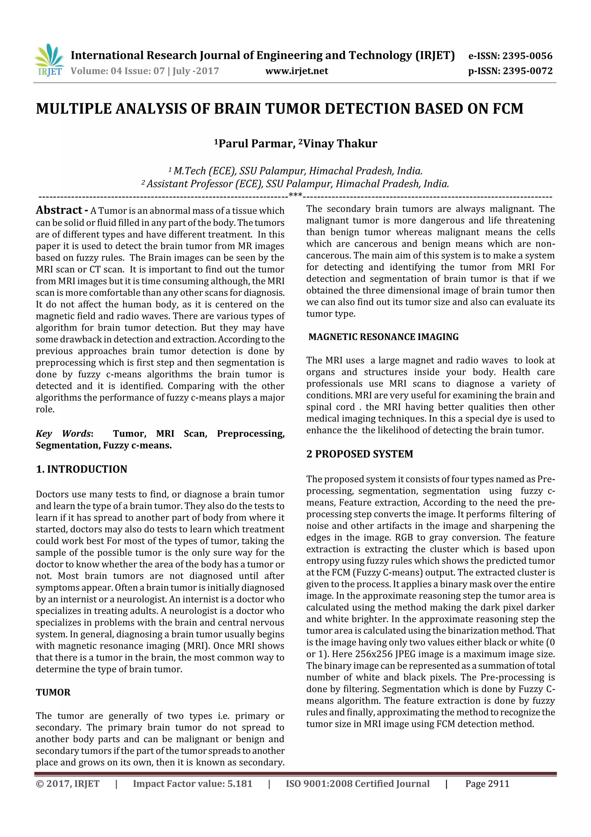
2.1 Flow Diagram For Proposed Method
Fig.1 Flow diagram of Proposed Method with all
processing steps.
2.2 Pre-processing
The pre-processing plays a very important role it is used for
processing the gray scale image by using different
techniques like brightness, filtering and many others. In this
process the white objects are defined from gray and light
items from dark objects. The RGB (red green and blue) to
grey conversion also takes place here. It includes a median
filter for noise deduction. In this process it changes the
brightness of the image and the tumor detection in the MRI
image got easier to identify.
Fig 2 pre-processing
3. Segmentation
In this process the clustering is the set of objects in the same
group which are similar then to the other groups. These are
basically unsupervised method of learning, the algorithm
Fcm are used to define the numberofclusters.Theclustering
are based on the features into k number of groupswhere k is
a positive integer. The clustering is done byentropybyusing
the fuzzy rules.
3.1 Fuzzy c-means
Fuzzy c-means (FCM) it is a method of clustering which
allows one piece of data to belong two or more clusters. In
this the number of clusters are found by the entropy it is
based on the fuzzy rules in this the median value should be
the same as near to the pixels.oneclustervalueand secondly
the cluster value should be subtracted the result should
come zero and then the rules in this the medianvalueshould
be the same as near to the pixels.one cluster value and are
found then the objective function will be zero and then
maximum value get morphed and the result when we get
one we will get a color of a tumor which is detected.in this
we get tumor region and tumor size.
Fig. 3 FCM resulted image
4. CONCLUSION
There are different types of tumors available. They may be
mass in the brain or malignant over the brain. The brain
image dataset is divided into two sets. Training dataset and
testing dataset. Thus, the pre-processing is done by filtering.
Segmentation is done by fuzzy c means algorithm and then
Feature extractions is done and finally the method scans the
RGB or grayscale, converts the image into binary image by
binarization technique and detects the edge of tumor pixels
in the binary image. Also, it calculates the size of the tumor
by calculating the number of white pixels (digit 0) in binary
image. The region of the tumor is based on thearea oftumor.
REFERENCES
[1] A.R. Kavitha, Dr. C.Chellamuthu, Ms. Kavin Rupa, ‟An
Efficient Approach for Brain Tumor Detection Based on
Modified Region Growing and Network in MRI Images”,
Information Forensics and Security, IEEE Transactions on,
Vol.9 (2), May 2012.
[2] R.B.Dubey, M.Hanmandlu, Sr. Member, Shantaram
Vasikarla, ‟ Evaluation of Three Methods for MRI Brain
Tumor segmentation”, IEEE Digital Object Identifier:
10.1109/ITNG.2011.92,2011.
[3] Shaheen Ahmed, Khan M. Iftekharuddin, ‟Efficacy of
Texture, Shape and Intensity Feature Fusion for Posterior
Pre-processing Segmentation Segmentation
using fuzzy
Feature
extraction](https://image.slidesharecdn.com/irjet-v4i7587-170913095649/75/Multiple-Analysis-of-Brain-Tumor-Detection-Based-on-FCM-2-2048.jpg)
![International Research Journal of Engineering and Technology (IRJET) e-ISSN: 2395-0056
Volume: 04 Issue: 07 | July -2017 www.irjet.net p-ISSN: 2395-0072
© 2017, IRJET | Impact Factor value: 5.181 | ISO 9001:2008 Certified Journal | Page 2913
Fossa Tumor Segmentation In MRI”, IEEE Vol (2), pag: 206-
13, Mar 2011.
[4] David Rivest-Henault, Mohamed Cheriet,‟ Unsupervised
MRI segmentation of brain tissuesUsinga local linear
model and set”, Elsevier,Vol 29, Issue 2, pag.243-259,
Mar2011.
[5] Vida Harati, Rasoul Khayati, Abdolreza Farzan, ‟Fully
automated tumor segmentation based on animproved fuzzy
connectedness Algorithm in BrainMR Images”, Elsevier
Ltd,Vol 7, pag: 483-92, May 2011.
[6] Ali Gooya, George Biros Christos Davatzikos, ‟An EM
Algorithm for BrainTumor ImagesRegistration: A Tumor
Growth Modling Based Approach”, IEEE, Vol 2, pag. 375-90,
May 2010.](https://image.slidesharecdn.com/irjet-v4i7587-170913095649/75/Multiple-Analysis-of-Brain-Tumor-Detection-Based-on-FCM-3-2048.jpg)

The document proposes a system to detect brain tumors in MRI images using multiple steps including pre-processing, segmentation using fuzzy c-means clustering, and feature extraction using fuzzy rules. It discusses how pre-processing improves tumor detection, fuzzy c-means segmentation identifies tumor regions and size, and prior approaches have limitations. The proposed system aims to better detect and identify brain tumors in MRI images as compared to other algorithms.