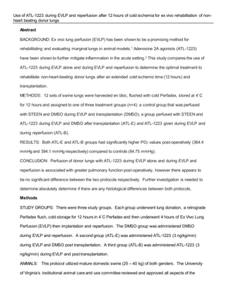
Use of ATL-1223 during EVLP and reperfusion after 12 hours of cold ischemia for ex vivo rehabilitation of non-
heart beating donor lungs
Abstract
BACKGROUND: Ex vivo lung perfusion (EVLP) has been shown to be a promising method for
rehabilitating and evaluating marginal lungs in animal models.1
Adenosine 2A agonists (ATL-1223)
have been shown to further mitigate inflammation in the acute setting.2
This study compares the use of
ATL-1223 during EVLP alone and during EVLP and reperfusion to determine the optimal treatment to
rehabilitate non-heart-beating donor lungs after an extended cold ischemic time (12 hours) and
transplantation.
METHODS: 12 sets of swine lungs were harvested en bloc, flushed with cold Perfadex, stored at 4˚C
for 12 hours and assigned to one of three treatment groups (n=4): a control group that was perfused
with STEEN and DMSO during EVLP and transplantation (DMSO), a group perfused with STEEN and
ATL-1223 during EVLP and DMSO after transplantation (ATL-E) and ATL-1223 given during EVLP and
during reperfusion (ATL-B).
RESULTS: Both ATL-E and ATL-B groups had significantly higher PO2 values post-operatively (364.4
mmHg and 394.1 mmHg respectively) compared to controls (84.75 mmHg).
CONCLUSION: Perfusion of donor lungs with ATL-1223 during EVLP alone and during EVLP and
reperfusion is associated with greater pulmonary function post-operatively, however there appears to
be no significant difference between the two protocols respectively. Further investigation is needed to
determine absolutely determine if there are any histological differences between both protocols.
Methods
STUDY GROUPS: There were three study groups. Each group underwent lung donation, a retrograde
Perfadex flush, cold storage for 12 hours in 4˚C Perfadex and then underwent 4 hours of Ex Vivo Lung
Perfusion (EVLP) then implantation and reperfusion. The DMSO group was administered DMSO
during EVLP and reperfusion. A second group (ATL-E) was administered ATL-1223 (3 ng/kg/min)
during EVLP and DMSO post transplantation. A third group (ATL-B) was administered ATL-1223 (3
ng/kg/min) during EVLP and post transplantation.
ANIMALS: This protocol utilized mature domestic swine (25 – 40 kg) of both genders. The University
of Virginia’s institutional animal care and use committee reviewed and approved all aspects of the
present study. Humane animal care was observed in accordance with the “Guide for Care and Use of
Laboratory Animals” (National Institutes of Health, publication no. 85-23, revised 1985).
CARDIOPULMONARY ARREST AND DONATION: This was an acute procedure, therefore sterile
technique was not necessary. The donor swine were initially anesthetized using Atropine (0.04 mg/kg
IM) and Telazol/Xylazine (4-6/2 mg/kg IM), shaved and transported to the OR. The donor was then
intubated and ventilated on room air. Anesthesia was maintained with 1-2% Isoflurane. A 5 lead EKG
was used to monitor cardioelectric function. Venous access was established by ear vein catheter. The
endotracheal tube was clamped causing pulmonary arrest. After cessation of cardioelectrical function a
15-minute period of warm ischemia was observed, after which ventilation was started again. A
sternotomy was performed. The pulmonary artery (PA) was cleaned and freed from overlying tissue
and a silastic vessel loop was placed around it. The posterior pulmonary ligaments were divided to free
the posterior aspect of the donor lungs. The pericardium was opened and a vessel loop placed around
the take off of the PA. The animal was then heparinized. A 14 gauge catheter was then placed in the
PA and the lung was flushed with clear solutions. The left atrium was then incised to allow the lungs to
flush clear. The thoracic cavity was then filled with frozen saline to cool the lung. The heart and lungs
were then excised and prepared for whole transplantation. The heart was removed and Vitrolife
cannulas were placed in the PA and left atrial (LA) cuff in preparation for EVLP. A retrograde flush was
performed with 1.5 L of Perfadex (4˚C). The lungs were then placed in cold storage for 12 hours in
Perfadex.
PORCINE EVLP: The EVLP circuit was composed of a hard-shell reservoir, membrane gas
exchanger, centrifugal pump and leukocyte depletion filter. It was primed with 1.5 L of Steen Solution,
10,000 IU sodium heparin, 500 mg cefazolin, and 500 mg of methylprednisolone. An endotracheal tube
was placed in the trachea and secured circumferentially with an umbilical tape. The lungs were
subjected to a slow retrograde flush to de-air the PA cannula. Antegrade flow was begun at 150 ml/min
with room temperature perfusate and then slowly warmed to 37˚C over 30 minutes. Lung ventilation
was begun at 32˚C (8 ml/kg, PEEP 5, rate 7 breaths/min, FiO2 21%) with the goal of reaching a flow of
40 ml/kg/min. Left atrial pressure was maintained at 3-5 mmHg. ATL-1223 was given in a bolus dose
of 1.5 g/kg with the initial Perfadex flush, followed by a constant infusion during the 4 hours of EVLP.
At the cessation of EVLP the perfusion was cooled to 20˚C and then stopped. The trachea was then
clamped and the lungs were flushed with 500 ml of Perfadex at 4˚C and stored in a bag with Perfadex
at 4˚C until implantation. During EVLP a tissue oximeter was placed on the lung to measure
oxygenation capacity.
PORCINE LEFT LUNG RECIPIENT TRANSPLANTATION: This was an acute procedure therefore
sterile technique was unnecessary. The recipient was prepared similarly to the donors. A venous cut
down was performed and catheters placed in the right carotid artery and left external jugular vein. The
carotid catheter was used to measure PA pressure via Swan-Ganz catheter. The recipient was
monitored by pulse oximetry, arterial blood pressure, blood gas analysis and temperature. The
recipient was laid on the table with the right chest down and a thoracotomy was performed using
electrocautery between the 4th
and 5th
ribs, posterior to the scapula. The hemiazygous vein was
isolated, sutured closed and removed to allow access to the PA and the bronchus. The PA, bronchus,
superior and inferior pulmonary veins were isolated. The animal was heparinized and the native left
lung was excised. The donor lung was then placed in the thoracic cavity. The bronchus was
anastomosed with 4-0 monofilament suture, the PA with 5-0 monofilament and the arterial cuff was
anastomosed with the left atrium using 5-0 monofilament suture. The recipient was monitored for 4
hours of reperfusion under anesthesia. At the end of this period blood gases were taken from the
superior and inferior pulmonary veins and the lungs were removed en bloc for histologic study. The
recipient was then euthanized with Euthanasia solution (5gr/mL 1 cc/10 lbs).
Results
The blood gas values for each subject were measured and used to calculate a treatment group
average. The mean PO2 (with the standard error) of each group was as follows: DMSO, 84.75 mmHg
(±8.85 mmHg); ATL-E, 364.43 mmHg (±17.81 mmHg); and ATL-B, 394.08 mmHg (±26.75 mmHg). The
following subjects’ were not included in the study due to medical complications discovered during
surgery: one DMSO candidate (due to severe lung
disease), three ATL-E subjects (one due to severe
pulmonary congestion, one to venous obstruction and
one to severe pulmonary disease) and two ATL-B
subjects (one due to venous congestion and another
because of a large occlusive thrombus in the left PA).
Further subjects were obtained to fill the treatment
groups.
Conclusions
Perfusion of donor lungs with ATL-1223 during EVLP alone and during EVLP and reperfusion is
associated with greater post operative pulmonary function however there appears to be no significant
difference between the ATL-E and ATL-B protocols respectively. It is suggested that histological
studies be conducted to further investigate whether there is a difference between the two treatment
groups at the microscopic level. It is also possible that 4 hours of reperfusion was not a sufficiently
great enough period of time to elucidate a difference between the ATL-E and B groups and a longer
reperfusion time could show a difference.
References
1. Mulloy DP, Stone ML, Crosby IK, et al. Ex vivo rehabilitation of non-heart-beating donor lungs in
preclinical porcine model: delayed perfusion results in superior lung function. J. Thorac.
Cardiovasc. Surg. 2012;144(5):1208-15. doi:10.1016/j.jtcvs.2012.07.056.
2. Emaminia A, Lapar DJ, Zhao Y, et al. Adenosine A₂A agonist improves lung function during ex
vivo lung perfusion. Ann. Thorac. Surg. 2011;92(5):1840-6.
doi:10.1016/j.athoracsur.2011.06.062.
0
100
200
300
400
500
DMSO ATL-E ATL-B
Pulmonary vein PO2 (mmHg)
**
Figure 1 displays the resulting mean blood gas values from
each of the treatmentgroups. ATL-E and ATL-B groups were
significantlygreater than the DMSO group.

MSSRP Final Report

  • 1.
    Use of ATL-1223during EVLP and reperfusion after 12 hours of cold ischemia for ex vivo rehabilitation of non- heart beating donor lungs Abstract BACKGROUND: Ex vivo lung perfusion (EVLP) has been shown to be a promising method for rehabilitating and evaluating marginal lungs in animal models.1 Adenosine 2A agonists (ATL-1223) have been shown to further mitigate inflammation in the acute setting.2 This study compares the use of ATL-1223 during EVLP alone and during EVLP and reperfusion to determine the optimal treatment to rehabilitate non-heart-beating donor lungs after an extended cold ischemic time (12 hours) and transplantation. METHODS: 12 sets of swine lungs were harvested en bloc, flushed with cold Perfadex, stored at 4˚C for 12 hours and assigned to one of three treatment groups (n=4): a control group that was perfused with STEEN and DMSO during EVLP and transplantation (DMSO), a group perfused with STEEN and ATL-1223 during EVLP and DMSO after transplantation (ATL-E) and ATL-1223 given during EVLP and during reperfusion (ATL-B). RESULTS: Both ATL-E and ATL-B groups had significantly higher PO2 values post-operatively (364.4 mmHg and 394.1 mmHg respectively) compared to controls (84.75 mmHg). CONCLUSION: Perfusion of donor lungs with ATL-1223 during EVLP alone and during EVLP and reperfusion is associated with greater pulmonary function post-operatively, however there appears to be no significant difference between the two protocols respectively. Further investigation is needed to determine absolutely determine if there are any histological differences between both protocols. Methods STUDY GROUPS: There were three study groups. Each group underwent lung donation, a retrograde Perfadex flush, cold storage for 12 hours in 4˚C Perfadex and then underwent 4 hours of Ex Vivo Lung Perfusion (EVLP) then implantation and reperfusion. The DMSO group was administered DMSO during EVLP and reperfusion. A second group (ATL-E) was administered ATL-1223 (3 ng/kg/min) during EVLP and DMSO post transplantation. A third group (ATL-B) was administered ATL-1223 (3 ng/kg/min) during EVLP and post transplantation. ANIMALS: This protocol utilized mature domestic swine (25 – 40 kg) of both genders. The University of Virginia’s institutional animal care and use committee reviewed and approved all aspects of the
  • 2.
    present study. Humaneanimal care was observed in accordance with the “Guide for Care and Use of Laboratory Animals” (National Institutes of Health, publication no. 85-23, revised 1985). CARDIOPULMONARY ARREST AND DONATION: This was an acute procedure, therefore sterile technique was not necessary. The donor swine were initially anesthetized using Atropine (0.04 mg/kg IM) and Telazol/Xylazine (4-6/2 mg/kg IM), shaved and transported to the OR. The donor was then intubated and ventilated on room air. Anesthesia was maintained with 1-2% Isoflurane. A 5 lead EKG was used to monitor cardioelectric function. Venous access was established by ear vein catheter. The endotracheal tube was clamped causing pulmonary arrest. After cessation of cardioelectrical function a 15-minute period of warm ischemia was observed, after which ventilation was started again. A sternotomy was performed. The pulmonary artery (PA) was cleaned and freed from overlying tissue and a silastic vessel loop was placed around it. The posterior pulmonary ligaments were divided to free the posterior aspect of the donor lungs. The pericardium was opened and a vessel loop placed around the take off of the PA. The animal was then heparinized. A 14 gauge catheter was then placed in the PA and the lung was flushed with clear solutions. The left atrium was then incised to allow the lungs to flush clear. The thoracic cavity was then filled with frozen saline to cool the lung. The heart and lungs were then excised and prepared for whole transplantation. The heart was removed and Vitrolife cannulas were placed in the PA and left atrial (LA) cuff in preparation for EVLP. A retrograde flush was performed with 1.5 L of Perfadex (4˚C). The lungs were then placed in cold storage for 12 hours in Perfadex. PORCINE EVLP: The EVLP circuit was composed of a hard-shell reservoir, membrane gas exchanger, centrifugal pump and leukocyte depletion filter. It was primed with 1.5 L of Steen Solution, 10,000 IU sodium heparin, 500 mg cefazolin, and 500 mg of methylprednisolone. An endotracheal tube was placed in the trachea and secured circumferentially with an umbilical tape. The lungs were subjected to a slow retrograde flush to de-air the PA cannula. Antegrade flow was begun at 150 ml/min with room temperature perfusate and then slowly warmed to 37˚C over 30 minutes. Lung ventilation was begun at 32˚C (8 ml/kg, PEEP 5, rate 7 breaths/min, FiO2 21%) with the goal of reaching a flow of
  • 3.
    40 ml/kg/min. Leftatrial pressure was maintained at 3-5 mmHg. ATL-1223 was given in a bolus dose of 1.5 g/kg with the initial Perfadex flush, followed by a constant infusion during the 4 hours of EVLP. At the cessation of EVLP the perfusion was cooled to 20˚C and then stopped. The trachea was then clamped and the lungs were flushed with 500 ml of Perfadex at 4˚C and stored in a bag with Perfadex at 4˚C until implantation. During EVLP a tissue oximeter was placed on the lung to measure oxygenation capacity. PORCINE LEFT LUNG RECIPIENT TRANSPLANTATION: This was an acute procedure therefore sterile technique was unnecessary. The recipient was prepared similarly to the donors. A venous cut down was performed and catheters placed in the right carotid artery and left external jugular vein. The carotid catheter was used to measure PA pressure via Swan-Ganz catheter. The recipient was monitored by pulse oximetry, arterial blood pressure, blood gas analysis and temperature. The recipient was laid on the table with the right chest down and a thoracotomy was performed using electrocautery between the 4th and 5th ribs, posterior to the scapula. The hemiazygous vein was isolated, sutured closed and removed to allow access to the PA and the bronchus. The PA, bronchus, superior and inferior pulmonary veins were isolated. The animal was heparinized and the native left lung was excised. The donor lung was then placed in the thoracic cavity. The bronchus was anastomosed with 4-0 monofilament suture, the PA with 5-0 monofilament and the arterial cuff was anastomosed with the left atrium using 5-0 monofilament suture. The recipient was monitored for 4 hours of reperfusion under anesthesia. At the end of this period blood gases were taken from the superior and inferior pulmonary veins and the lungs were removed en bloc for histologic study. The recipient was then euthanized with Euthanasia solution (5gr/mL 1 cc/10 lbs). Results The blood gas values for each subject were measured and used to calculate a treatment group average. The mean PO2 (with the standard error) of each group was as follows: DMSO, 84.75 mmHg (±8.85 mmHg); ATL-E, 364.43 mmHg (±17.81 mmHg); and ATL-B, 394.08 mmHg (±26.75 mmHg). The following subjects’ were not included in the study due to medical complications discovered during
  • 4.
    surgery: one DMSOcandidate (due to severe lung disease), three ATL-E subjects (one due to severe pulmonary congestion, one to venous obstruction and one to severe pulmonary disease) and two ATL-B subjects (one due to venous congestion and another because of a large occlusive thrombus in the left PA). Further subjects were obtained to fill the treatment groups. Conclusions Perfusion of donor lungs with ATL-1223 during EVLP alone and during EVLP and reperfusion is associated with greater post operative pulmonary function however there appears to be no significant difference between the ATL-E and ATL-B protocols respectively. It is suggested that histological studies be conducted to further investigate whether there is a difference between the two treatment groups at the microscopic level. It is also possible that 4 hours of reperfusion was not a sufficiently great enough period of time to elucidate a difference between the ATL-E and B groups and a longer reperfusion time could show a difference. References 1. Mulloy DP, Stone ML, Crosby IK, et al. Ex vivo rehabilitation of non-heart-beating donor lungs in preclinical porcine model: delayed perfusion results in superior lung function. J. Thorac. Cardiovasc. Surg. 2012;144(5):1208-15. doi:10.1016/j.jtcvs.2012.07.056. 2. Emaminia A, Lapar DJ, Zhao Y, et al. Adenosine A₂A agonist improves lung function during ex vivo lung perfusion. Ann. Thorac. Surg. 2011;92(5):1840-6. doi:10.1016/j.athoracsur.2011.06.062. 0 100 200 300 400 500 DMSO ATL-E ATL-B Pulmonary vein PO2 (mmHg) ** Figure 1 displays the resulting mean blood gas values from each of the treatmentgroups. ATL-E and ATL-B groups were significantlygreater than the DMSO group.