This study examines the effects of the antibiotic chloramphenicol (CAP) on brain metabolism in freely moving rats using picosecond time-resolved fluorescence spectroscopy. The researchers found that CAP, in addition to inhibiting mitochondrial protein synthesis, also influences redox processes in the respiratory chain by producing a marked increase in fluorescent signal in the nucleus raphe dorsalis, indicating a rise in NADH concentration. This suggests CAP efficiently inhibits complex I of the respiratory chain, which could explain why it suppresses paradoxical sleep. The approach provides a novel method for evaluating drug effects on deep brain structures in vivo.
![2 S. Mottin et al.
and Arendzen 1972; Abou-Khalil et al. 1980) further pointed with a special VUV window (StreakScope from Hamamatsu, Japan)
out that this compound is an inhibitor of the mitochondria and a 270-M spectrograph (Spex Jobin-Yvon, France) were also
complex I (NADH-ubiquinone oxidoreductase, EC 1.6.5.3) used in the second generation of the optical design. Images obtained
on isolated mitochondria preparations. They allowed the with the streak camera were registered through a two-dimensional
single-photon counting mode (Watanabe et al. 1994).
conclusion that the concentration of CAP necessary for the
inhibition of complex I is far superior to the concentration
TRES methodology in vivo
necessary for the mitochondria protein synthesis inhibition. When the measurements are limited in intensity, without a real-time
Since this period, CAP has been used, even in vivo, as a TRES analysis, the link existing between the photo-electron counts
ÔpureÕ mitochondria protein synthesis inhibitor. Its ability to and the fluorophore concentration variation cannot be defined
inhibit the mitochondria complex I has been neglected since (Mottin et al. 1993). The optical signal is proportional to the NADH
considered as effective only in vitro at strong overdoses concentration variation only if the spectrum and the decay-time
(Abou-Khalil et al. 1980; Yunis 1988; Holt et al. 1993; remain unchanged. Thus, if the NADH quantum efficiency changes,
Bories and Cravendi 1994). CAP presents, nevertheless, an if another emission overlaps the NADH fluorescence or if the inner
unusual excellent accessibility to the cerebro-spinal fluid and filters absorb NADH emission, then the conventional fluorimetric
brain tissue where its accumulation may reach a concentra- methods fail. TRES imagery avoids these inconveniences and
allows a more objective analysis of tissue optics. In order to add
tion efficient enough to inhibit mitochondria respiration
strength to our methodology, we also introduced the control of the
(Meulemans et al. 1986).
photon counting rate. For this purpose, the laser intensity was set at
Throughout this report, we provide answers to the a low level: 0.15 mW, 30 Hz, 5 lJ/pulse. The excitation wave-
unsolved problems attached to the adverse effects of CAP lengths were (i) 337 nm, with a nitrogen laser at a repetition rate of
not related to the inhibition of mitochondria protein synthe- 30 Hz and a FWHM (full width at half max) of 300 ps (LN 100,
sis. For this purpose, NADH/NAD+ redox processes taking Laser Photonics, USA) and (ii) 355 nm with a tripled YAG laser at a
place in the nucleus raphe dorsalis (nRD) of the freely repetition rate of 30 Hz and a FWHM of 3.5 ns (OPO901,
moving rat were first monitored with a picosecond time- BMIndustrie, France). Despite this long FWHM, the 355 nm
resolved fluorescence method. The nRD target was chosen wavelength was of a great interest with regard to the recent
because of its involvement in sleep triggering (Cespuglio picosecond YAG microchip laser developments. For the 337 and
et al. 1992) and CAP was employed on the basis of its ability 355 nm wavelengths, we used a time window of 10 ns and 20 ns,
respectively, the integration time being set at one minute. In the
to suppress PS (Petitjean et al. 1979). Afterwards, the effect
487–508 nm emission wavelength window, the magnitude of the
of CAP on the NADH/NAD+ redox balance was checked.
noise (measured in deionized water) was 2% and 8.5% of the basal
nRD fluorescent signal for 337 and 335 excitation wavelengths,
respectively.
Materials and methods Significance of the increase in fluorescence observed after CAP
administration can be analysed in using different statistical tests. As
Experimental procedure our in vivo results are time series data we used a paired t-test well
In 15 OFA male rats (IFFA CREDO, France) weighing 280–300 g adapted to evaluate the significance of the changes observed.
and anaesthetised with chloral hydrate [400 mg/kg, intraperitoneal In pharmacology, temporal distributions of such time-dependent
(i.p.)], a guide canula was implanted in the nRD according to a variables are usually studied by non-linear regression analysis. Thus,
procedure previously described (Mottin et al. 1997). After 10 days in order to quantify all aspects of the mean increase in the
of recovery (12 h)12 h light/dark, temperature at 24 ± 0.5°C, food autofluorescence induced by CAP, a mathematical pharmacokinetic
and water ad libitum) time-resolved fluorescence measurements model was defined, i.e. y ¼ a + b {1 ) exp[– (t ) d )/c]}, y being
were carried out (daily sessions of 4–8 h). At the end of the the value of the fluorescent signal expressed in single photo-electron
experimental sessions, the animals were killed with a lethal dose of count units (SPE) and t the temporal scale in minutes. Coefficients a,
nembutal and the position of the working sensor checked. CAP b, c and d represent, respectively, the basic autofluorescence level
hemisuccinate (SolnicolÓ, Synthelabo, France) and saline solution (in SPE count), its increase (in SPE count), the time lapse covering
were administered i.p. For the 337 nm excitation wavelength this variation (min) and the delay (min) existing between the
experiments, two CAP doses were used, i.e. 200 and 400 mg/kg. injection procedure and the beginning of the signal increase. To
With the 355 nm excitation wavelength, experiments were conduc- assess the validity of the model, the regression coefficient (R), was
ted with a 300-mg/kg dose. always set above 0.95.
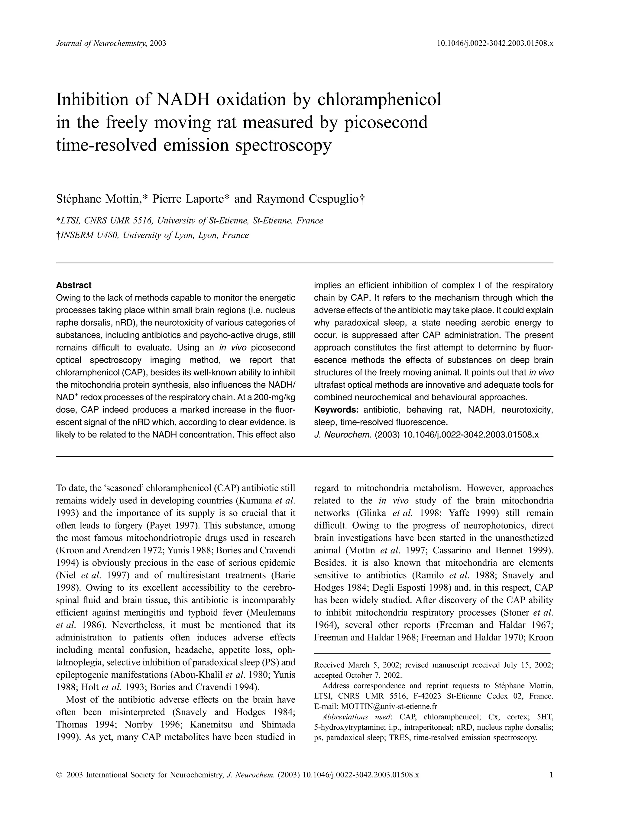
Time-resolved emission spectroscopy (TRES)
An application of ultrafast neurophotonics enabling both spectral
Results
and temporal analysis of tissue fluorescence in behaving animals has
been achieved in this study. The first generation of the set-up used
was described before (Mottin et al. 1997). Briefly, delivery and TRES imagery in vivo
collection of the optical signals (laser excitation and emission) were A typical TRES image, derived from the nRD, is illustrated
performed through a thin optical fibre allowing a good anatomical in Fig. 1(a). The autofluorescence spectrum is measured in
resolution (core diameter ¼ 200 lm). A streak camera equipped the 377–554 nm window (Fig. 1b). The temporal analysis of
Ó 2003 International Society for Neurochemistry, J. Neurochem. (2003) 10.1046/j.0022-3042.2003.01508.x](https://image.slidesharecdn.com/mottinlaportecespuglio2003-111025041213-phpapp01/75/Mottin-laporte-cespuglio-2003-2-2048.jpg)
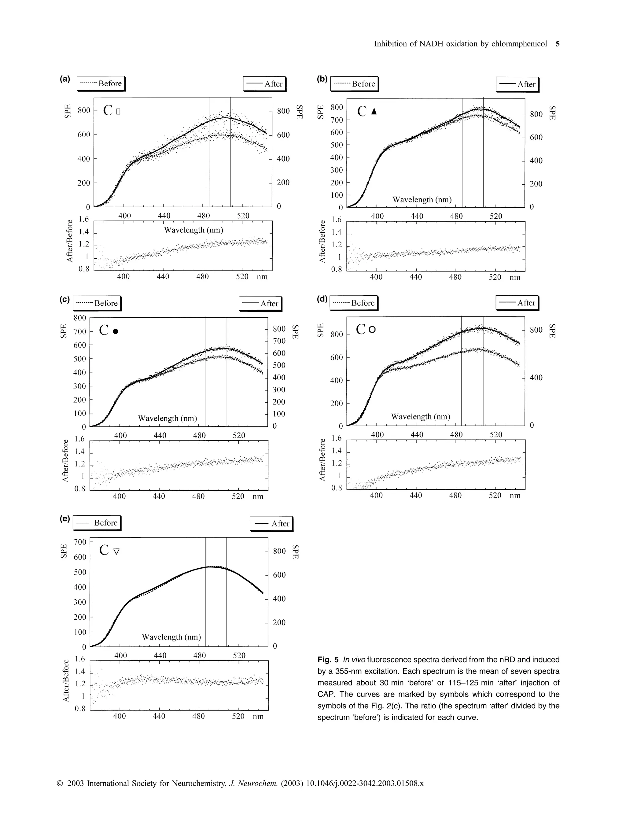
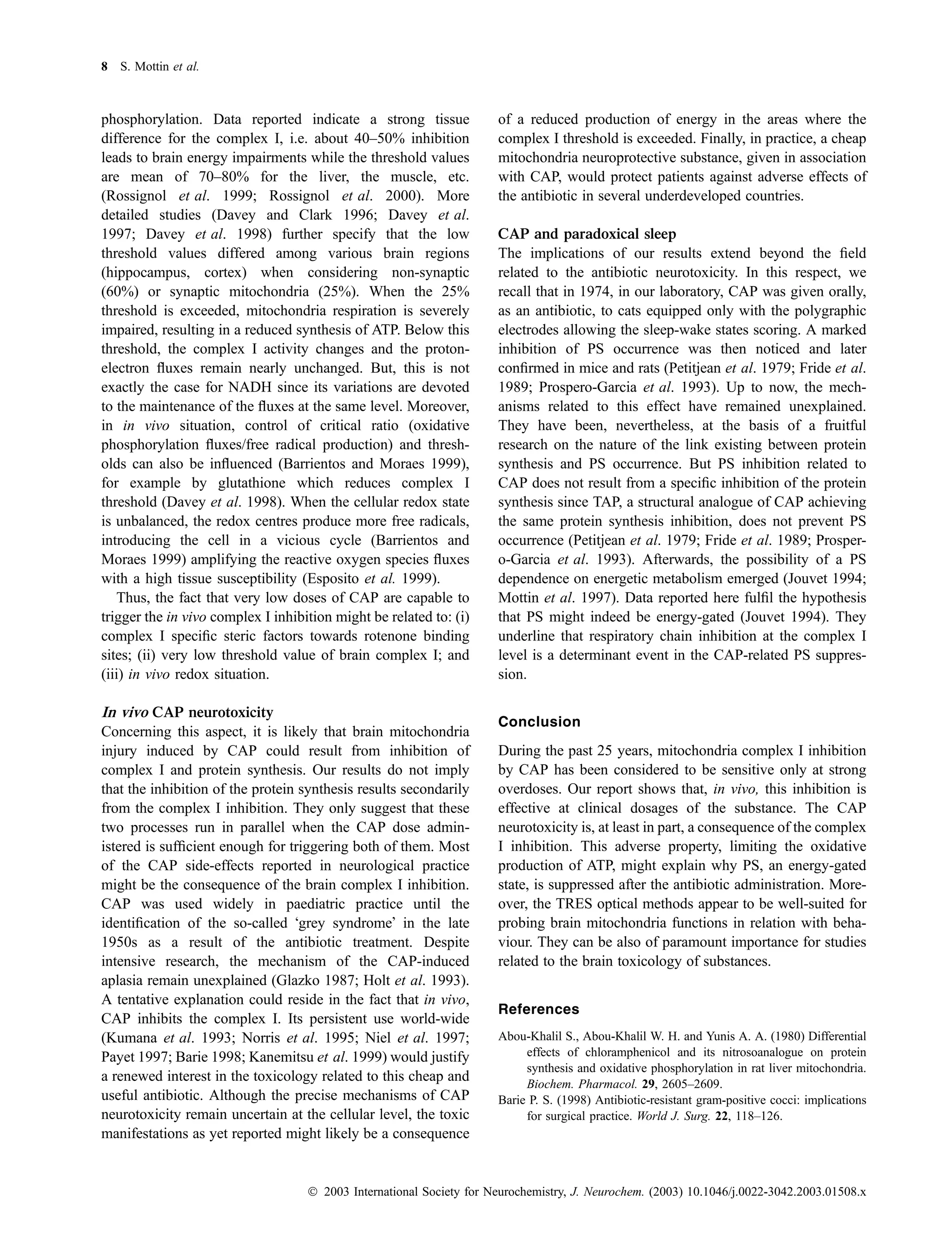
![4 S. Mottin et al.
Fig. 4 In vivo fluorescence spectra derived from the nRD and induced
by a nitrogen laser excitation. Each spectrum is the mean of seven
spectra measured about 30 min before injection.
are significant. The differences existing between the CAP
doses between 200 mg/kg and 300 mg/kg are significant.
Between 300 mg/kg and 400 mg/kg or between 200 mg/kg
and 400 mg/kg the differences are highly significant.
Regarding the 337 nm excitation, spectra obtained before
CAP injection exhibited a high variability in the UV-purple
part. Below 450 nm, several patterns of the spectra and decay
times were also measured. This variability might be due to the
presence or the absence of a UV-purple shoulder coming
probably from different endogeneous fluorophores also com-
bined with the Soret band of the haemoglobins (inner effect).
Concerning again the above variability, the 450–480 nm
window was in an intermediate position while above 480 nm,
the UV-purple shoulder was less sensitive (Fig. 4).
In the case of the 355 nm excitation, basal spectra were
Fig. 2 Time-resolved spectroscopic measurements achieved in the more reproducible. Figure 5 illustrates the variations induced
nRD. (a), (b) and (c) are, respectively, devoted to the 200 mg/kg, by a CAP injection on the whole spectral window. In the
400 mg/kg and 300 mg/kg dose. Each point represents the sum of
450–550 nm window, the increase in fluorescence obtained
single photo-electron counts performed in time-resolved emission
was greater than in the 380–440 nm window (four positive
spectroscopy within the nRD (windows: 488–507 nm). The pharma-
effects/five trials).
cokinetic exponential fitting y ¼ a + b{1 ) exp[– (t ) d )/c]} is shown.
Some data are missing due to data processing and the back-up pro-
Finally, we also checked that, for a 300-mg/kg dose of
cedure. Symbols are used for clarity. CAP, the overall CAP pharmacokinetics (increase and
decrease down to the basic fluorescence level) occurred
within 6–7 h.
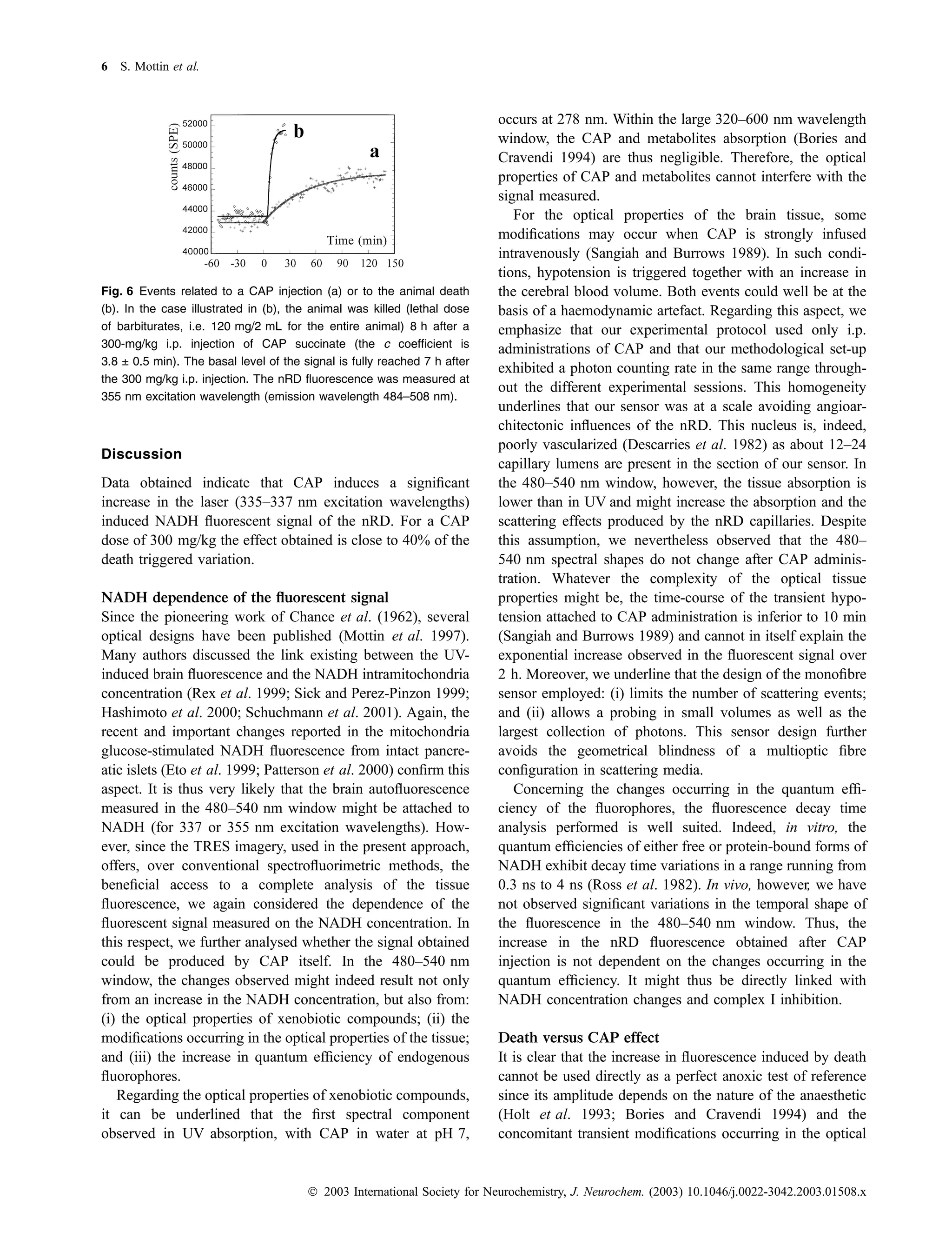
Changes induced by the animal death
Concerning the ability of CAP (or metabolites) to inhibit the
NADH/NAD+ redox processes of the respiratory chain in
the nRD, it is hard to give an absolute evaluation of the
inhibition strength. In order to overcome this difficulty, we
compared the changes occurring in the signal during the
animal death (lethal dose of barbiturates: 120 mg/kg) with
those obtained after a CAP injection. The lethal dose was
used when the basal level fluorescent signal before CAP
Fig. 3 The mean NADH fluorescence increment induced by CAP
injection was fully reached. Results obtained indicate that
injections is quantified by (b). A paired t-test comparison indicates that during death the increase in the fluorescent signal is faster
significant differences exist between the CAP doses (between 200 and and higher than after a CAP dose of 300 mg/kg (Fig. 6). The
300 mg/kg; between 300 and 400 mg/kg and between 200 and magnitude of the NADH fluorescence increase induced by
400 mg/kg). the CAP is close to 40% of the death effect.
Ó 2003 International Society for Neurochemistry, J. Neurochem. (2003) 10.1046/j.0022-3042.2003.01508.x](https://image.slidesharecdn.com/mottinlaportecespuglio2003-111025041213-phpapp01/75/Mottin-laporte-cespuglio-2003-4-2048.jpg)
![Inhibition of NADH oxidation by chloramphenicol 9
Barrientos A. and Moraes T. (1999) Titrating the effects of mitochondrial Glinka Y., Tipton K. F. and Youdim M. B. H. (1998) Mechanism of
complex I impairment in the cell physiology. J. Biol. Chem. 274, inhibition of mitochondrial respiratory complex I by 6-hydroxy-
16188–16197. dopamine and its prevention by desferrioxamine. Eur. J. Phar-
Bories G. F. and Cravendi J.-P. (1994) Metabolism of chloramphenicol. macol. 351, 121–129.
A story of nearly 50 years. Drug Metabol. Rev. 26, 767–783. Hashimoto M., Takeda Y., Sato T., Kawahara H., Nagano O. and Hir-
Cassarino D. S. and Bennet J. P. (1999) An evaluation of the role of akawa M. (2000) Dynamic changes of NADH fluorescence images
mitochondria in neurodegenerative diseases: mitochondrial muta- and NADH content during spreading depression in the cerebral
tions and oxidative pathology, protective nuclear responses, and cortex of gerbils. Brain Res. 872, 294–300.
cell death in neurodegeneration. Brain Res. Rev. 29, 1–25. Higgins D. S. and Greenamyre J. T. (1996) [3H]Dihydrorotenone binding
Cespuglio R., Houdouin F., Oulerich M., El Mansari M. and Jouvet M. to NADH: ubiquinone reductase (complex I) of the electron trans-
(1992) Axonal and somato-dendritic modalities of serotonin port chain: an autoradiographic study. J. Neurosci. 16, 3807–3816.
release: their involvment in sleep preparation, triggering and Holt D., Harvey D. and Hurley R. (1993) Chloramphenicol toxicity. Adv.
maintenance. J. Sleep Res. 1, 150–156. Drug. React. Rev. Toxicol. Rev. 12, 83–95.
Chance B., Cohen P., Jobsis F. and Schoener B. (1962) Intracellular Jouvet M. (1994) Paradoxical sleep mechanisms. Sleep 17, S77–S83.
oxidation-reduction states in vivo. Science 137, 499–508. Kanemitsu K. and Shimada J. (1999) Neurotoxicity of antibacterial
Chazal G. and Ralston H. J. (1987) Serotonin-containing structures in the agents (tetracyclines, chloramphenicol, aminoglycosides, beta-lac-
nucleus raphe dorsalis of the cat: an ultrastructural analysis of tams and others). Ryoikibetsu Shokogun Shirizu 27, 545–551.
dendrites, presynaptic dentrites and axon terminals. J. Comp. Kroon A. M. and Arendzen A. J. (1972) Inhibition of mitochondrial
Neurol. 259, 317. biogenesis. FEBS Meeting 28, 71–83.
Davey G. P. and Clark J. B. (1996) Threshold effects and control of Kroon A. M. and de Jong L. (1979) The use of antibiotics to study
oxidative phosphorylation in nonsynaptic rat brain mitochondria. mitochondrial protein synthesis, in Methods in Enzymology,
J. Neurochem. 66, 1617–1624. (Fleischer S. and Packer L., eds). Academic Press, New York.
Davey G. P., Canevari L. and Clark J. B. (1997) Threshold effects in Kumana C. R., Li K. Y. and Kou M. (1993) Do chloramphenicol blood
synaptosomal and nonsynaptic mitochondria from hippocampal dyscrasias occur in Hong Kong? Adv. Drug. React. Rev. Toxicol.
CA1 and paramedian neocortex brain regions. J. Neurochem. 69, Rev. 12, 97–106.
2564–2570. Meulemans A., Vicard P., Mohler J., Vulpillat M. and Pocidalo J. J.
Davey G. P., Peuchen S. and Clark J. B. (1998) Energy thresholds in (1986) Continuous sampling for determination of pharmacokinet-
brain mitochondria. Potential involvement in neurodegeneration. ics in rat cerebrospinal fluid. Antimicrob. Agents Chemother. 30,
J. Biol. Chem. 273, 12753–12757. 888–891.
Degli Esposti M. (1998) Inhibitors of NADH-ubiquinone reductase: an Mottin S., Tran-Minh C., Laporte P., Cespuglio R. and Jouvet M. (1993)
overview. Biochim. Biophys. Acta 1364, 222–235. Fiber optic time-resolved fluorescence sensor for in vitro serotonin
Delpy D. T., Cope M., van der Zee P., Arridge S., Wray S. and Wray J. determination. Appl. Spectroscopy 47, 590–597.
(1988) Estimation of optical pathlength through tissue from Mottin S., Laporte P., Cespuglio R. and Jouvet M. (1997) Determination
direct time of flight measurement. Phys. Med. Biol. 33, 1433– of NADH in the rat brain during sleep wake states with an optic
1442. fibre sensor and time resolved fluorescence procedures. Neuro-
Descarries L., Watkins K. C., Garcia S. and Beaudet A. J. (1982) The science 79, 683–693.
serotonin neurons in nucleus raphe dorsalis of adult rat: a light and Niel L., Lamarque D., Coue J. C., Soares J. L., Milleliri J. M., Boutin
´ `
electron microscope radioautographic study. Comp. Neurol. 207, J. P., Merouze F. and Rey J. L. (1997) Chronique d’une epidemie
´ ´ ´
239–254. annoncee de meningite a meningocoque (Goma, Za). Bull. Soc.
´ ´ ` ´
Dorph-Petersen K.-A. (1999) Stereological estimation using vertical Pathol. Exot. 90, 299–302.
sections in a complex tissue. J. Microsc. 195, 79–86. Norrby S. R. (1996) Neurotoxicity of carbapenem antibacterials. Drug
Esposito L. A., Melov S., Panov A., Cottrell B. A. and Wallace D. C. Saf. 15, 87–90.
(1999) Mitochondrial disease in mouse results in increased Norris A. H., Reilly J. P., Edelstein P. H., Brennan P. J. and Schuster M.
oxidative stress. Proc. Natl Acad. Sci. USA 96, 4820–4825. G. (1995) Chloramphenicol for the treatment of vancomycin-
Eto K., Tsubamoto Y., Teraushi Y., Sugiyama T., Kishimoto T., resistant enterococcal infections. Clin. Infect. Dis. 20, 1137–1142.
Takahashi N., Yamauchi N., Kubota N., Murayama S., Aizawa T., Patterson G. H., Knobel S. M., Arkhammar P., Thastrup O. and Piston D.
Akanuma Y., Aizawa S., Kasai H., Yazaki Y. and Kadowaki T. W. (2000) Separation of the glucose-stimulated cytoplasmic and
(1999) Role of NADH shuttle system in glucose-induced activation mitochondrial NAD(P)H responses in pancreatic islet beta cells.
of mitochondrial metabolism and insulin secretion. Science 283, Proc. Natl Acad. Sci. USA 97, 5203–5207.
981–985. Payet M. (1997) Trafics sur ordonnance. Croissance 401, 14–18.
Freeman K. B. and Haldar D. (1967) The inhibition of NADH oxidation Petitjean F., Buda C., Janin M., David M. and Jouvet M. (1979) Effets du
in mammalian mitochondria by chloramphenicol. Biochem. Bio- chloramphenicol sur le sommeil du chat. psychopharmacol. 66,
phys. Res. Comm. 28, 8–12. 147–153.
Freeman K. B. and Haldar D. (1968) The inhibition of mammalian Prospero-Garcia O., Jimenez-Anguiano A. and Drucker-Colin R. (1993)
mitochondrial NADH oxidation by chloramphenicol and its iso- Chloramphenicol prevents carbachol-induced REM sleep in cats.
mers and analogues. Can. J. Biochem. 46, 1003–1008. Neurosci. Lett. 154, 168–170.
Freeman K. B. and Haldar D. (1970) Effects of chloramphenicol and its Ramilo O., Kinane B. T. and McCracken G. H. (1988) Chloramphenicol
isomers and analogues on the mitochondrial respiratory chain. Can. neurotoxicity. Ped. Infect. Dis. J. 7, 358–359.
J. Biochem. 48, 469–485. Rex A., Pfeifer L., Fink F. and Fink H. (1999) Cortical NADH during
Fride E., Ben-Or S. and Allweis C. (1989) Mitochondrial protein syn- pharmacological manipulations of the respiratory chain and
thesis may be involved in long-term memory formation. Pharma- spreading depression in vivo. J. Neurosci. Res. 57, 359–370.
col. Biochem. Behav. 32, 873–878. Ross J. B. A., Subramanian S. and Brand L. (1982) Spectroscopic
Glazko A. J. (1987) Early adventures in drug metabolism – chloram- studies of the pyridine nucleotide coenzymes and their complexes
phenicol. Ther. Drug Monit. 9, 320–330. with dehydrogenases, in The Pyridine Nucleotide Coenzymes
Ó 2003 International Society for Neurochemistry, J. Neurochem. (2003) 10.1046/j.0022-3042.2003.01508.x](https://image.slidesharecdn.com/mottinlaportecespuglio2003-111025041213-phpapp01/75/Mottin-laporte-cespuglio-2003-9-2048.jpg)


![Coded Agents – with UiPath SDK + LangGraph [Virtual Hands-on Workshop]](https://cdn.slidesharecdn.com/ss_thumbnails/codedagentsdeck-251215155422-5497c599-thumbnail.jpg?width=640&height=640&fit=bounds)


