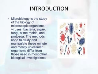
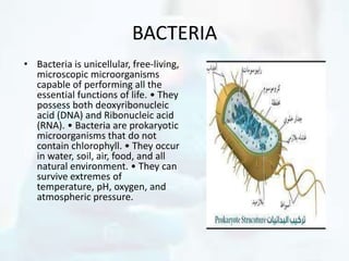
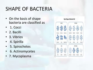
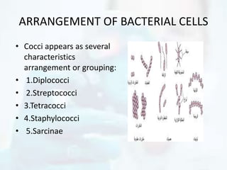
Tyler was a blast he was a very funny kid, he had me laughing the whole time, which made it very hard to take notes. He was a fourth generation Mexican American, and the first of his family to go to college. His parents taught him well that life is harder in America for a Mexican. Margaret E. Montoya states a similar experience: “Her lessons about combing, washing, and doing homework frequently relayed a different message: be prepared, because you will be judged by your skin color, your names, your accents. They will see you as ugly, lazy dumb and dirty.” (Montoya 436) Montoya’s experience is very closely related to that of Tyler’s childhood, his parents would try their very best to instill good qualities of hard work and determination into their child, because they knew he was going to need it. Tyler has done well for himself, he is an honors student and he has a 3.5 GPA, he works very hard to be the best he can be, but is still seen as inferior. Tyler’s job just became harder, after the passing of this bill, all of the Mexicans are presumed to be illegal until proven innocent. Thanks to this new law, upon first sight he is looked at as a nuisance, a criminal, and a foreigner. However, he is an excellent student, a volunteer who donates ten hours a week of his time at homeless shelters, and a resident, whose family has been on this land longer that John McCain has been alive. The fact that this racism still happens today, and is seemingly getting worse, just adds to what Montoya’s mother taught her “be prepared, because you will be judged” (Montoya 436)
These three people have many things in common with all Americans, including; they want to live their life, be successful, and be judged on their effort and accomplishments like everyone else. These are people just trying to live the American Dream, which is just made harder by this law that has been passed which just increases the active racism in this country. The 1070 law has done it’s job: making it harder for Mexican Americans to be successful and be equal to whites, through increasing active racism Tyler was a blast he was a very funny kid, he had me laughing the whole time, which made it very hard to take notes. He was a fourth generation Mexican American, and the first of his family to go to college. His parents taught him well that life is harder in America for a Mexican. Margaret E. Montoya states a similar experience: “Her lessons about combing, washing, and doing homework frequently relayed a different message: be prepared, because you will be judged by your skin color, your names, your accents. They will see you as ugly, lazy dumb and dirty.” (Montoya 436) Montoya’s experience is very closely related to that of Tyler’s childhood, his parents would try their very best to instill good qualities of hard work and determination into their child, because they knew he was going to need it. Tyler has done well for himself, he is an honors student and he has a 3.5 GPA, he works very hard to b
The document provides information about the morphology and classification of bacteria. It discusses the key differences between prokaryotic and eukaryotic cells, including that prokaryotes lack a nucleus and membrane-bound organelles while eukaryotes have these structures. It also describes the shapes and arrangements of bacterial cells, and the various microscopy techniques used to study bacterial morphology, including sample preparation methods like fixation and staining. Common staining techniques like Gram staining and acid-fast staining are also outlined.
Prokaryotes include bacteria and archaea. Bacteria were first observed in 1674 and include diverse species found in many habitats. Prokaryotes are single-celled without internal membranes and reproduce through binary fission. Major groups include eubacteria like E. coli and archaea found in extreme environments like hot springs or salt lakes. Bacteria play important roles in ecosystems through processes like nitrogen fixation and photosynthesis.
1. The first person to observe living microorganisms using a microscope he invented was Antony van Leeuwenhoek in 1674, when he saw bacteria in dental plaque.
2. Bacteria are the most common and diverse type of prokaryotes, ranging in size from 0.2 to 2.0 μm and having a variety of shapes.
3. Bacterial cell walls are composed of peptidoglycan, while archaea cell walls contain other components like glycoproteins or polysaccharides.
- Prokaryotic cells like bacteria have a simple cellular structure without organelles and their genetic material exists freely in the cytoplasm. Eukaryotic cells like humans have membrane-bound organelles and their genetic material is contained within the nucleus.
- The genetic material of prokaryotes is a single circular chromosome while eukaryotes have multiple linear chromosomes. Prokaryotic DNA replication and transcription occur simultaneously in the cytoplasm, while in eukaryotes transcription occurs in the nucleus and translation in the cytoplasm.
- Both prokaryotes and eukaryotes have DNA as their genetic material but it is organized differently - prokaryotes have a compact circular chromosome while eukaryotes package their DNA
The document provides an overview of cell biology, including:
- Differences between prokaryotic and eukaryotic cells and their organelles. Prokaryotes lack a nucleus and organelles while eukaryotes have membrane-bound organelles.
- Characteristics of plant and animal cells including organelles like the nucleus, mitochondria, chloroplasts, cell wall, and vacuoles.
- Chemical composition and basic components of cells including proteins, nucleic acids, lipids, and carbohydrates.
- Structures of prokaryotic cells like the cell wall, plasma membrane, flagella, and pili. Eukaryotic cell structures like the nucleus, cytoplasm, mitochondria, Gol
Introduction to Environmental Microbiology (by- Meenu Malik)meenumalik3
This document provides an introduction to environmental microbiology. It discusses microscopic organisms such as bacteria, fungi, protozoans, algae, and viruses. It notes that bacteria were some of the earliest life on Earth and can be found nearly everywhere. The document outlines the history of microbiology including early pioneers like Van Leeuwenhoek, Hooke, Pasteur and Jenner. It describes prokaryotic and eukaryotic cells and some of their key structures. Overall, the document provides a high-level overview of microorganisms and their role in environmental microbiology.
Bacteria have a simple structure compared to eukaryotic cells, lacking organelles. Their small size allows rapid growth and inhabitation of diverse environments. Bacterial cells contain a cytoplasm surrounded by a cell membrane and cell wall. The cytoplasm holds the circular chromosome, ribosomes for protein production, and storage structures. Some bacteria have flagella for mobility or pili for attachment. Gram-positive bacteria have a thick peptidoglycan cell wall, while Gram-negatives have a thin wall and an outer membrane. This membrane structure contributes to differences in antibiotic susceptibility between Gram-positive and Gram-negative bacteria.
Living beings have 7 main characteristics:
1. They are composed of cells, which can be single-celled or multicellular.
2. They have a chemical composition and are highly organized from primary bioelements like carbon, hydrogen, nitrogen, and oxygen.
3. Living things use energy, either producing their own through photosynthesis (autotrophs) or consuming organic molecules (heterotrophs).
This summarizes the key points about the characteristics of living beings according to the document.
The document provides information about cell structure and organelles. It discusses:
- Cells as the basic structural and functional units of organisms.
- The cell theory proposed by Schleiden and Schwann stating that cells are the basic unit of life, all cells come from preexisting cells, and organisms are made of cells.
- Key organelles like the nucleus, endoplasmic reticulum, ribosomes, Golgi apparatus, mitochondria, and lysosomes and their functions.
Bacteria are unicellular prokaryotic organisms that can exist as single cells or in clusters. They have a simple cell structure without organelles, and their genetic material is contained in a single loop of DNA. Bacteria come in various shapes including cocci, bacilli, vibrios, spirilla, and spirochetes. Their size typically ranges from 0.5-2.0 μm. Bacteria can be identified through their shape, size, aggregation properties, staining characteristics, and imaging methods such as light, phase contrast, dark field, and electron microscopy. Their cell walls are composed of peptidoglycan and they are either Gram-positive or Gram-negative. Bacteria have a
The document discusses the five kingdom classification system proposed by Whittaker in 1969. It outlines the key criteria used for classification including cell structure, organism complexity, nutrition mode, and lifestyle. The five kingdoms are Monera, Protista, Fungi, Plantae, and Animalia. Details are provided on the defining characteristics of each kingdom, including whether cells are prokaryotic or eukaryotic, unicellular or multicellular, autotrophic or heterotrophic. Examples of organisms from each kingdom are also listed.
This document summarizes the ultrastructure of bacterial cells. It describes that bacteria are unicellular and microscopic. They have characteristic shapes including cocci, bacilli, vibrio and spirals. Their structures include a cell wall, plasma membrane, flagella for motility, pili for adhesion, capsules for protection, ribosomes for protein synthesis, and some can form endospores. The cell wall structure differs between gram-positive and gram-negative bacteria. The cytoplasm contains ribosomes and DNA but not membrane-bound organelles.
This document summarizes the structure and morphology of bacterial cells. It discusses that bacteria are unicellular and microscopic, between 0.4-1.5 micrometers in size. Bacteria have characteristic shapes including cocci (spherical), bacilli (rod-shaped), and spirals. They contain DNA, RNA, ribosomes, and in some cases plasmids and mesosomes. Bacteria have a cell wall, plasma membrane, flagella or pili for motility, and may contain a capsule or endospores. The cell wall structure differs between gram-positive and gram-negative bacteria.
1. The document discusses the structure and function of cells. It provides background on the history of cell discovery from Hooke to Virchow and defines key terms like cytology.
2. The basic components of plant and animal cells are described along with the differences between them. Unicellular and multicellular organisms are also defined.
3. The key characteristics of prokaryotic and eukaryotic cells are summarized, including whether they have a nucleus, organelles, and how they reproduce. Diagrams of sample prokaryotic and eukaryotic cell structures are also included.
Bacteria have a simple cellular structure compared to eukaryotes. They lack internal membrane-bound organelles. Their cellular components include a cytoplasm containing a nucleoid, plasmids, ribosomes and other structures. The cell envelope includes a plasma membrane, cell wall, and in some cases an outer membrane. Some bacteria have external structures like flagella, fimbriae or pili that allow movement or attachment. Gram staining distinguishes bacteria based on differences in their cell wall composition and thickness.
Bacteria have a simple cellular structure compared to eukaryotes. They lack membrane-bound organelles but carry out all necessary functions within the cell. Bacterial cells contain a plasma membrane, cell wall, cytoplasmic inclusions, ribosomes, circular chromosome, and may contain extrachromosomal DNA like plasmids. The structures allow bacteria to rapidly grow and divide while inhabiting diverse environments.
The document discusses the basic building blocks of life including cells, chromosomes, genes, and nucleic acids. It provides details on the structure and components of prokaryotic and eukaryotic cells. Chromosomes are described as thread-like structures in the nucleus that contain DNA and genes which carry genetic information. The chemical composition and importance of studying chromosomes is also summarized.
Bacteria are unicellular prokaryotic organisms that are studied in medical microbiology. They have distinct cell structures including a cell wall, cell membrane, cytoplasm, and nucleic material. Bacteria can be visualized under light, phase contrast, or electron microscopes and stained using simple, differential, or acid-fast staining techniques. Gram staining divides bacteria into Gram-positive and Gram-negative categories based on cell wall structure. Bacteria exhibit a variety of external structures such as flagella, pili, capsules, and endospores, and follow a defined growth curve with lag, log, stationary, and death phases.
This document compares and contrasts prokaryotic and eukaryotic cells. It begins by introducing that all living things are made up of cells, which are the smallest living units. It then describes that cells are divided into two main categories: prokaryotic cells, which lack a nucleus, and eukaryotic cells, which contain a nucleus enclosed within a membrane. The document proceeds to list and describe the various organelles contained within each cell type, including their locations and functions. It concludes by outlining some key differences in how prokaryotic and eukaryotic cells reproduce.
The term "biochemistry" originated from combining the words "bios," meaning life, and "chemistry."
Biochemistry is defined as the branch of science that deals with the study of chemical reactions that take place inside a living organism.
The word "biochemistry" was first introduced by a German chemist, Carl Neuberg, in 1903.
This document provides an overview of cells. It begins by defining a cell as the fundamental unit of life and describes how Robert Hooke first observed cells in cork through a microscope in 1665. It then discusses the main instruments used for studying cells - light microscopes and electron microscopes. The document outlines the key differences between plant and animal cells and describes some of the main cell organelles like the cell wall, nucleus, chloroplasts, and mitochondria. It provides comparisons between different types of cells and cell components.
This document discusses the classification of microorganisms. It describes the three domain system proposed by Carl Woese which divides organisms into Archaea, Bacteria and Eukarya. It then provides details on the characteristics of fungi, algae, protozoa, viruses and bacteria; and discusses methods used to identify bacteria including biochemical tests and serological tests.
This document discusses the classification of microorganisms. It describes the differences between bacteria, archaea, and eukarya. It discusses the binominal nomenclature system developed by Carolus Linnaeus to classify organisms. It also describes Woese's three domain system that divides organisms into the domains of Archaea, Bacteria, and Eukarya. Finally, it provides details on the classification and characteristics of fungi, algae, protozoa, viruses, and bacteria.
This document provides information about different types of cells including prokaryotic, eukaryotic, bacterial, fungal, protozoan, algal, and viral cells. It discusses their key distinguishing characteristics such as whether they have a nucleus, cell wall composition, mode of nutrition, reproduction, and more. Examples are provided of medically important microorganisms from each category like bacteria (Staphylococcus, Streptococcus), viruses (influenza, HIV), fungi (Candida), protozoa (Plasmodium, Toxoplasma), and algae (diatoms, dinoflagellates). The document also covers cell staining techniques including gram staining and acid-fast staining used to classify
ABDOMINAL TRAUMA in pediatrics part one.drhasanrajab
Abdominal trauma in pediatrics refers to injuries or damage to the abdominal organs in children. It can occur due to various causes such as falls, motor vehicle accidents, sports-related injuries, and physical abuse. Children are more vulnerable to abdominal trauma due to their unique anatomical and physiological characteristics. Signs and symptoms include abdominal pain, tenderness, distension, vomiting, and signs of shock. Diagnosis involves physical examination, imaging studies, and laboratory tests. Management depends on the severity and may involve conservative treatment or surgical intervention. Prevention is crucial in reducing the incidence of abdominal trauma in children.
More Related Content
Similar to MORPHOLOGY-AND-CLASSIFICATION-OF-BACTERI(1).pptx
The document provides information about cell structure and organelles. It discusses:
- Cells as the basic structural and functional units of organisms.
- The cell theory proposed by Schleiden and Schwann stating that cells are the basic unit of life, all cells come from preexisting cells, and organisms are made of cells.
- Key organelles like the nucleus, endoplasmic reticulum, ribosomes, Golgi apparatus, mitochondria, and lysosomes and their functions.
Bacteria are unicellular prokaryotic organisms that can exist as single cells or in clusters. They have a simple cell structure without organelles, and their genetic material is contained in a single loop of DNA. Bacteria come in various shapes including cocci, bacilli, vibrios, spirilla, and spirochetes. Their size typically ranges from 0.5-2.0 μm. Bacteria can be identified through their shape, size, aggregation properties, staining characteristics, and imaging methods such as light, phase contrast, dark field, and electron microscopy. Their cell walls are composed of peptidoglycan and they are either Gram-positive or Gram-negative. Bacteria have a
The document discusses the five kingdom classification system proposed by Whittaker in 1969. It outlines the key criteria used for classification including cell structure, organism complexity, nutrition mode, and lifestyle. The five kingdoms are Monera, Protista, Fungi, Plantae, and Animalia. Details are provided on the defining characteristics of each kingdom, including whether cells are prokaryotic or eukaryotic, unicellular or multicellular, autotrophic or heterotrophic. Examples of organisms from each kingdom are also listed.
This document summarizes the ultrastructure of bacterial cells. It describes that bacteria are unicellular and microscopic. They have characteristic shapes including cocci, bacilli, vibrio and spirals. Their structures include a cell wall, plasma membrane, flagella for motility, pili for adhesion, capsules for protection, ribosomes for protein synthesis, and some can form endospores. The cell wall structure differs between gram-positive and gram-negative bacteria. The cytoplasm contains ribosomes and DNA but not membrane-bound organelles.
This document summarizes the structure and morphology of bacterial cells. It discusses that bacteria are unicellular and microscopic, between 0.4-1.5 micrometers in size. Bacteria have characteristic shapes including cocci (spherical), bacilli (rod-shaped), and spirals. They contain DNA, RNA, ribosomes, and in some cases plasmids and mesosomes. Bacteria have a cell wall, plasma membrane, flagella or pili for motility, and may contain a capsule or endospores. The cell wall structure differs between gram-positive and gram-negative bacteria.
1. The document discusses the structure and function of cells. It provides background on the history of cell discovery from Hooke to Virchow and defines key terms like cytology.
2. The basic components of plant and animal cells are described along with the differences between them. Unicellular and multicellular organisms are also defined.
3. The key characteristics of prokaryotic and eukaryotic cells are summarized, including whether they have a nucleus, organelles, and how they reproduce. Diagrams of sample prokaryotic and eukaryotic cell structures are also included.
Bacteria have a simple cellular structure compared to eukaryotes. They lack internal membrane-bound organelles. Their cellular components include a cytoplasm containing a nucleoid, plasmids, ribosomes and other structures. The cell envelope includes a plasma membrane, cell wall, and in some cases an outer membrane. Some bacteria have external structures like flagella, fimbriae or pili that allow movement or attachment. Gram staining distinguishes bacteria based on differences in their cell wall composition and thickness.
Bacteria have a simple cellular structure compared to eukaryotes. They lack membrane-bound organelles but carry out all necessary functions within the cell. Bacterial cells contain a plasma membrane, cell wall, cytoplasmic inclusions, ribosomes, circular chromosome, and may contain extrachromosomal DNA like plasmids. The structures allow bacteria to rapidly grow and divide while inhabiting diverse environments.
The document discusses the basic building blocks of life including cells, chromosomes, genes, and nucleic acids. It provides details on the structure and components of prokaryotic and eukaryotic cells. Chromosomes are described as thread-like structures in the nucleus that contain DNA and genes which carry genetic information. The chemical composition and importance of studying chromosomes is also summarized.
Bacteria are unicellular prokaryotic organisms that are studied in medical microbiology. They have distinct cell structures including a cell wall, cell membrane, cytoplasm, and nucleic material. Bacteria can be visualized under light, phase contrast, or electron microscopes and stained using simple, differential, or acid-fast staining techniques. Gram staining divides bacteria into Gram-positive and Gram-negative categories based on cell wall structure. Bacteria exhibit a variety of external structures such as flagella, pili, capsules, and endospores, and follow a defined growth curve with lag, log, stationary, and death phases.
This document compares and contrasts prokaryotic and eukaryotic cells. It begins by introducing that all living things are made up of cells, which are the smallest living units. It then describes that cells are divided into two main categories: prokaryotic cells, which lack a nucleus, and eukaryotic cells, which contain a nucleus enclosed within a membrane. The document proceeds to list and describe the various organelles contained within each cell type, including their locations and functions. It concludes by outlining some key differences in how prokaryotic and eukaryotic cells reproduce.
The term "biochemistry" originated from combining the words "bios," meaning life, and "chemistry."
Biochemistry is defined as the branch of science that deals with the study of chemical reactions that take place inside a living organism.
The word "biochemistry" was first introduced by a German chemist, Carl Neuberg, in 1903.
This document provides an overview of cells. It begins by defining a cell as the fundamental unit of life and describes how Robert Hooke first observed cells in cork through a microscope in 1665. It then discusses the main instruments used for studying cells - light microscopes and electron microscopes. The document outlines the key differences between plant and animal cells and describes some of the main cell organelles like the cell wall, nucleus, chloroplasts, and mitochondria. It provides comparisons between different types of cells and cell components.
This document discusses the classification of microorganisms. It describes the three domain system proposed by Carl Woese which divides organisms into Archaea, Bacteria and Eukarya. It then provides details on the characteristics of fungi, algae, protozoa, viruses and bacteria; and discusses methods used to identify bacteria including biochemical tests and serological tests.
This document discusses the classification of microorganisms. It describes the differences between bacteria, archaea, and eukarya. It discusses the binominal nomenclature system developed by Carolus Linnaeus to classify organisms. It also describes Woese's three domain system that divides organisms into the domains of Archaea, Bacteria, and Eukarya. Finally, it provides details on the classification and characteristics of fungi, algae, protozoa, viruses, and bacteria.
This document provides information about different types of cells including prokaryotic, eukaryotic, bacterial, fungal, protozoan, algal, and viral cells. It discusses their key distinguishing characteristics such as whether they have a nucleus, cell wall composition, mode of nutrition, reproduction, and more. Examples are provided of medically important microorganisms from each category like bacteria (Staphylococcus, Streptococcus), viruses (influenza, HIV), fungi (Candida), protozoa (Plasmodium, Toxoplasma), and algae (diatoms, dinoflagellates). The document also covers cell staining techniques including gram staining and acid-fast staining used to classify
ABDOMINAL TRAUMA in pediatrics part one.drhasanrajab
Abdominal trauma in pediatrics refers to injuries or damage to the abdominal organs in children. It can occur due to various causes such as falls, motor vehicle accidents, sports-related injuries, and physical abuse. Children are more vulnerable to abdominal trauma due to their unique anatomical and physiological characteristics. Signs and symptoms include abdominal pain, tenderness, distension, vomiting, and signs of shock. Diagnosis involves physical examination, imaging studies, and laboratory tests. Management depends on the severity and may involve conservative treatment or surgical intervention. Prevention is crucial in reducing the incidence of abdominal trauma in children.
Osteoporosis - Definition , Evaluation and Management .pdfJim Jacob Roy
Osteoporosis is an increasing cause of morbidity among the elderly.
In this document , a brief outline of osteoporosis is given , including the risk factors of osteoporosis fractures , the indications for testing bone mineral density and the management of osteoporosis
Basavarajeeyam is an important text for ayurvedic physician belonging to andhra pradehs. It is a popular compendium in various parts of our country as well as in andhra pradesh. The content of the text was presented in sanskrit and telugu language (Bilingual). One of the most famous book in ayurvedic pharmaceutics and therapeutics. This book contains 25 chapters called as prakaranas. Many rasaoushadis were explained, pioneer of dhatu druti, nadi pareeksha, mutra pareeksha etc. Belongs to the period of 15-16 century. New diseases like upadamsha, phiranga rogas are explained.
Here is the updated list of Top Best Ayurvedic medicine for Gas and Indigestion and those are Gas-O-Go Syp for Dyspepsia | Lavizyme Syrup for Acidity | Yumzyme Hepatoprotective Capsules etc
Adhd Medication Shortage Uk - trinexpharmacy.comreignlana06
The UK is currently facing a Adhd Medication Shortage Uk, which has left many patients and their families grappling with uncertainty and frustration. ADHD, or Attention Deficit Hyperactivity Disorder, is a chronic condition that requires consistent medication to manage effectively. This shortage has highlighted the critical role these medications play in the daily lives of those affected by ADHD. Contact : +1 (747) 209 – 3649 E-mail : sales@trinexpharmacy.com
NVBDCP.pptx Nation vector borne disease control programSapna Thakur
NVBDCP was launched in 2003-2004 . Vector-Borne Disease: Disease that results from an infection transmitted to humans and other animals by blood-feeding arthropods, such as mosquitoes, ticks, and fleas. Examples of vector-borne diseases include Dengue fever, West Nile Virus, Lyme disease, and malaria.
TEST BANK For Community Health Nursing A Canadian Perspective, 5th Edition by...Donc Test
TEST BANK For Community Health Nursing A Canadian Perspective, 5th Edition by Stamler, Verified Chapters 1 - 33, Complete Newest Version Community Health Nursing A Canadian Perspective, 5th Edition by Stamler, Verified Chapters 1 - 33, Complete Newest Version Community Health Nursing A Canadian Perspective, 5th Edition by Stamler Community Health Nursing A Canadian Perspective, 5th Edition TEST BANK by Stamler Test Bank For Community Health Nursing A Canadian Perspective, 5th Edition Pdf Chapters Download Test Bank For Community Health Nursing A Canadian Perspective, 5th Edition Pdf Download Stuvia Test Bank For Community Health Nursing A Canadian Perspective, 5th Edition Study Guide Test Bank For Community Health Nursing A Canadian Perspective, 5th Edition Ebook Download Stuvia Test Bank For Community Health Nursing A Canadian Perspective, 5th Edition Questions and Answers Quizlet Test Bank For Community Health Nursing A Canadian Perspective, 5th Edition Studocu Test Bank For Community Health Nursing A Canadian Perspective, 5th Edition Quizlet Test Bank For Community Health Nursing A Canadian Perspective, 5th Edition Stuvia Community Health Nursing A Canadian Perspective, 5th Edition Pdf Chapters Download Community Health Nursing A Canadian Perspective, 5th Edition Pdf Download Course Hero Community Health Nursing A Canadian Perspective, 5th Edition Answers Quizlet Community Health Nursing A Canadian Perspective, 5th Edition Ebook Download Course hero Community Health Nursing A Canadian Perspective, 5th Edition Questions and Answers Community Health Nursing A Canadian Perspective, 5th Edition Studocu Community Health Nursing A Canadian Perspective, 5th Edition Quizlet Community Health Nursing A Canadian Perspective, 5th Edition Stuvia Community Health Nursing A Canadian Perspective, 5th Edition Test Bank Pdf Chapters Download Community Health Nursing A Canadian Perspective, 5th Edition Test Bank Pdf Download Stuvia Community Health Nursing A Canadian Perspective, 5th Edition Test Bank Study Guide Questions and Answers Community Health Nursing A Canadian Perspective, 5th Edition Test Bank Ebook Download Stuvia Community Health Nursing A Canadian Perspective, 5th Edition Test Bank Questions Quizlet Community Health Nursing A Canadian Perspective, 5th Edition Test Bank Studocu Community Health Nursing A Canadian Perspective, 5th Edition Test Bank Quizlet Community Health Nursing A Canadian Perspective, 5th Edition Test Bank Stuvia
Recomendações da OMS sobre cuidados maternos e neonatais para uma experiência pós-natal positiva.
Em consonância com os ODS – Objetivos do Desenvolvimento Sustentável e a Estratégia Global para a Saúde das Mulheres, Crianças e Adolescentes, e aplicando uma abordagem baseada nos direitos humanos, os esforços de cuidados pós-natais devem expandir-se para além da cobertura e da simples sobrevivência, de modo a incluir cuidados de qualidade.
Estas diretrizes visam melhorar a qualidade dos cuidados pós-natais essenciais e de rotina prestados às mulheres e aos recém-nascidos, com o objetivo final de melhorar a saúde e o bem-estar materno e neonatal.
Uma “experiência pós-natal positiva” é um resultado importante para todas as mulheres que dão à luz e para os seus recém-nascidos, estabelecendo as bases para a melhoria da saúde e do bem-estar a curto e longo prazo. Uma experiência pós-natal positiva é definida como aquela em que as mulheres, pessoas que gestam, os recém-nascidos, os casais, os pais, os cuidadores e as famílias recebem informação consistente, garantia e apoio de profissionais de saúde motivados; e onde um sistema de saúde flexível e com recursos reconheça as necessidades das mulheres e dos bebês e respeite o seu contexto cultural.
Estas diretrizes consolidadas apresentam algumas recomendações novas e já bem fundamentadas sobre cuidados pós-natais de rotina para mulheres e neonatos que recebem cuidados no pós-parto em unidades de saúde ou na comunidade, independentemente dos recursos disponíveis.
É fornecido um conjunto abrangente de recomendações para cuidados durante o período puerperal, com ênfase nos cuidados essenciais que todas as mulheres e recém-nascidos devem receber, e com a devida atenção à qualidade dos cuidados; isto é, a entrega e a experiência do cuidado recebido. Estas diretrizes atualizam e ampliam as recomendações da OMS de 2014 sobre cuidados pós-natais da mãe e do recém-nascido e complementam as atuais diretrizes da OMS sobre a gestão de complicações pós-natais.
O estabelecimento da amamentação e o manejo das principais intercorrências é contemplada.
Recomendamos muito.
Vamos discutir essas recomendações no nosso curso de pós-graduação em Aleitamento no Instituto Ciclos.
Esta publicação só está disponível em inglês até o momento.
Prof. Marcus Renato de Carvalho
www.agostodourado.com
Histololgy of Female Reproductive System.pptxAyeshaZaid1
Dive into an in-depth exploration of the histological structure of female reproductive system with this comprehensive lecture. Presented by Dr. Ayesha Irfan, Assistant Professor of Anatomy, this presentation covers the Gross anatomy and functional histology of the female reproductive organs. Ideal for students, educators, and anyone interested in medical science, this lecture provides clear explanations, detailed diagrams, and valuable insights into female reproductive system. Enhance your knowledge and understanding of this essential aspect of human biology.
These lecture slides, by Dr Sidra Arshad, offer a quick overview of the physiological basis of a normal electrocardiogram.
Learning objectives:
1. Define an electrocardiogram (ECG) and electrocardiography
2. Describe how dipoles generated by the heart produce the waveforms of the ECG
3. Describe the components of a normal electrocardiogram of a typical bipolar lead (limb II)
4. Differentiate between intervals and segments
5. Enlist some common indications for obtaining an ECG
6. Describe the flow of current around the heart during the cardiac cycle
7. Discuss the placement and polarity of the leads of electrocardiograph
8. Describe the normal electrocardiograms recorded from the limb leads and explain the physiological basis of the different records that are obtained
9. Define mean electrical vector (axis) of the heart and give the normal range
10. Define the mean QRS vector
11. Describe the axes of leads (hexagonal reference system)
12. Comprehend the vectorial analysis of the normal ECG
13. Determine the mean electrical axis of the ventricular QRS and appreciate the mean axis deviation
14. Explain the concepts of current of injury, J point, and their significance
Study Resources:
1. Chapter 11, Guyton and Hall Textbook of Medical Physiology, 14th edition
2. Chapter 9, Human Physiology - From Cells to Systems, Lauralee Sherwood, 9th edition
3. Chapter 29, Ganong’s Review of Medical Physiology, 26th edition
4. Electrocardiogram, StatPearls - https://www.ncbi.nlm.nih.gov/books/NBK549803/
5. ECG in Medical Practice by ABM Abdullah, 4th edition
6. Chapter 3, Cardiology Explained, https://www.ncbi.nlm.nih.gov/books/NBK2214/
7. ECG Basics, http://www.nataliescasebook.com/tag/e-c-g-basics
2. INTRODUCTION
• Microbiology is the study
of the biology of
microscopic organisms -
viruses, bacteria, algae,
fungi, slime molds, and
protozoa. The methods
used to study and
manipulate these minute
and mostly unicellular
organisms differ from
those used in most other
biological investigations.
3. Prokaryotes
• Prokaryotes are organisms whose
cells lack a nucleus and other
organelles. Prokaryotes are
divided into two distinct groups:
the bacteria and the archaea,
which scientists believe have
unique evolutionary lineages.
• Most prokaryotes are small,
single-celled organisms that have
a relatively simple structure.
Prokaryotic cells are surrounded
by a plasma membrane, but they
have no internal membrane-bound
organelles within their cytoplasm.
The absence of a nucleus and
other membrane-bound organelles
differentiates prokaryotes from
another class of organisms called
eukaryotes.
4. A typical prokaryotic cell might contain th
following parts:
• Cell wall: the membrane
surrounding and protecting
the cell
• Cytoplasm: all of the material
inside a cell except the nucleus
• Flagella and pili: protein-based
filaments found on the outside
of some prokaryotic cells
• Nucleoid: a nucleus-like region
of the cell where genetic
material is kept
• Plasmid: a small molecule of
DNA that can reproduce
independently
5. Eukaryotics
• Eukaryotics or
eukarytic cell is acell
that contains
membrane-bound
structures, is the basis
for every multicellular
organism, including
animals, plants, and
humans as well as
some unicellular
organisms (organisms
with a single cell), such
as protozoa.
6. A typical eukaryotic cell
• is surrounded by a plasma membrane and contains many different
structures and organelles with a variety of functions. Examples
include the chromosomes (a structure of nucleic acids and protein
which carry genetic information in the form of genes.
• mitochondria (often described as the "powerhouse of the cell")
• Cell wall
• Ribosomes
• Flagella
• Cytoplasmic membrane
• lipids
• Lysosomes
• Golgi apparatus
• Endoplasmic Reticulum
7. How are prokaryotic and eukaryotic
cells similar?
• All prokaryotic and eukaryotic cells have some
similar features as they both contain ribosomes,
genetic material, a cytoplasm, and plasma
membranes.
• The cytoplasm is made up of cytosol, which is
the intracellular fluid in which the organic
material inside the cell is suspended and the
place where most cellular activity occurs.
• Plasma membranes protect the cell and allow for
transportation of materials in or out of the cells.
8. What is the difference between prokaryotic
and eukaryotic cells?
• nucleus and other membrane-bound
organelles are only present in eukaryotic
cells.
• Prokaryotes are always unicellular, while
eukaryotes are often multi-celled organisms.
Additionally, eukaryotic cells are more than 100
to 10,000 times larger than prokaryotic cells and
are much more complex.
• The DNA in eukaryotes is stored within the
nucleus, while DNA is stored in the cytoplasm of
prokaryotes.
9. • DNA in eukaryotic cells is stored in double-
stranded chromosomes that are condensed by
histones. In contrast, prokaryotic cells have
one primary circular chromosome and various
plasmids, which are small rings of DNA.
• The ribosomes in eukaryotic cells are 80S,
with 40S and 60S subunits, and in prokaryotic
cells: 70S with 30 and 50S subunits.
10. • In eukaryotic cells, flagella are microtubule
bundles composed of dynein and a plasma
membrane that is powered by ATP to make a
blending motion. Prokaryotic locomotive
structures are instead composed of repeated
flagellin, a hook, and a motor complex
attached to the cellular membrane that is
powered by protons to make a rotator motion.
11. • Prokaryotes can undergo binary, All eukaryotes
undergo a similar but more complicated process
called mitosis. In both binary fission and mitosis,
the parent cells have the exact same number of
chromosomes as their daughter cells.
• However, in sexually reproducing eukaryotic
organisms, they can also undergo meiosis during
which re-assortment creates genetically unique
reproductive cells called gametes or sex cells,
which have half the number of chromosomes as
the parent cells, so they are known as haploids.
12.
13. BACTERIA
• Bacteria is unicellular, free-living,
microscopic microorganisms
capable of performing all the
essential functions of life. • They
possess both deoxyribonucleic
acid (DNA) and Ribonucleic acid
(RNA). • Bacteria are prokaryotic
microorganisms that do not
contain chlorophyll. • They occur
in water, soil, air, food, and all
natural environment. • They can
survive extremes of
temperature, pH, oxygen, and
atmospheric pressure.
14. SIZE OF BACTERIA
• Bacteria are very small microorganisms which
are visible under the microscope.
• They are having the size range in microns.
• Bacteria are stained by staining reagents and
then visualised under high power of
magnification (1000X) of compound microscope.
• An electron microscope is used for clear
visualization of internal structure of bacteria.
15. SHAPE OF BACTERIA
• On the basis of shape
bacteria are classified as
• 1. Cocci
• 2. Bacilli
• 3. Vibrios
• 4. Spirilla
• 5. Spirochetes
• 6. Actinomycetes
• 7. Mycoplasma
16. ARRANGEMENT OF BACTERIAL CELLS
• Cocci appears as several
characteristics
arrangement or grouping:
• 1.Diplococci
• 2.Streptococci
• 3.Tetracocci
• 4.Staphylococci
• 5.Sarcinae
17. Microscopy
• The morphological study of
bacteria requires the use of
microscopes. Microscopy has
come a long way since
Leeuwenhoek first observed
bacteria using handground
lenses. The types of
microscope are
• (1) Light or optical microscope
• (2) Phase contrast microscope
• (3) Dark field/ Dark ground
microscope
• (4) Electron microscope
18. Sample Preparation for the Study
through Microscope
• Living bacteria are
difficult to observe under
microscope directly for
the reason that they are
of very small dimensions
and most bacteria are
colorless hence there is
utmost need to prepare
bacterial samples in such
a way so as to make them
visible under microscope.
19. Fixation
• It is the first step in sample preparation
and has the aim of preserving tissue in its
original state. Specimens for light and
electron microscopy are commonly fixed
with a solution containing chemical that
crosslink most proteins and nucleic acid.
Fixatives are acids and aldehydes such as
acetic acid, picric acid, formaldehyde, and
glutaraldehyde. OsO4 vapours used for
preparing samples for SEM analysis
20. Staining
• Most biological materials show little contrast
with their surrounding unless they are strained.
In the case of light microscopy, contrast can be
enhanced by using colored stains which
selectively absorbed certain wavelength.
Specimens for light microscopy are stained to
visualize the structural features. Many chemical
stains bind to specific molecules present in the
specimen. For example, hematoxylin, bind to
basic amino acid (lysine and arginine) of
different proteins, whereas eosin binds to acidic
molecules such as DNA and side chains of
asparatate and glutamate)
21. Types of staining:
• Simple Stains :
Dyes such as methylene blue or basic fuchsin are used for simple
staining. They provide colour contrast, but impart the same colour to
all bacteria.
• Negative Staining :
Bacteria are mixed with dyes such as Indian ink or nigrosin that provide
a uniformly coloured background against which the unstained bacteria
stand out in contrast. Very slender bacteria like spirochetes that
cannot be demonstrated by simple staining methods can be viewed by
negative staining.
• Impregnation Methods:
Cells and structures too thin to be seen under ordinary microscope
may be rendered visible if they are impregnated with silver on the
surface. These are used for demonstration of spirochetes and bacterial
flagella.
22. • Differential Stains:
These stains impart different colours to different bacteria
or bacterial structures, the two most widely used
differential stains are the Gram stain and Acid fast stain.
The gram stain was devised by histologist Christian Gram
as a method of staining bacteria in tissues. Gram positive
cells are simpler chemical structure with a acidic
protoplasm. It has a thick peptidoglycan layer. Teichoic
acids are intertwined among the peptidoglycan and the
teichoic acids are the major surface antigen determinants
• The acid fast stain:
was discovered by Ehrlich, who found that after staining
with aniline dyes, tubercle bacilli resist decolourisation
with acids. The method as modified by Ziehl and Neelsen,
is in common use now.
24. • “Bacteria and Viruses.” FoodSafety.gov.
Updated 21 Nov. 2019.
• Linares, Daniel M., et al. “Beneficial Microbes:
The Pharmacy in the Gut.” Bioengineered,
Taylor & Francis, 28 Dec. 2015