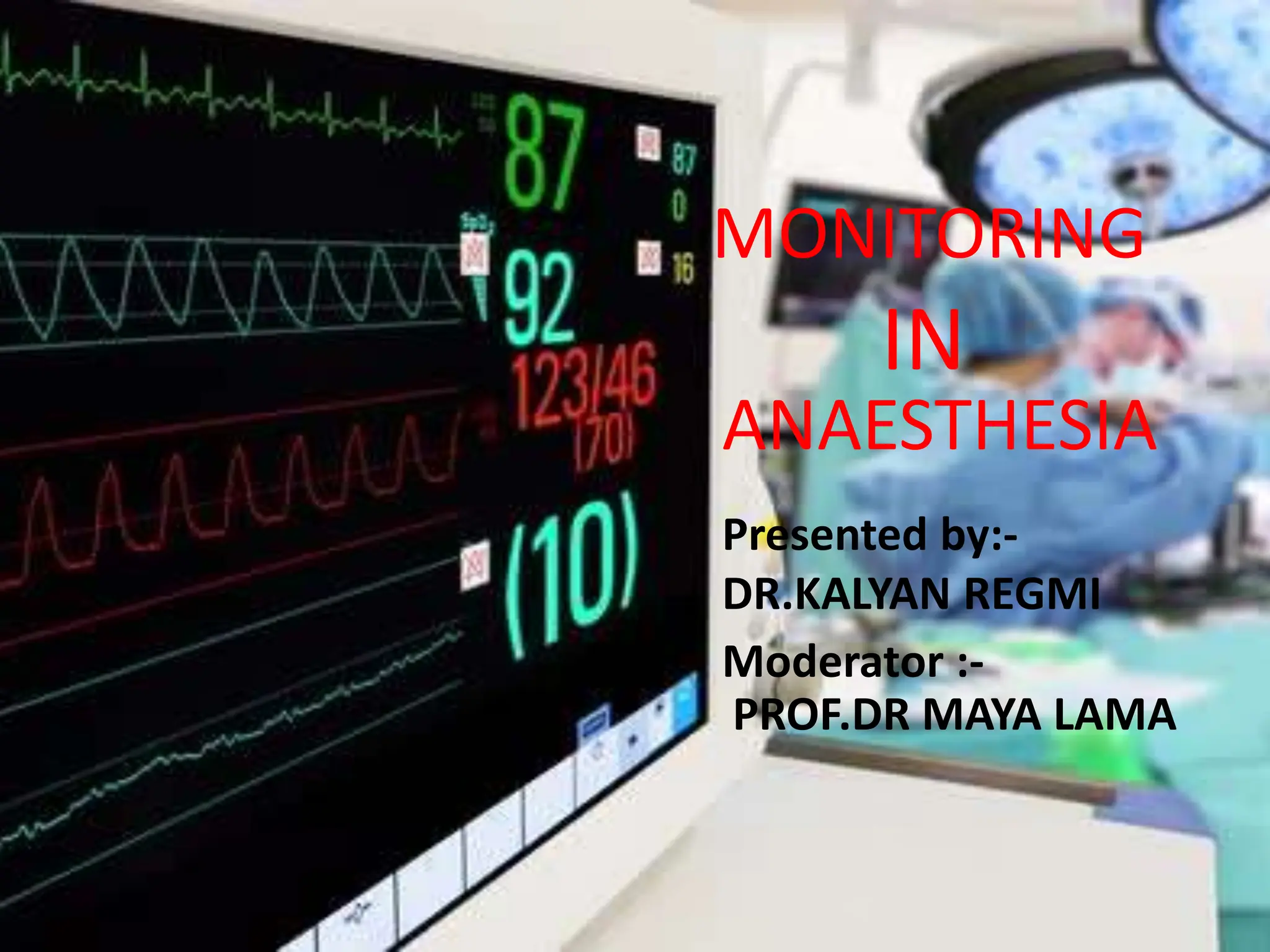
2. OBJECTIVES
• To know the important of monitoring in anesthesia.
• ASA Standards guideline for Basic Anesthetic Monitoring.
• Different types of monitoring device and there uses.
3. -Monitoring is an essential part of anesthesia care
- The term is derived from monere,Latin word- means - to
warn, remind.
- In anesthesia, monitoring means using both our senses
& electronic device to repeatedly or continuously
measure important variables in anaesthesized pt.
MONITORING
4. WHY MONITORING IS
IMPORTANT ??
1. To monitor physiological homeostasis ,prompt recognition
of adverse changes
2. To look responses to therapeutic intervention
3. To know either anesthetic equipment is Properly
functioning or not
4. Prompt response to adverse changes
5. -Standard I -Qualified anesthesia provider will be present
with the patient throughout the anesthetic procedure
-Standard II –Patient oxygenation, ventilation, circulation,
and temperature should be continually monitored.
The ASA Standards for Basic
Anesthetic Monitoring
6. -Assessment of oxygenation involves two parts:
measurement of inspired gas with an oxygen analyzer and
assessment of hemoglobin saturation with a pulse oximeter
and observation of skin color
- Assessment of ventilation is by clinical assessment and
preferably capnography
7. MONITORING DURING
ANESTHESIA
1. BASIC MONITORING
Basic monitoring is based on
• visual inspection
• Auscultation
• Palpation
which primary determine patient safety.
Any changes in clinical signs often precede
abnormalities in parameters measured by monitoring
devices.
12. -NON-INVASIVE BLOOD PRESSURE MONITORING:
• Measure blood pressure at set interval automatically
by automated oscillometry.
• Cuff sized should cover 2/3rd of arm
13. 2. SEMI-INVASIVE
-TRANESOPHAGEAL ECHOCARDIOGRAPHY:
• A transesophageal echocardiogram is done by inserting a
probe with a transducer down the esophagus.
• This provides a clearer image of the heart because the
sound waves do not have to pass through skin, muscle,
or bone tissue.
• Detect ischemia, air embolism ,valvular dysfunction etc
14. 3. INVASIVE
-INVASIVE BLOOD PRESSURE :
Beat to beat monitoring of blood pressure via
catheterization of artery i.e radial artery, ulnar artery,
femoral artery , brachial artery etc
-CENTRAL VENOUS PRESSURE :
• Measured by catheterization over the right internal
jugular vein.
• Normal value 2-6 mmHg.
• Other sites: subclavian vein ,femoral vein
15. Indication:-
• Continuous central venous pressure monitoring
• Open heart surgery
• Fluid management in shock
• Parenteral nutrition
• Venous access
21. RESPIRATORY MONITORING
1.PULSE OXIMETRY:-
-Measure oxygen saturation of hemoglobin and pulse rate on
non-invasive and continuous basis.
-Based on Beer-Lambert law
-Pulse oximetry combines the technology of
plethysmography and spectrophotometry.
-Pulse oximetry helps to detect hypoxia.
22. LIMITATION OF PULSE OXIMETRY
False high spo2 seen in
•Presence of abnormal Hb like
carboxyhemoglobin,
methemoglobin
False low spo2 seen in
•Nail polish
• Dark skin
•High venous pressure
23. 2. CAPNOGRAPHY
-Non-invasive measurement of partial pressure of CO2 in
exhaled breath expressed as CO2 concentration over time.
-Determination of end-tidal CO2 (ETCO2 ) concentration.
-Surest sign for confirming right placement of tube
29. 4.OXYGEN ANALYZER
-Fitted in inspiratory in limb of breathing circuit
- Monitor acutal value of oxygen delivered
- Used in closed circuit
30. 5.AIRWAY PRESSURE MONITORING
-Airway pressure should be less than30 cm of h20
-Low pressure : indicated disconection
-High pressure :indicated obstrution
31. 6. APNEA MONITORING
-Cessation of respiration >10 sec
INTUBATED PATIENT
-Capnography
-Airway pressure monitoring
-Pulseoximeter
NON-INTUBATED PATIENT
-Airflow at nostril
-Chest movement
-Pulse oximeter
32. NEUROMUSCULAR MONITORING
-Detecting the onset of paralysis during induction
- Adequacy of block
- Plan for extubation
1. TRAIN OF FOUR
-4 stimuli ,each of 2Hz for 2 sec are given and recorded.
-Normal: amplitude height of 4th and 1st twitches will be
same i.e T4/T1=1
-Twitches fade as nondepolarizing muscle relaxant block
increase.
36. CENTRAL NERVOUS SYSTEM
MONITORING
1.ELECTROENCEPHALOGRAPHY (EEG)
Indications-
-Cerebrovascular surgery to confirm the adequacy of cerebral
oxygenation.
-symmetry between the left and right hemispheres.
-To detect areas of cerebral ischemia
39. Examine four components-
(1) Low frequency, as found during deep anesthesia
(2) High-frequency beta activation found during “light”
anesthesia
(3) Suppressed EEG waves
(4) Burst suppression.
2.BISPECTRAL INDEX (BIS)
40.
41. TEMPERATURE MONITORING
Indications
1)Hypothermia -is associated with delayed drug
metabolism, increased blood glucose, vasoconstriction,
impaired coagulation, and impaired resistance to surgical
infections.
2)Hyperthermia - have deleterious effects perioperatively,
leading to tachycardia, vasodilation, and neurological injury.
44. URINE OUTPUT MONITORING
-Urinary bladder catheterization is the most reliable
method of monitoring urinary output
-Catheterization is routine in cardiac surgery, aortic or
renal vascular surgery, craniotomy, major abdominal
surgery in which large fluid shifts are expected
-Lengthy surgeries and intraoperative diuretic
administration