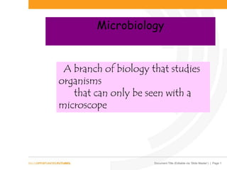
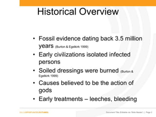
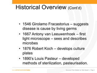
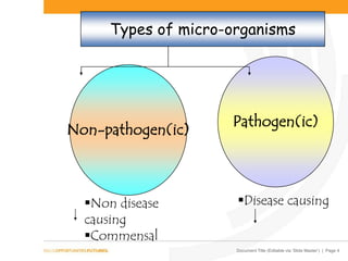
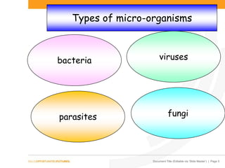
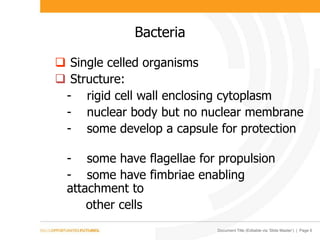
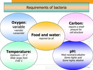
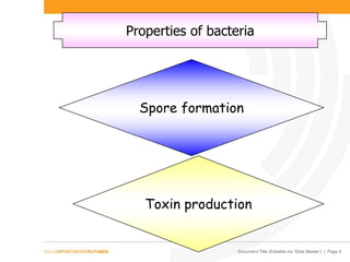
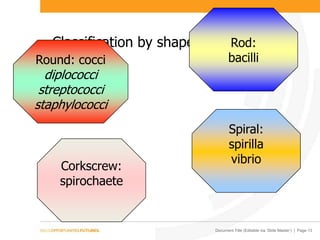
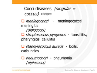
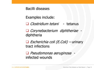
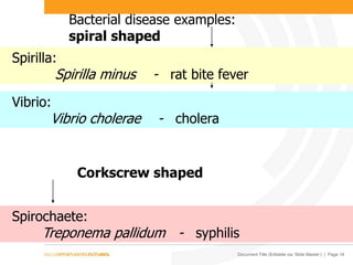
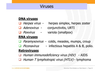
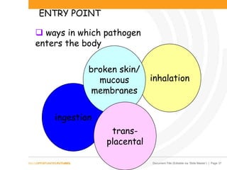
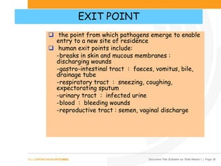
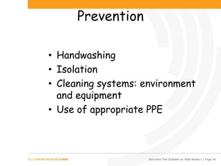
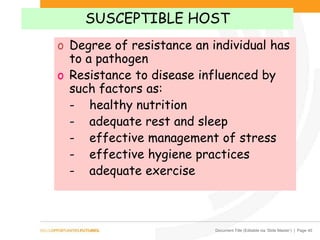
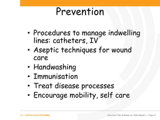
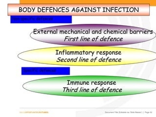
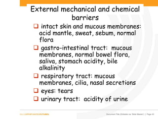
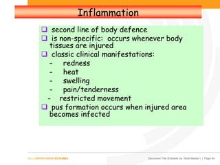
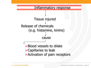
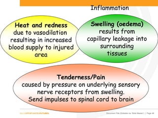
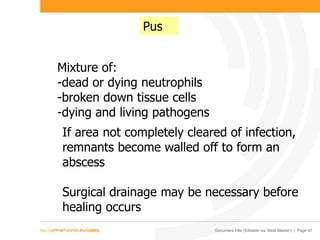
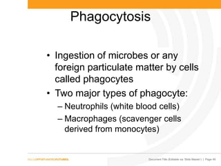
Document Title (Editable via ‘Slide Master’) | Page 1
Microbiology
A branch of biology that studies
organisms
that can only be seen with a
microscope
Document Title (Editable via ‘Slide Master’) | Page 2
Historical Overview
• Fossil evidence dating back 3.5 million
years (Burton & Egelkirk 1999)
• Early civilizations isolated infected
persons
• Soiled dressings were burned (Burton &
Egelkirk 1999)
• Causes believed to be the action of
gods
• Early treatments – leeches, bleeding
Document Title (Editable via ‘Slide Master’) | Page 3
Historical Overview (Cont’d)
• 1546 Girolamo Fracastorius – suggests
disease is cause by living germs
• 1667 Antony van Leeuwenhoek – first
light microscope – sees and describes
microbes
• 1876 Robert Koch – develops culture
plates
• 1890’s Louis Pasteur – developed
methods of sterilization, pasteurisation.
Document Title (Editable via ‘Slide Master’) | Page 4
Types of micro-organisms
Non-pathogen(ic)
Pathogen(ic)
Non disease
causing
Commensal
Disease causing
Document Title (Editable via ‘Slide Master’) | Page 5
Types of micro-organisms
bacteria
parasites
viruses
fungi
Document Title (Editable via ‘Slide Master’) | Page 6
Bacteria
 Single celled organisms
 Structure:
- rigid cell wall enclosing cytoplasm
- nuclear body but no nuclear membrane
- some develop a capsule for protection
- some have flagellae for propulsion
- some have fimbriae enabling
attachment to
other cells
Document Title (Editable via ‘Slide Master’) | Page 7
Requirements of bacteria
Oxygen:
variable
•aerobic
•anaerobic
pH:
Most neutral/sl.alkaline
Some highly acid
Some highly alkaline
Carbon:
require a small
amount for
cell structure
Temperature:
Optimum – 37 C
Wide range from
5-60 C
Food and water:
required by all
Document Title (Editable via ‘Slide Master’) | Page 8
Properties of bacteria
Spore formation
Toxin production
Document Title (Editable via ‘Slide Master’) | Page 9
Bacterial spores formation
Protective mechanism developed by
some bacteria to survive in adverse
conditions
Highly resistant to:
- high temperatures
- freezing
- sunlight
- disinfectants
Some spores (e.g. anthrax) have been
found to be active after 100 years of
being in inactive form
Document Title (Editable via ‘Slide Master’) | Page 10
ANTHRAX
Document Title (Editable via ‘Slide Master’) | Page 11
Production of toxins
2 types:
Exotoxins:
Living bacteria
released into and
cause damage to
surrounding
tissues
Endotoxins:
Remain in bacterial
wall
released on death
of bacterium
Document Title (Editable via ‘Slide Master’) | Page 12
Classification of bacteria
1. Staining properties
Gram positive
bacteria:
retain
violet
colour
Gram negative
bacteria:
retain
red
colour
Acid fast bacteria:
stain unable to penetrate due to
waxy envelope
Reactions of
bacteria to a
staining
technique
Document Title (Editable via ‘Slide Master’) | Page 13
Classification by shape
Round: cocci
diplococci
streptococci
staphylococci
Rod:
bacilli
Spiral:
spirilla
vibrio
Corkscrew:
spirochaete
Document Title (Editable via ‘Slide Master’) | Page 14
Cocci diseases (singular =
coccus)
 meningococci - meningococcal
meningitis
(diplococci)
 streptococcus pyogenes - tonsillitis,
pharyngitis, cellulitis
 staphylococcus aureus - boils,
carbuncles
 pneumococci - pneumonia
(diplococci)
Examples:
Document Title (Editable via ‘Slide Master’) | Page 15
Bacilli diseases
Examples include:
 Clostridium tetani - tetanus
 Corynebacterium diphtheriae -
diphtheria
 Escherichia coli (E.Coli) – urinary
tract infections
 Pseudomonas aeruginosa -
infected wounds
Document Title (Editable via ‘Slide Master’) | Page 16
Bacterial disease examples:
spiral shaped
Spirilla:
Spirilla minus - rat bite fever
Vibrio:
Vibrio cholerae - cholera
Spirochaete:
Treponema pallidum - syphilis
Corkscrew shaped
Document Title (Editable via ‘Slide Master’) | Page 17
CHOLERA
Document Title (Editable via ‘Slide Master’) | Page 18
Specialised bacteria
Rickettsiae and chlamydiae
o smaller than most bacteria
o can only reproduce in living cells - are
therefore parasites
o rickettsiae often carried by fleas, ticks,
lice (vectors)
Disease examples:
 Rickettsia australis - Queensland tick
typhus
 Chlamydia trachomatis - trachoma,
salpingitis
Document Title (Editable via ‘Slide Master’) | Page 19
Specialised bacteria
Mycoplasma
• ultramicroscopic bacteria
• do not have a cell wall
Diseases caused:
• certain respiratory and genital tract
diseases
Document Title (Editable via ‘Slide Master’) | Page 20
Viruses
• ultramicroscopic
• no cell structure
• no rigid cell wall
• are intracellular parasites
• can only reproduce within a host cell
• composed of either DNA or RNA but
not both
• can lie dormant in cells with
activation occurring at a later time
Document Title (Editable via ‘Slide Master’) | Page 21
VIRUS
Document Title (Editable via ‘Slide Master’) | Page 22
Viruses
DNA viruses
 Herpes virus - herpes simplex, herpes zoster
 Adenovirus - conjunctivitis, URTI
 Poxvirus - variola (smallpox)
RNA viruses
 Paramyxovirus - colds, measles, mumps, croup
 Piconavirus - infectious hepatitis A & B, polio.
Retroviruses
 Human immunodeficiency virus (HIV) - AIDS
 Human T lymphotropic virus (HTLV) - lymphoma
Document Title (Editable via ‘Slide Master’) | Page 23
“BIRD FLU” VIRUS
Document Title (Editable via ‘Slide Master’) | Page 24
SMALLPOX VIRUS
Document Title (Editable via ‘Slide Master’) | Page 25
VIRUS
Document Title (Editable via ‘Slide Master’) | Page 26
Fungi
• Plant organisms: mainly moulds
and yeasts
• Do not contain chlorophyll
• Present in soil, air, water
• Multiply by producing spores
• Most non-pathogenic
• A few are pathogenic
Document Title (Editable via ‘Slide Master’) | Page 27
Pathogenic fungi (mycoses)
Superficial:
skin, mucous
membranes,
hair,
nails
Intermediate:
subcutaneous
tissue
Systemic:
deep tissues
organs
3 types
of
mycotic
infections
Document Title (Editable via ‘Slide Master’) | Page 28
Fungi
Diseases caused
Dermatophytes:
o Tinea pedis - athlete’s foot
o Tinea unguium – toenail infections
Candidiases (moniliasis):
o Candida species - oral thrush, skin
infections
o Aspergillus - aspergillosis
(respiratory disease)
Document Title (Editable via ‘Slide Master’) | Page 29
TINEA PEDIS
Document Title (Editable via ‘Slide Master’) | Page 30
Parasites
• Survival requirements depend on a
living organism – the host
• Parasite-host effects variable: from little
damage to death
• Classification includes:
helminths – multicellular animals
(worms) e.g. flatworms (platyhelminths)
: roundworms (nematodes) : flukes
(trematodes)
protozoa - single celled animal
organisms transmitted to humans by
insects
Document Title (Editable via ‘Slide Master’) | Page 31
Pathogenic parasites
o Trematodes (flukes) : liver, lungs or
intestinal infestation
o Nematodes – round worm infestation
o Echinococcus granulosis (dog tapeworm) –
hydatid cysts
o Protozoa – malaria, toxoplasmosis
Document Title (Editable via ‘Slide Master’) | Page 32
PEDICULI CAPITUS
Document Title (Editable via ‘Slide Master’) | Page 33PEDICULI
Document Title (Editable via ‘Slide Master’) | Page 34
Prevention
• Immunisation
• Cleaning processes: environment &
equipment
• Health status of staff, visitors etc
• Provision of clean water, food &
sanitation
Document Title (Editable via ‘Slide Master’) | Page 35
Prevention
• PPE
• Management of waste and body
fluids
• Handwashing
Document Title (Editable via ‘Slide Master’) | Page 36
Transmission modes
 the mode of movement of pathogen from exit point to new
host
Airborne:
droplets
dust particles
Contact:
direct
indirect
Ingestion:
infected food/
water/utensils/
objects
hands
Vectors:
Flies, rats, mosquitos
Document Title (Editable via ‘Slide Master’) | Page 37
ENTRY POINT
 ways in which pathogen
enters the body
ingestion
inhalation
broken skin/
mucous
membranes
trans-
placental
Document Title (Editable via ‘Slide Master’) | Page 38
EXIT POINT
 the point from which pathogens emerge to enable
entry to a new site of residence
 human exit points include:
-breaks in skin and mucous membranes :
discharging wounds
-gastro-intestinal tract : faeces, vomitus, bile,
drainage tube
-respiratory tract : sneezing, coughing,
expectorating sputum
-urinary tract : infected urine
-blood : bleeding wounds
-reproductive tract : semen, vaginal discharge
Document Title (Editable via ‘Slide Master’) | Page 39
Prevention
• Handwashing
• Isolation
• Cleaning systems: environment
and equipment
• Use of appropriate PPE
Document Title (Editable via ‘Slide Master’) | Page 40
SUSCEPTIBLE HOST
o Degree of resistance an individual has
to a pathogen
o Resistance to disease influenced by
such factors as:
- healthy nutrition
- adequate rest and sleep
- effective management of stress
- effective hygiene practices
- adequate exercise
Document Title (Editable via ‘Slide Master’) | Page 41
Prevention
• Procedures to manage indwelling
lines: catheters, IV
• Aseptic techniques for wound
care
• Handwashing
• Immunisation
• Treat disease processes
• Encourage mobility, self care
Document Title (Editable via ‘Slide Master’) | Page 42
BODY DEFENCES AGAINST INFECTION
External mechanical and chemical barriers
First line of defence
Inflammatory response
Second line of defence
Immune response
Third line of defence
Non-specific defences
Specific defences
Document Title (Editable via ‘Slide Master’) | Page 43
External mechanical and chemical
barriers
 intact skin and mucous membranes:
acid mantle, sweat, sebum, normal
flora
 gastro-intestinal tract: mucous
membranes, normal bowel flora,
saliva, stomach acidity, bile
alkalinity
 respiratory tract: mucous
membranes, cilia, nasal secretions
 eyes: tears
 urinary tract: acidity of urine
Document Title (Editable via ‘Slide Master’) | Page 44
Inflammation
 second line of body defence
 is non-specific: occurs whenever body
tissues are injured
 classic clinical manifestations:
- redness
- heat
- swelling
- pain/tenderness
- restricted movement
 pus formation occurs when injured area
becomes infected
Document Title (Editable via ‘Slide Master’) | Page 45
Inflammatory response
Tissue injured
Release of chemicals
(e.g. histamine, kinins)
Blood vessels to dilate
Capillaries to leak
Activation of pain receptors
cause
Document Title (Editable via ‘Slide Master’) | Page 46
Inflammation
Heat and redness
due to vasodilation
resulting in increased
blood supply to injured
area
Tenderness/Pain
caused by pressure on underlying sensory
nerve receptors from swelling.
Send impulses to spinal cord to brain
Swelling (oedema)
results from
capillary leakage into
surrounding
tissues
Document Title (Editable via ‘Slide Master’) | Page 47
Pus
Mixture of:
-dead or dying neutrophils
-broken down tissue cells
-dying and living pathogens
If area not completely cleared of infection,
remnants become walled off to form an
abscess
Surgical drainage may be necessary before
healing occurs
Document Title (Editable via ‘Slide Master’) | Page 48
Other non-specific defences
• Complement fixation
- 22 plasma proteins
-attach to foreign cells
• Interferons
- proteins secreted by virus infected cells
• Natural Killer Cells
– Lymphocytes that destroy infectious microbes plus
certain spontaneously arising tumour cells
• Fever
- temperature rise may kill certain pathogens
Document Title (Editable via ‘Slide Master’) | Page 49
Phagocytosis
• Ingestion of microbes or any
foreign particulate matter by cells
called phagocytes
• Two major types of phagocyte:
– Neutrophils (white blood cells)
– Macrophages (scavenger cells
derived from monocytes)
Document Title (Editable via ‘Slide Master’) | Page 50
Mechanism of Phagocytosis
• Three phases:
– Chemotaxis – activated complement
proteins cause chemical attraction of
phagocytes to a particular location
– Adherence – attachment of the plasma
membrane of the phagocyte to the
surface of the foreign material.
– Ingestion – the cell membrane of the
phagocyte extends projections that engulf
the micro-organism
Document Title (Editable via ‘Slide Master’) | Page 51
Immune Response
o Third line of body defence
o Response is specific, systematic
and has a memory
o Cells of the immune system are:
- B lymphocytes
- T lymphocytes
- Macrophages
Document Title (Editable via ‘Slide Master’) | Page 52
Immune response
o Two kinds – closely allied
o Both triggered by antigens
o Cell-mediated (cellular) immune
responses (CMI)
o T cells proliferate into “killer” cells and
directly attack the invading antigen
o Antibody-mediated (humoral) immune
responses (AMI)
o B cells transform into plasma cells which
synthesize and secrete specific proteins
called antibodies or immunoglobulin's
Document Title (Editable via ‘Slide Master’) | Page 53
Formation of T Cells and B Cells
• Both develop from haemopoietic
stem cells in red bone marrow
• B cells complete maturity in bone
marrow
• T cells leave as pre-T cells and
migrate to the thymus gland to
mature
Document Title (Editable via ‘Slide Master’) | Page 54
Immune system cells
B lymphocytes:
-produce antibodies
(immunoglobulins)
T lymphocytes:
-non-antibody
producing
Macrophages:
-engulf foreign
particles
Humoral (antibody
mediated) immunity
Cell mediated and
humoral immunity
Cell-mediated (cellular)
immunity
Document Title (Editable via ‘Slide Master’) | Page 55
Immunity
Any substance capable
of
stimulating the
immune system and
causing an
immune response
Are immunoglobulins
(Igs)
produced in the body
in response to
Antigens
Five classes:
-IgM, IgA, IgD,
-IgG, IgE
Antigens:
(Ags) Antibodies: (Ab)
Document Title (Editable via ‘Slide Master’) | Page 56
Functions of Antibodies
• Neutralizing antigen
• Immobilization of bacteria
• Agglutination and precipitation of
antigen
• Activation of complement
• Enhancing phagocytosis
• Providing foetal and newborn
immunity
Document Title (Editable via ‘Slide Master’) | Page 57
Immunity
Innate
(genetic, inborn)
Acquired
Naturally acquired Artificially acquired
Active
Protection acquired by
getting the disease
Passive
Antibodies passively cross
from mother to baby
via placenta or breast milk
Active
Protection acquired by
immunisation with vaccine
that stimulates body to
actively produce own
antibodies
Passive
Protection acquired
by direct injection
of antibodies
(immunoglobulins)
Ref: Tabbner, 2005, p.331 (Adapted from Herlihy and Maebius: 2000, p.354)
Document Title (Editable via ‘Slide Master’) | Page 58
Immunological Memory
• Memory for certain antigens
triggers immune responses
• Immune responses are much
quicker and more intense after a
second exposure to an antigen
• Basis for immunization
Document Title (Editable via ‘Slide Master’) | Page 59
References
• Burton, G., and Engelkirk, P., (1999).
Microbiology for the Health Sciences,
Lippincott Williams & Wilkins, Philadelphia
• TAFE Frontiers, (2003). Learners Resource:
Microbiology and Wound Management,
Dept. of Education, Employment and
Training. Victoria
• Marieb, E., (2006). Essentials of Human
Anatomy & Physiology (8E). Pearson. San
Francisco
• Tortora, G. J., & Grabowski, S., (1996).
Principles of Anatomy and Physiology (8 E).
Harper Collins. New York

Microbiology 2014

  • 1.
    Document Title (Editablevia ‘Slide Master’) | Page 1 Microbiology A branch of biology that studies organisms that can only be seen with a microscope
  • 2.
    Document Title (Editablevia ‘Slide Master’) | Page 2 Historical Overview • Fossil evidence dating back 3.5 million years (Burton & Egelkirk 1999) • Early civilizations isolated infected persons • Soiled dressings were burned (Burton & Egelkirk 1999) • Causes believed to be the action of gods • Early treatments – leeches, bleeding
  • 3.
    Document Title (Editablevia ‘Slide Master’) | Page 3 Historical Overview (Cont’d) • 1546 Girolamo Fracastorius – suggests disease is cause by living germs • 1667 Antony van Leeuwenhoek – first light microscope – sees and describes microbes • 1876 Robert Koch – develops culture plates • 1890’s Louis Pasteur – developed methods of sterilization, pasteurisation.
  • 4.
    Document Title (Editablevia ‘Slide Master’) | Page 4 Types of micro-organisms Non-pathogen(ic) Pathogen(ic) Non disease causing Commensal Disease causing
  • 5.
    Document Title (Editablevia ‘Slide Master’) | Page 5 Types of micro-organisms bacteria parasites viruses fungi
  • 6.
    Document Title (Editablevia ‘Slide Master’) | Page 6 Bacteria  Single celled organisms  Structure: - rigid cell wall enclosing cytoplasm - nuclear body but no nuclear membrane - some develop a capsule for protection - some have flagellae for propulsion - some have fimbriae enabling attachment to other cells
  • 7.
    Document Title (Editablevia ‘Slide Master’) | Page 7 Requirements of bacteria Oxygen: variable •aerobic •anaerobic pH: Most neutral/sl.alkaline Some highly acid Some highly alkaline Carbon: require a small amount for cell structure Temperature: Optimum – 37 C Wide range from 5-60 C Food and water: required by all
  • 8.
    Document Title (Editablevia ‘Slide Master’) | Page 8 Properties of bacteria Spore formation Toxin production
  • 9.
    Document Title (Editablevia ‘Slide Master’) | Page 9 Bacterial spores formation Protective mechanism developed by some bacteria to survive in adverse conditions Highly resistant to: - high temperatures - freezing - sunlight - disinfectants Some spores (e.g. anthrax) have been found to be active after 100 years of being in inactive form
  • 10.
    Document Title (Editablevia ‘Slide Master’) | Page 10 ANTHRAX
  • 11.
    Document Title (Editablevia ‘Slide Master’) | Page 11 Production of toxins 2 types: Exotoxins: Living bacteria released into and cause damage to surrounding tissues Endotoxins: Remain in bacterial wall released on death of bacterium
  • 12.
    Document Title (Editablevia ‘Slide Master’) | Page 12 Classification of bacteria 1. Staining properties Gram positive bacteria: retain violet colour Gram negative bacteria: retain red colour Acid fast bacteria: stain unable to penetrate due to waxy envelope Reactions of bacteria to a staining technique
  • 13.
    Document Title (Editablevia ‘Slide Master’) | Page 13 Classification by shape Round: cocci diplococci streptococci staphylococci Rod: bacilli Spiral: spirilla vibrio Corkscrew: spirochaete
  • 14.
    Document Title (Editablevia ‘Slide Master’) | Page 14 Cocci diseases (singular = coccus)  meningococci - meningococcal meningitis (diplococci)  streptococcus pyogenes - tonsillitis, pharyngitis, cellulitis  staphylococcus aureus - boils, carbuncles  pneumococci - pneumonia (diplococci) Examples:
  • 15.
    Document Title (Editablevia ‘Slide Master’) | Page 15 Bacilli diseases Examples include:  Clostridium tetani - tetanus  Corynebacterium diphtheriae - diphtheria  Escherichia coli (E.Coli) – urinary tract infections  Pseudomonas aeruginosa - infected wounds
  • 16.
    Document Title (Editablevia ‘Slide Master’) | Page 16 Bacterial disease examples: spiral shaped Spirilla: Spirilla minus - rat bite fever Vibrio: Vibrio cholerae - cholera Spirochaete: Treponema pallidum - syphilis Corkscrew shaped
  • 17.
    Document Title (Editablevia ‘Slide Master’) | Page 17 CHOLERA
  • 18.
    Document Title (Editablevia ‘Slide Master’) | Page 18 Specialised bacteria Rickettsiae and chlamydiae o smaller than most bacteria o can only reproduce in living cells - are therefore parasites o rickettsiae often carried by fleas, ticks, lice (vectors) Disease examples:  Rickettsia australis - Queensland tick typhus  Chlamydia trachomatis - trachoma, salpingitis
  • 19.
    Document Title (Editablevia ‘Slide Master’) | Page 19 Specialised bacteria Mycoplasma • ultramicroscopic bacteria • do not have a cell wall Diseases caused: • certain respiratory and genital tract diseases
  • 20.
    Document Title (Editablevia ‘Slide Master’) | Page 20 Viruses • ultramicroscopic • no cell structure • no rigid cell wall • are intracellular parasites • can only reproduce within a host cell • composed of either DNA or RNA but not both • can lie dormant in cells with activation occurring at a later time
  • 21.
    Document Title (Editablevia ‘Slide Master’) | Page 21 VIRUS
  • 22.
    Document Title (Editablevia ‘Slide Master’) | Page 22 Viruses DNA viruses  Herpes virus - herpes simplex, herpes zoster  Adenovirus - conjunctivitis, URTI  Poxvirus - variola (smallpox) RNA viruses  Paramyxovirus - colds, measles, mumps, croup  Piconavirus - infectious hepatitis A & B, polio. Retroviruses  Human immunodeficiency virus (HIV) - AIDS  Human T lymphotropic virus (HTLV) - lymphoma
  • 23.
    Document Title (Editablevia ‘Slide Master’) | Page 23 “BIRD FLU” VIRUS
  • 24.
    Document Title (Editablevia ‘Slide Master’) | Page 24 SMALLPOX VIRUS
  • 25.
    Document Title (Editablevia ‘Slide Master’) | Page 25 VIRUS
  • 26.
    Document Title (Editablevia ‘Slide Master’) | Page 26 Fungi • Plant organisms: mainly moulds and yeasts • Do not contain chlorophyll • Present in soil, air, water • Multiply by producing spores • Most non-pathogenic • A few are pathogenic
  • 27.
    Document Title (Editablevia ‘Slide Master’) | Page 27 Pathogenic fungi (mycoses) Superficial: skin, mucous membranes, hair, nails Intermediate: subcutaneous tissue Systemic: deep tissues organs 3 types of mycotic infections
  • 28.
    Document Title (Editablevia ‘Slide Master’) | Page 28 Fungi Diseases caused Dermatophytes: o Tinea pedis - athlete’s foot o Tinea unguium – toenail infections Candidiases (moniliasis): o Candida species - oral thrush, skin infections o Aspergillus - aspergillosis (respiratory disease)
  • 29.
    Document Title (Editablevia ‘Slide Master’) | Page 29 TINEA PEDIS
  • 30.
    Document Title (Editablevia ‘Slide Master’) | Page 30 Parasites • Survival requirements depend on a living organism – the host • Parasite-host effects variable: from little damage to death • Classification includes: helminths – multicellular animals (worms) e.g. flatworms (platyhelminths) : roundworms (nematodes) : flukes (trematodes) protozoa - single celled animal organisms transmitted to humans by insects
  • 31.
    Document Title (Editablevia ‘Slide Master’) | Page 31 Pathogenic parasites o Trematodes (flukes) : liver, lungs or intestinal infestation o Nematodes – round worm infestation o Echinococcus granulosis (dog tapeworm) – hydatid cysts o Protozoa – malaria, toxoplasmosis
  • 32.
    Document Title (Editablevia ‘Slide Master’) | Page 32 PEDICULI CAPITUS
  • 33.
    Document Title (Editablevia ‘Slide Master’) | Page 33PEDICULI
  • 34.
    Document Title (Editablevia ‘Slide Master’) | Page 34 Prevention • Immunisation • Cleaning processes: environment & equipment • Health status of staff, visitors etc • Provision of clean water, food & sanitation
  • 35.
    Document Title (Editablevia ‘Slide Master’) | Page 35 Prevention • PPE • Management of waste and body fluids • Handwashing
  • 36.
    Document Title (Editablevia ‘Slide Master’) | Page 36 Transmission modes  the mode of movement of pathogen from exit point to new host Airborne: droplets dust particles Contact: direct indirect Ingestion: infected food/ water/utensils/ objects hands Vectors: Flies, rats, mosquitos
  • 37.
    Document Title (Editablevia ‘Slide Master’) | Page 37 ENTRY POINT  ways in which pathogen enters the body ingestion inhalation broken skin/ mucous membranes trans- placental
  • 38.
    Document Title (Editablevia ‘Slide Master’) | Page 38 EXIT POINT  the point from which pathogens emerge to enable entry to a new site of residence  human exit points include: -breaks in skin and mucous membranes : discharging wounds -gastro-intestinal tract : faeces, vomitus, bile, drainage tube -respiratory tract : sneezing, coughing, expectorating sputum -urinary tract : infected urine -blood : bleeding wounds -reproductive tract : semen, vaginal discharge
  • 39.
    Document Title (Editablevia ‘Slide Master’) | Page 39 Prevention • Handwashing • Isolation • Cleaning systems: environment and equipment • Use of appropriate PPE
  • 40.
    Document Title (Editablevia ‘Slide Master’) | Page 40 SUSCEPTIBLE HOST o Degree of resistance an individual has to a pathogen o Resistance to disease influenced by such factors as: - healthy nutrition - adequate rest and sleep - effective management of stress - effective hygiene practices - adequate exercise
  • 41.
    Document Title (Editablevia ‘Slide Master’) | Page 41 Prevention • Procedures to manage indwelling lines: catheters, IV • Aseptic techniques for wound care • Handwashing • Immunisation • Treat disease processes • Encourage mobility, self care
  • 42.
    Document Title (Editablevia ‘Slide Master’) | Page 42 BODY DEFENCES AGAINST INFECTION External mechanical and chemical barriers First line of defence Inflammatory response Second line of defence Immune response Third line of defence Non-specific defences Specific defences
  • 43.
    Document Title (Editablevia ‘Slide Master’) | Page 43 External mechanical and chemical barriers  intact skin and mucous membranes: acid mantle, sweat, sebum, normal flora  gastro-intestinal tract: mucous membranes, normal bowel flora, saliva, stomach acidity, bile alkalinity  respiratory tract: mucous membranes, cilia, nasal secretions  eyes: tears  urinary tract: acidity of urine
  • 44.
    Document Title (Editablevia ‘Slide Master’) | Page 44 Inflammation  second line of body defence  is non-specific: occurs whenever body tissues are injured  classic clinical manifestations: - redness - heat - swelling - pain/tenderness - restricted movement  pus formation occurs when injured area becomes infected
  • 45.
    Document Title (Editablevia ‘Slide Master’) | Page 45 Inflammatory response Tissue injured Release of chemicals (e.g. histamine, kinins) Blood vessels to dilate Capillaries to leak Activation of pain receptors cause
  • 46.
    Document Title (Editablevia ‘Slide Master’) | Page 46 Inflammation Heat and redness due to vasodilation resulting in increased blood supply to injured area Tenderness/Pain caused by pressure on underlying sensory nerve receptors from swelling. Send impulses to spinal cord to brain Swelling (oedema) results from capillary leakage into surrounding tissues
  • 47.
    Document Title (Editablevia ‘Slide Master’) | Page 47 Pus Mixture of: -dead or dying neutrophils -broken down tissue cells -dying and living pathogens If area not completely cleared of infection, remnants become walled off to form an abscess Surgical drainage may be necessary before healing occurs
  • 48.
    Document Title (Editablevia ‘Slide Master’) | Page 48 Other non-specific defences • Complement fixation - 22 plasma proteins -attach to foreign cells • Interferons - proteins secreted by virus infected cells • Natural Killer Cells – Lymphocytes that destroy infectious microbes plus certain spontaneously arising tumour cells • Fever - temperature rise may kill certain pathogens
  • 49.
    Document Title (Editablevia ‘Slide Master’) | Page 49 Phagocytosis • Ingestion of microbes or any foreign particulate matter by cells called phagocytes • Two major types of phagocyte: – Neutrophils (white blood cells) – Macrophages (scavenger cells derived from monocytes)
  • 50.
    Document Title (Editablevia ‘Slide Master’) | Page 50 Mechanism of Phagocytosis • Three phases: – Chemotaxis – activated complement proteins cause chemical attraction of phagocytes to a particular location – Adherence – attachment of the plasma membrane of the phagocyte to the surface of the foreign material. – Ingestion – the cell membrane of the phagocyte extends projections that engulf the micro-organism
  • 51.
    Document Title (Editablevia ‘Slide Master’) | Page 51 Immune Response o Third line of body defence o Response is specific, systematic and has a memory o Cells of the immune system are: - B lymphocytes - T lymphocytes - Macrophages
  • 52.
    Document Title (Editablevia ‘Slide Master’) | Page 52 Immune response o Two kinds – closely allied o Both triggered by antigens o Cell-mediated (cellular) immune responses (CMI) o T cells proliferate into “killer” cells and directly attack the invading antigen o Antibody-mediated (humoral) immune responses (AMI) o B cells transform into plasma cells which synthesize and secrete specific proteins called antibodies or immunoglobulin's
  • 53.
    Document Title (Editablevia ‘Slide Master’) | Page 53 Formation of T Cells and B Cells • Both develop from haemopoietic stem cells in red bone marrow • B cells complete maturity in bone marrow • T cells leave as pre-T cells and migrate to the thymus gland to mature
  • 54.
    Document Title (Editablevia ‘Slide Master’) | Page 54 Immune system cells B lymphocytes: -produce antibodies (immunoglobulins) T lymphocytes: -non-antibody producing Macrophages: -engulf foreign particles Humoral (antibody mediated) immunity Cell mediated and humoral immunity Cell-mediated (cellular) immunity
  • 55.
    Document Title (Editablevia ‘Slide Master’) | Page 55 Immunity Any substance capable of stimulating the immune system and causing an immune response Are immunoglobulins (Igs) produced in the body in response to Antigens Five classes: -IgM, IgA, IgD, -IgG, IgE Antigens: (Ags) Antibodies: (Ab)
  • 56.
    Document Title (Editablevia ‘Slide Master’) | Page 56 Functions of Antibodies • Neutralizing antigen • Immobilization of bacteria • Agglutination and precipitation of antigen • Activation of complement • Enhancing phagocytosis • Providing foetal and newborn immunity
  • 57.
    Document Title (Editablevia ‘Slide Master’) | Page 57 Immunity Innate (genetic, inborn) Acquired Naturally acquired Artificially acquired Active Protection acquired by getting the disease Passive Antibodies passively cross from mother to baby via placenta or breast milk Active Protection acquired by immunisation with vaccine that stimulates body to actively produce own antibodies Passive Protection acquired by direct injection of antibodies (immunoglobulins) Ref: Tabbner, 2005, p.331 (Adapted from Herlihy and Maebius: 2000, p.354)
  • 58.
    Document Title (Editablevia ‘Slide Master’) | Page 58 Immunological Memory • Memory for certain antigens triggers immune responses • Immune responses are much quicker and more intense after a second exposure to an antigen • Basis for immunization
  • 59.
    Document Title (Editablevia ‘Slide Master’) | Page 59 References • Burton, G., and Engelkirk, P., (1999). Microbiology for the Health Sciences, Lippincott Williams & Wilkins, Philadelphia • TAFE Frontiers, (2003). Learners Resource: Microbiology and Wound Management, Dept. of Education, Employment and Training. Victoria • Marieb, E., (2006). Essentials of Human Anatomy & Physiology (8E). Pearson. San Francisco • Tortora, G. J., & Grabowski, S., (1996). Principles of Anatomy and Physiology (8 E). Harper Collins. New York