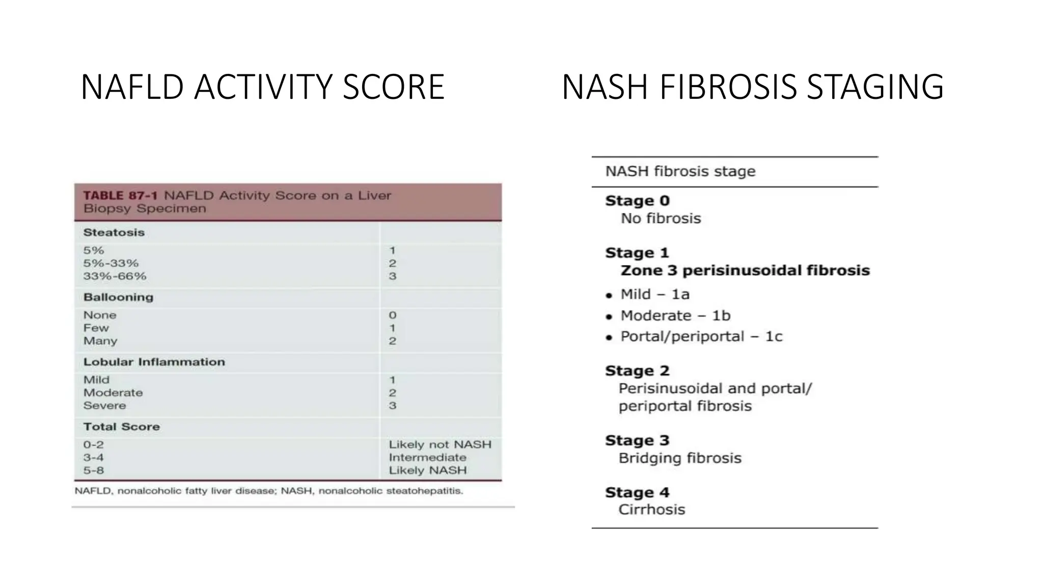
NON ALCOHOLIC FATTY LIVER
DISEASE AND NON ALCOHOLIC
STEATOHEPATITIS
PRESENTOR : DR BHARATH S J MODERATOR : DR PARIKSHITH J
JUNIOR RESIDENT ASSISTANT PROFFESSOR
GENERAL MEDICINE GENERAL MEDICINE
OUTLINE
• MASLD NOMENCLATURE
• INTRODUCTION
• SPECTRUM OF STEATOTIC LIVER DISEASES.
• CRITERIA FOR MASLD.
• PREVALENCE
• ETIOPATHOGENESIS.
• CLINICAL FEATURES
• APPROACH TO NAFLD AND NASH
• MANAGEMENT
Metabolic dysfunction Associated Steatotic
Liver Disease(MASLD).
INTRODUCTION.
• A global delphi consensus process co-led by American Association for
the study of Liver Diseases (AASLD) and the European Association for
the study of Liver (EASL) in collaboration with the Latin American
Association for the study of Liver (ALEH) recommended renaming of
NAFLD to MASLD on June 24, 2023.
• Steatotic Liver Disease (SLD) was chosen as an overarching term to
classify individuals with hepatic steatosis(>5% fat in liver) due to
various etiologies.
• The term SLD includes MASLD, MetALD, ALD, Specific etiology SLD,
Cryptogenic SLD.
SPECTRUM OF STEATOTIC LIVER DISEASES
• MASLD is defined as presence of hepatic steatosis with one or more
cardiometabolic risk factors (CMRF), and no other identifiable cause
of steatosis.
• Similarly NASH was replaced by MASH (Metabolic dysfunction
Associated Steatohepatitis) and NAFL was replaced by MAFL
(Metabolic dysfunction associated steatotic liver).
• Patients with hepatic steatosis, CMRFs and alcohol use are classified
as having MetALD.
• Patients with steatosis consuming alcohol in excess of 50g/day in
females or 60g/day in males or weekly equivalent are classified as
Alcohol associated liver disease(ALD).
CRITERIA FOR MASLD
• Studies suggest a near complete overlap (99%) between MASLD
defined populations and the historical NAFLD populations.
• All recommendations in the AASLD Practice guidance on clinical
assessment and management of NAFLD can be applied to patients
with MASLD and MASH.
PREVALENCE OF NAFLD
• NAFLD is the most common cause of Chronic liver disease in US and
worldwide.
• NAFLD risk is 4-10 times higher in patients with metabolic syndrome.
• NAFLD is further classified as NAFL (isolated steatosis) and NASH
(steatosis complicated by liver cell injury and accumulation of
inflammatory cells).
• Around 20-25% of NAFLD patients develop NASH.
• Around 6% of NASH patients develop cirrhosis and 1-2% individuals
will progress to hepatocellular carcinoma.
• NAFLD is more common in males than females in premenopausal age
group, followed by later peak in postmenopausal women.
ETIOPATHOGENESIS
• RISK FACTORS
COMMON RISK FACTORS OTHER RISK
FACTORS
GENETIC
POLYMORPHISM
Obesity Hypothyroidism PNPLA3 gene
variants
Diabetes mellitus Obstructive sleep
apnea
TM6SF2-
cholesterol
metabolism
Dyslipidemia Hypopituitary,
hypogonadism
MBOAT7-
phospholipid
metabolism
Metabolic syndrome Alterations in gut
microbiome.
CIDEB
hypertension. PCOD HSD17B13
ALTERNATIVE CAUSES OF HEPATIC STEATOSIS
MICROVESICULAR STEATOSIS MACROVESICULAR STEATOSIS DRUGS CAUSING STEATOSIS
Pregnancy - AFLP Chronic viral hepatitis – HCV Tetracyclines
Reyes syndrome Wilsons disease Valproate
Eclampsia, HELLP syndrome NASH Zidovudine
Tetracyclines Amiodarone, bleomycin
Valproate toxicity. Estrogens, steroids
Alcohol Metals-chromium barium antimony
Acid lipase deficiency.
• TWO HIT HYPOTHESIS
• First hit in the form of sedentary lifestyle, obesity, high fat diet, diabetes
mellitus, insulin resistance lead to accumulation of fats within hepatocytes.
• Obesity lead to altered gut microbiota and increased hepatic exposure to
gut derived products as well adipokines together are responsible for insulin
resistance.
• Second hit in the form of hepatocyte lipotoxicity (due to diacylglycerols,
fatty acids), oxidative stress (reactive oxygen species) lead to activation of
inflammatory cytokines and hepatocyte cell death.
• This lead to activation of myofibroblasts, progenitor cells that lead to
progressive accumulation of wound healing cells, fibrous matrix, abnormal
vasculature resulting in irreversible fibrosis.
• Cirrhosis and hepatocellular carcinoma are the potential outcomes of
NASH.
CLINICAL FEATURES
• Most patients with NAFLD are asymptomatic
• Some present with vague RUQ abdominal pain fatigue malaise.
• Hepatomegaly can be found on abdominal examination.
• Signs of chronic liver disease like splenomegaly palmar erythema
spider angiomata ascites can be seen in some patients.
• Most of the patients will be associated with obesity diabetes
hypertension dyslipidemia cardiovascular disease.
APPROACH TO NAFLD AND NASH
BIOCHEMICAL PARAMETERS
• LFT shows ALT and AST elevations 2 to 5 times upper limit of normal
(30-150IU/L).
• AST/ALT ratio is less than 1 unlike that of alcoholic liver disease.
• Serum ferritin can be elevated
• S Bilirubin albumin prothrombin time are usually normal in NAFLD
except in patients with cirrhosis.
• In Isolated fatty liver (NAFL) liver may not be enlarged and
aminotransferases and LFT may be completely normal.
• Risk factors for NAFLD should be evaluated – body mass index,
diabetes, lipid profile, PCOD, thyroid function tests etc.
• Other causes of fatty liver like viral hepatitis autoimmune hepatitis
Wilson’s disease hemochromatosis etc should be excluded.
• NAFLD can coexist in patients with HCV infection
• Serum and hepatic iron levels to be checked to rule out
hemochromatosis
• Serum levels of copper ceruloplasmin to rule out Wilson’s disease
• About one fourth of patients with NAFLD have antinuclear antibodies
positivity in low titres (<1:320).
• SCREENING FOR ADVANCED FIBROSIS IN HIGH RISK POPULATION.
NON INVASIVE TESTS
IMAGING STUDIES
1. Ultrasound abdomen
• It is the first line imaging choice in NAFLD assessment.
• USG shows fatty liver (macrovesicular steatosis), features of cirrhosis
and its complications.
• It has low sensitivity across NAFLD spectrum.
• USG can’t differentiate between NAFLD and NASH and can’t detect
fibrosis.
2. CT and MRI enhances sensitivity for liver fat detection but adds
expense.
• MRI-PDFF (proton density fat fraction)
• It is helpful in quantification of liver fat (>5% fat detection).
FIBROSIS-4 SCORE
• ELF (enhanced liver fibrosis) score
• It is a blood test involving 3 elements-
o Hyaluronic acid
o Tissue inhibitor of metalloproteinase 1
o N terminal procollagen 3 peptide.
• Score < 7.7 – unlikely cirrhosis
• Score > 9.8 – at risk of progression to cirrhosis and liver related
complications
• >11.3 – indicates cirrhosis.
ELASTOGRAPHY
1. VIBRATION CONTROLLED ELASTOGRAPHY (VCTE)– FIBROSCAN.
• Most common method used to assess liver stiffness and to detect
fibrosis.
• A low amplitude wave is used that propagates through the liver
parenchyma.
• A low liver stiffness score excludes cirrhosis.
• Liver stiffness measurement (LSM)
o< 8kpa - rules out fibrosis.
o8-12 kpa - Fibrotic NASH
o>12 kpa – Advanced fibrosis.
o>20 kpa – cirrhosis
• MAGNETIC RESONSNCE ELASTOGRAPHY (MRE).
• It combines MRI with elastography
• It is more accurate non invasive imaging based biomarker of fibrosis
in NAFLD.
• It has sensitivity of 86% and specificity of 85% and is better than
FIBROSCAN.
• LSM >5% kpa – indicates cirrhosis.
BIOPSY
• It is the gold standard for diagnosing NAFLD and establishing severity
of liver injury and fibrosis.
• It is an invasive procedure with some serious but rare complications.
• 16G needle is used to take 2-2.5 cm core size tissue sections.
INDICATIONS
• In patients with unclear diagnosis
• Persistently elevated ALT and AST levels.
• When non invasive tests suggest significant fibrosis(>F2),
• When additional/alternate diagnosis is suspected.
• COMPLICATIONS include hemorrhage, pain, puncture of adjacent
abdominal organs biliary leak, bile stasis.
• LIMITATIONS
• Tissue sampling errors can be seen unless tissue cores of 2cm or more
are taken.
• Examining at single point does not determine whether disease is
progressing or regressing.
• Repeated biopsy procedures are generally not acceptable by patients.
HISTOLOGY OF NAFLD
• A) hepatic steatosis
• B) multiple ballooned hepatocytes
with Mallory hyaline bodies with
mild lobular inflammation
• C) ballooned hepatocytes with
moderate lobular inflammation.
• D)interface hepatitis
• E) periportal and perisinusoidal
fibrosis.
• F)cirrhosis
NAFLD ACTIVITY SCORE NASH FIBROSIS STAGING
MANAGEMENT
• DIET AND EXERCISE
• PHARMACOTHERAPY
• DIABETES MANAGEMENT
• HYPERTENSION MANAGEMENT
• DYSLIPIDEMIA MANAGEMENT
• BARIATRIC SURGERY
• LIVER TRANSPLANTATION.
DIET AND EXERCISE
• Reduce sedentary lifestyle and increase physical activity
• Promote weight loss – 3-5% weight loss improves steatosis
• >7-10% weight loss over 6 months improves steatohepatitis and fibrosis.
• Low carbohydrate, low fats diet, increase PUFA, omega 3 fatty acids, high
fibre intake and reduce saturated fatty acids.
• Mediterranean diet has role in NASH and liver fibrosis independent of
weight loss.
• Coffee intake has shown to be beneficial in reducing risk of fibrosis.
• Intermittent fasting.
• Avoid alcohol intake.
• Exercise improves steatosis as well as insulin sensitivity
• Regular exercise – moderate activity for 30-60 mins for 3-5 days/
week
• Intense activity for 20-30 minutes for 2-3 days/week improves NASH
and fibrosis.
• OBESITY treatment by phentermine, orlistat, topiramate GLP -1
receptor agonists can be considered for weight control although there
use is not yet approved for NAFLD management.
• THIAZOLIDINEDIONES(PPAR gamma agonists).
• Pioglitazone, Rosiglitazone
• These drugs improve systemic insulin resistance.
• Drug of choice in patients with diabetes with NASH.
• They improve aminotransferases, liver histology, but no effect on
fibrosis.
• PIVENS study done showed patients treated with pioglitazone
(30mg/day) showed histologic regression of NASH compared to those
treated with placebo for 18 months.
• Adverse effects – Weight gain, osteoporosis in post menopausal
women, risk of bladder carcinoma.
• GLP -1 RECEPTOR AGONISTS.
• These are approved for diabetes and obesity management but not yet
approved for NASH treatment.
• These drugs improve steatosis, NASH resolution but no effect on fibrosis.
• LIRAGLUTIDE- 1.8 mg s/c daily for diabetes.
• 0.6 – 3mg daily for obesity.
• SEMAGLUTIDE – 0.4mg s/c daily or 0.25-2.4mg weekly showed reduction in
NASH patients compared to those treated with placebo.
• TIRZEPATIDE – recently approved GLP-1 RA for T2DM treatment.
• Has shown possible benefit in reducing weight in around 20.9% and
absolute reduction in liver fat in 8.1% individuals of NASH.
• Adverse effects - gallstones, pancreatitis
• SGLT2 INHIBITORS
• Dapaglifozin, Empaglifozin.
• These improve hyperglycemia by blocking renal absorption of glucose
• These are approved for use in patients with diabetes and NASH.
• They improve serum liver enzymes but have no effect on histology or
fibrosis.
• Metformin – improves hepatic insulin sensitivity and has a role in
improving liver enzymes.
• It has no role in improving liver histology or fibrosis.
• ANTIOXIDANTS
• VITAMIN E at a dose of 800mg/day has been accepted for use in NASH
patients.
• DOC in patients without diabetes.
• PIVENS Study done showed improvement in liver enzymes, hepatic
steatosis, and histology of NASH in 43% of patients treated with VITAMIN E
compared to 34% in pioglitazone group and 19% in placebo group.
• TONIC trial – vit E also improved liver histology in pediatric patients with
NASH.
• ADVERSE effects (long term therapy) – hemorrhagic stroke, cardiovascular
mortality, prostate cancer in patients treated for long duration.
DYSLIPIDEMIA MANAGEMENT
• Statins are first line drugs to treat dyslipidemias and to reduce
cardiovascular risk.
• Use moderate to high grade statins as initial therapy.
• If LDL levels are not under desired range add 2nd line agents like PCSK9
Inhibitors like alirocumab, evolocumab, ezetimibe, fibrates or icosapent
ethyl.
• Fibrates, omega 3 fatty acids to be used in case of increased triglyceride
levels.
• Icosapent ethyl is recommended as an adjunct to statin in high risk
patients.
• Statins are safe in patients with NAFLD across disease spectrum including
compensated cirrhosis.
BARIATRIC SURGERY
• INDICATIONS
oBMI > 40 irrespective of metabolic comorbid conditions.
oBMI> 35 in patients with metabolic comorbidities, NAFLD and NASH.
oIn patients with well compensated chronic liver disease.
• CONTRAINDICATIONS
• In patients with NAFLD – CIRRHOSIS.
• CLD with portal hypertension.
LIVER TRANSPLANTATION
• NAFLD patients with end stage liver disease can be considered for
liver transplantation
• It is 3rd most common cause for liver transplantation after cirrhosis
caused by HCV and alcoholic liver disease.
• Outcomes are good in patients with no comorbid conditions.
• Hepatic steatosis in donor grafts is common and are associated with
primary graft non function and poor overall outcomes.
• Grafts with less than 30% steatosis are acceptable for use and grafts
with more than 60% steatosis are not acceptable.
• NAFLD may recur after transplantation.
• The risk factors for recurrent or de novo NAFLD after liver
transplantation are multifactorial and include cardiometabolic risk
factors and immunosuppressive therapies particularly steroids.
• MARKERS OF HISTOLOGICAL TREATMENT RESPONSE
• ALT levels
• Reduction of ALT – improves histology
• Normalization of ALT – NASH resolution.
• FLINT trial – reduction in ALT by 17IU/L.
• Improvement in NIT’S like FIB-4 INDEX, ELF VCTE indicates reduction
of hepatic fat and suggests histological resolution.
ASSOCIATIONS OF NAFLD AND MORTALITY
INDICATORS
• Cause of death in NAFLD is cardiovascular diseases more than liver
disease
• Strong association occurs between atherosclerotic heart disease,
heart failure, and arrythmias.
• Other associations include colonic adenomas, CKD, VIT D deficiency
• Mortality predictors – male age, cirrhosis, metabolic parameters carry
increased risk of liver disease related mortality and morbidity.
REFERENCES
• Harrisons principles of internal medicine 21st edition
• AASLD Practice guidance on clinical assessment and management of
NAFLD
• Metabolic associated steatotic liver disease (MASLD): update and
impact of new nomenclature on NAFLD.
• Sherlocks diseases of liver and biliary system 12th edition.
THANK YOU

metabolic dysfunction associated steatotic liver disease -1.pptx

  • 1.
    NON ALCOHOLIC FATTYLIVER DISEASE AND NON ALCOHOLIC STEATOHEPATITIS PRESENTOR : DR BHARATH S J MODERATOR : DR PARIKSHITH J JUNIOR RESIDENT ASSISTANT PROFFESSOR GENERAL MEDICINE GENERAL MEDICINE
  • 2.
    OUTLINE • MASLD NOMENCLATURE •INTRODUCTION • SPECTRUM OF STEATOTIC LIVER DISEASES. • CRITERIA FOR MASLD. • PREVALENCE • ETIOPATHOGENESIS. • CLINICAL FEATURES • APPROACH TO NAFLD AND NASH • MANAGEMENT
  • 3.
    Metabolic dysfunction AssociatedSteatotic Liver Disease(MASLD).
  • 4.
    INTRODUCTION. • A globaldelphi consensus process co-led by American Association for the study of Liver Diseases (AASLD) and the European Association for the study of Liver (EASL) in collaboration with the Latin American Association for the study of Liver (ALEH) recommended renaming of NAFLD to MASLD on June 24, 2023. • Steatotic Liver Disease (SLD) was chosen as an overarching term to classify individuals with hepatic steatosis(>5% fat in liver) due to various etiologies. • The term SLD includes MASLD, MetALD, ALD, Specific etiology SLD, Cryptogenic SLD.
  • 5.
    SPECTRUM OF STEATOTICLIVER DISEASES
  • 6.
    • MASLD isdefined as presence of hepatic steatosis with one or more cardiometabolic risk factors (CMRF), and no other identifiable cause of steatosis. • Similarly NASH was replaced by MASH (Metabolic dysfunction Associated Steatohepatitis) and NAFL was replaced by MAFL (Metabolic dysfunction associated steatotic liver). • Patients with hepatic steatosis, CMRFs and alcohol use are classified as having MetALD. • Patients with steatosis consuming alcohol in excess of 50g/day in females or 60g/day in males or weekly equivalent are classified as Alcohol associated liver disease(ALD).
  • 7.
  • 8.
    • Studies suggesta near complete overlap (99%) between MASLD defined populations and the historical NAFLD populations. • All recommendations in the AASLD Practice guidance on clinical assessment and management of NAFLD can be applied to patients with MASLD and MASH.
  • 9.
  • 10.
    • NAFLD isthe most common cause of Chronic liver disease in US and worldwide. • NAFLD risk is 4-10 times higher in patients with metabolic syndrome. • NAFLD is further classified as NAFL (isolated steatosis) and NASH (steatosis complicated by liver cell injury and accumulation of inflammatory cells). • Around 20-25% of NAFLD patients develop NASH. • Around 6% of NASH patients develop cirrhosis and 1-2% individuals will progress to hepatocellular carcinoma. • NAFLD is more common in males than females in premenopausal age group, followed by later peak in postmenopausal women.
  • 11.
    ETIOPATHOGENESIS • RISK FACTORS COMMONRISK FACTORS OTHER RISK FACTORS GENETIC POLYMORPHISM Obesity Hypothyroidism PNPLA3 gene variants Diabetes mellitus Obstructive sleep apnea TM6SF2- cholesterol metabolism Dyslipidemia Hypopituitary, hypogonadism MBOAT7- phospholipid metabolism Metabolic syndrome Alterations in gut microbiome. CIDEB hypertension. PCOD HSD17B13
  • 12.
    ALTERNATIVE CAUSES OFHEPATIC STEATOSIS MICROVESICULAR STEATOSIS MACROVESICULAR STEATOSIS DRUGS CAUSING STEATOSIS Pregnancy - AFLP Chronic viral hepatitis – HCV Tetracyclines Reyes syndrome Wilsons disease Valproate Eclampsia, HELLP syndrome NASH Zidovudine Tetracyclines Amiodarone, bleomycin Valproate toxicity. Estrogens, steroids Alcohol Metals-chromium barium antimony Acid lipase deficiency.
  • 13.
    • TWO HITHYPOTHESIS • First hit in the form of sedentary lifestyle, obesity, high fat diet, diabetes mellitus, insulin resistance lead to accumulation of fats within hepatocytes. • Obesity lead to altered gut microbiota and increased hepatic exposure to gut derived products as well adipokines together are responsible for insulin resistance. • Second hit in the form of hepatocyte lipotoxicity (due to diacylglycerols, fatty acids), oxidative stress (reactive oxygen species) lead to activation of inflammatory cytokines and hepatocyte cell death. • This lead to activation of myofibroblasts, progenitor cells that lead to progressive accumulation of wound healing cells, fibrous matrix, abnormal vasculature resulting in irreversible fibrosis. • Cirrhosis and hepatocellular carcinoma are the potential outcomes of NASH.
  • 15.
    CLINICAL FEATURES • Mostpatients with NAFLD are asymptomatic • Some present with vague RUQ abdominal pain fatigue malaise. • Hepatomegaly can be found on abdominal examination. • Signs of chronic liver disease like splenomegaly palmar erythema spider angiomata ascites can be seen in some patients. • Most of the patients will be associated with obesity diabetes hypertension dyslipidemia cardiovascular disease.
  • 16.
  • 17.
    BIOCHEMICAL PARAMETERS • LFTshows ALT and AST elevations 2 to 5 times upper limit of normal (30-150IU/L). • AST/ALT ratio is less than 1 unlike that of alcoholic liver disease. • Serum ferritin can be elevated • S Bilirubin albumin prothrombin time are usually normal in NAFLD except in patients with cirrhosis. • In Isolated fatty liver (NAFL) liver may not be enlarged and aminotransferases and LFT may be completely normal.
  • 18.
    • Risk factorsfor NAFLD should be evaluated – body mass index, diabetes, lipid profile, PCOD, thyroid function tests etc. • Other causes of fatty liver like viral hepatitis autoimmune hepatitis Wilson’s disease hemochromatosis etc should be excluded. • NAFLD can coexist in patients with HCV infection • Serum and hepatic iron levels to be checked to rule out hemochromatosis • Serum levels of copper ceruloplasmin to rule out Wilson’s disease • About one fourth of patients with NAFLD have antinuclear antibodies positivity in low titres (<1:320).
  • 19.
    • SCREENING FORADVANCED FIBROSIS IN HIGH RISK POPULATION.
  • 20.
    NON INVASIVE TESTS IMAGINGSTUDIES 1. Ultrasound abdomen • It is the first line imaging choice in NAFLD assessment. • USG shows fatty liver (macrovesicular steatosis), features of cirrhosis and its complications. • It has low sensitivity across NAFLD spectrum. • USG can’t differentiate between NAFLD and NASH and can’t detect fibrosis.
  • 21.
    2. CT andMRI enhances sensitivity for liver fat detection but adds expense. • MRI-PDFF (proton density fat fraction) • It is helpful in quantification of liver fat (>5% fat detection).
  • 22.
  • 23.
    • ELF (enhancedliver fibrosis) score • It is a blood test involving 3 elements- o Hyaluronic acid o Tissue inhibitor of metalloproteinase 1 o N terminal procollagen 3 peptide. • Score < 7.7 – unlikely cirrhosis • Score > 9.8 – at risk of progression to cirrhosis and liver related complications • >11.3 – indicates cirrhosis.
  • 24.
    ELASTOGRAPHY 1. VIBRATION CONTROLLEDELASTOGRAPHY (VCTE)– FIBROSCAN. • Most common method used to assess liver stiffness and to detect fibrosis. • A low amplitude wave is used that propagates through the liver parenchyma. • A low liver stiffness score excludes cirrhosis. • Liver stiffness measurement (LSM) o< 8kpa - rules out fibrosis. o8-12 kpa - Fibrotic NASH o>12 kpa – Advanced fibrosis. o>20 kpa – cirrhosis
  • 25.
    • MAGNETIC RESONSNCEELASTOGRAPHY (MRE). • It combines MRI with elastography • It is more accurate non invasive imaging based biomarker of fibrosis in NAFLD. • It has sensitivity of 86% and specificity of 85% and is better than FIBROSCAN. • LSM >5% kpa – indicates cirrhosis.
  • 26.
    BIOPSY • It isthe gold standard for diagnosing NAFLD and establishing severity of liver injury and fibrosis. • It is an invasive procedure with some serious but rare complications. • 16G needle is used to take 2-2.5 cm core size tissue sections. INDICATIONS • In patients with unclear diagnosis • Persistently elevated ALT and AST levels. • When non invasive tests suggest significant fibrosis(>F2), • When additional/alternate diagnosis is suspected.
  • 27.
    • COMPLICATIONS includehemorrhage, pain, puncture of adjacent abdominal organs biliary leak, bile stasis. • LIMITATIONS • Tissue sampling errors can be seen unless tissue cores of 2cm or more are taken. • Examining at single point does not determine whether disease is progressing or regressing. • Repeated biopsy procedures are generally not acceptable by patients.
  • 28.
    HISTOLOGY OF NAFLD •A) hepatic steatosis • B) multiple ballooned hepatocytes with Mallory hyaline bodies with mild lobular inflammation • C) ballooned hepatocytes with moderate lobular inflammation. • D)interface hepatitis • E) periportal and perisinusoidal fibrosis. • F)cirrhosis
  • 29.
    NAFLD ACTIVITY SCORENASH FIBROSIS STAGING
  • 31.
    MANAGEMENT • DIET ANDEXERCISE • PHARMACOTHERAPY • DIABETES MANAGEMENT • HYPERTENSION MANAGEMENT • DYSLIPIDEMIA MANAGEMENT • BARIATRIC SURGERY • LIVER TRANSPLANTATION.
  • 32.
    DIET AND EXERCISE •Reduce sedentary lifestyle and increase physical activity • Promote weight loss – 3-5% weight loss improves steatosis • >7-10% weight loss over 6 months improves steatohepatitis and fibrosis. • Low carbohydrate, low fats diet, increase PUFA, omega 3 fatty acids, high fibre intake and reduce saturated fatty acids. • Mediterranean diet has role in NASH and liver fibrosis independent of weight loss. • Coffee intake has shown to be beneficial in reducing risk of fibrosis. • Intermittent fasting. • Avoid alcohol intake.
  • 34.
    • Exercise improvessteatosis as well as insulin sensitivity • Regular exercise – moderate activity for 30-60 mins for 3-5 days/ week • Intense activity for 20-30 minutes for 2-3 days/week improves NASH and fibrosis. • OBESITY treatment by phentermine, orlistat, topiramate GLP -1 receptor agonists can be considered for weight control although there use is not yet approved for NAFLD management.
  • 36.
    • THIAZOLIDINEDIONES(PPAR gammaagonists). • Pioglitazone, Rosiglitazone • These drugs improve systemic insulin resistance. • Drug of choice in patients with diabetes with NASH. • They improve aminotransferases, liver histology, but no effect on fibrosis. • PIVENS study done showed patients treated with pioglitazone (30mg/day) showed histologic regression of NASH compared to those treated with placebo for 18 months. • Adverse effects – Weight gain, osteoporosis in post menopausal women, risk of bladder carcinoma.
  • 37.
    • GLP -1RECEPTOR AGONISTS. • These are approved for diabetes and obesity management but not yet approved for NASH treatment. • These drugs improve steatosis, NASH resolution but no effect on fibrosis. • LIRAGLUTIDE- 1.8 mg s/c daily for diabetes. • 0.6 – 3mg daily for obesity. • SEMAGLUTIDE – 0.4mg s/c daily or 0.25-2.4mg weekly showed reduction in NASH patients compared to those treated with placebo. • TIRZEPATIDE – recently approved GLP-1 RA for T2DM treatment. • Has shown possible benefit in reducing weight in around 20.9% and absolute reduction in liver fat in 8.1% individuals of NASH. • Adverse effects - gallstones, pancreatitis
  • 38.
    • SGLT2 INHIBITORS •Dapaglifozin, Empaglifozin. • These improve hyperglycemia by blocking renal absorption of glucose • These are approved for use in patients with diabetes and NASH. • They improve serum liver enzymes but have no effect on histology or fibrosis. • Metformin – improves hepatic insulin sensitivity and has a role in improving liver enzymes. • It has no role in improving liver histology or fibrosis.
  • 39.
    • ANTIOXIDANTS • VITAMINE at a dose of 800mg/day has been accepted for use in NASH patients. • DOC in patients without diabetes. • PIVENS Study done showed improvement in liver enzymes, hepatic steatosis, and histology of NASH in 43% of patients treated with VITAMIN E compared to 34% in pioglitazone group and 19% in placebo group. • TONIC trial – vit E also improved liver histology in pediatric patients with NASH. • ADVERSE effects (long term therapy) – hemorrhagic stroke, cardiovascular mortality, prostate cancer in patients treated for long duration.
  • 42.
    DYSLIPIDEMIA MANAGEMENT • Statinsare first line drugs to treat dyslipidemias and to reduce cardiovascular risk. • Use moderate to high grade statins as initial therapy. • If LDL levels are not under desired range add 2nd line agents like PCSK9 Inhibitors like alirocumab, evolocumab, ezetimibe, fibrates or icosapent ethyl. • Fibrates, omega 3 fatty acids to be used in case of increased triglyceride levels. • Icosapent ethyl is recommended as an adjunct to statin in high risk patients. • Statins are safe in patients with NAFLD across disease spectrum including compensated cirrhosis.
  • 43.
    BARIATRIC SURGERY • INDICATIONS oBMI> 40 irrespective of metabolic comorbid conditions. oBMI> 35 in patients with metabolic comorbidities, NAFLD and NASH. oIn patients with well compensated chronic liver disease. • CONTRAINDICATIONS • In patients with NAFLD – CIRRHOSIS. • CLD with portal hypertension.
  • 44.
    LIVER TRANSPLANTATION • NAFLDpatients with end stage liver disease can be considered for liver transplantation • It is 3rd most common cause for liver transplantation after cirrhosis caused by HCV and alcoholic liver disease. • Outcomes are good in patients with no comorbid conditions. • Hepatic steatosis in donor grafts is common and are associated with primary graft non function and poor overall outcomes.
  • 45.
    • Grafts withless than 30% steatosis are acceptable for use and grafts with more than 60% steatosis are not acceptable. • NAFLD may recur after transplantation. • The risk factors for recurrent or de novo NAFLD after liver transplantation are multifactorial and include cardiometabolic risk factors and immunosuppressive therapies particularly steroids.
  • 46.
    • MARKERS OFHISTOLOGICAL TREATMENT RESPONSE • ALT levels • Reduction of ALT – improves histology • Normalization of ALT – NASH resolution. • FLINT trial – reduction in ALT by 17IU/L. • Improvement in NIT’S like FIB-4 INDEX, ELF VCTE indicates reduction of hepatic fat and suggests histological resolution.
  • 47.
    ASSOCIATIONS OF NAFLDAND MORTALITY INDICATORS • Cause of death in NAFLD is cardiovascular diseases more than liver disease • Strong association occurs between atherosclerotic heart disease, heart failure, and arrythmias. • Other associations include colonic adenomas, CKD, VIT D deficiency • Mortality predictors – male age, cirrhosis, metabolic parameters carry increased risk of liver disease related mortality and morbidity.
  • 48.
    REFERENCES • Harrisons principlesof internal medicine 21st edition • AASLD Practice guidance on clinical assessment and management of NAFLD • Metabolic associated steatotic liver disease (MASLD): update and impact of new nomenclature on NAFLD. • Sherlocks diseases of liver and biliary system 12th edition.
  • 49.

Editor's Notes

  • #5 Reasons – stigmata of fatty liver, non alcoholic terms , exclusion nature of diagnosis, lack of recognition for root cause of condition. 236 panelists , 56 countries
  • #7 Daily – 20-50g/30-60g in females/males. Weekly – 140-350g/210-420g in females/males.
  • #12 Gene encodes an enzyme involved in intracellular trafficking of lipids. Patatin like phospholipase domain containing protein 3
  • #17 Android – truncal and visceral increase risk of nafld by IR CVDS FIBROSIS Gynoid – hip buttock fat protective
  • #20 AT RISK NASH – NAFLD SCORE >4, NASH FIBROSIS STAGE >2
  • #21 Absence of steatosis doesn’t rule out nash or fibrosis. Imaging to assess hepatic steatosis
  • #23 FIRST line assessment of steatosis Not useful in age< 35years, acutely ill patients.
  • #24 FIB 4 AND ELF – TO assess advanced fibrosis
  • #25 ELF FIB4 VCTE MRE to assess cirrhosis.
  • #28 Multiple biopsies is unacceptable.
  • #36 No single fda approved drug for nash rx But drugs approved for rx of diabetes obesity have a potential benefit in nafld.
  • #37 Pioglitazone 30-45mg/day
  • #38 Larger phase 3 trial of semaglutide is underway.
  • #39 A/E – genitourinary infections – yeast infections, bone loss, volume depletion.
  • #40 Alpha tocopherol Rx duration 96 weeks.
  • #43 Including advanced disease compensated cirrhosis
  • #44 COCKRANE REVIEW. Lack of rct or adequate clinical trials prevents benefits and assessment of benefits and harms of beriatric sx