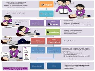
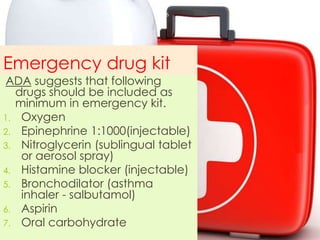
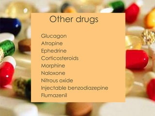
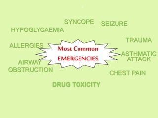
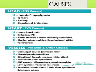
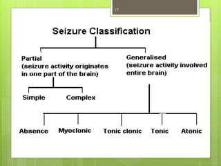
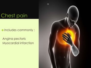
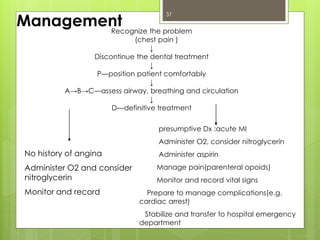
MEDICAL EMERGENCIES
IN COMMUNITY
 Presented by:
Jigyasha timsina
Batch 2011
1
Emergency
An unforeseen combination of circumstances or the
resulting state that calls for immediate action
2
WHY FOCUS ON MEDICAL EMERGENCIES???
Does not allow time for orderly information
gathering and formulation of a narrow differential
diagnosis before the initiation of therapy.
“When you prepare for emergency, the emercency ceases to exist”
3
APPROACH TO A MEDICAL EMERGENCY
 Comprehensive medical history
 Vigilant observation & prompt PREVENTION
recognition of symptoms of an emergency
 Basic life support PREPARATION
 Affiliation to definitive medical care
Did you know ???
A person who receives BLS has
20%increase in survival rate than one who
does not…so just act..
4
5
Emergency drug kit
ADA suggests that following
drugs should be included as
minimum in emergency kit.
1. Oxygen
2. Epinephrine 1:1000(injectable)
3. Nitroglycerin (sublingual tablet
or aerosol spray)
4. Histamine blocker (injectable)
5. Bronchodilator (asthma
inhaler - salbutamol)
6. Aspirin
7. Oral carbohydrate
6
Other drugs
Glucagon
Atropine
Ephedrine
Corticosteroids
Morphine
Naloxone
Nitrous oxide
Injectable benzodiazepine
Flumazenil
7
Most Common
EMERGENCIES
SYNCOPE SEIZURE
TRAUMA
ASTHMATIC
ATTACK
HYPOGLYCAEMIA
AIRWAY
OBSTRUCTION
ALLERGIES
CHEST PAIN
8
DRUG TOXICITY
SYNCOPE
 Defined as a short loss of consciousness
and muscle strength, characterized by a
fast onset, short duration, and
spontaneous recovery
9
CAUSES10
Clinical symptoms
 Presyncope
 Syncope
 Postsyncope
11
Presyncope
Early
 Feeling of warmth
 Loss of skin colour
 Heavy perspiration
 Complaints of feeling ill
 Nausea
 Hypotension
 Tachycardia
Late
 Pupillary dilatation
 Hyperpnea
 Cold hands and
feet
 Hypotension
 Bradycardia
 Visual disturbances
 Dizziness
12
Syncope
 Breathing
 Irregular, jerky and gasping
 Dilated pupils – death like appaerance
 Convulsive movements
 Bradycardia < 50 beats/min
 Weak thready pulse
 Loss of consciousness
 Partial or complete airway obstruction
13
Postsyncope
 Pallor
 Nausea
 Weakness
 Sweating
14
MANAGEMENT
 Position: supine position with brain and heart at same
level with feet elevated slightly (10 to 15 degree)
 ABC – Basic life support as needed
 Definitive management : Monitor vital signs
Administer aromatic ammonia
Administration of atropine (0.1mg/ml)
If delayed recovery seek medical assistance
15
SEIZURE
• A paroxysmal disorder of cerebral
function characterized by an attack
involving changes in the state of
consciousness ,motor activity or sensory
phenomena.
• Usually sudden in onset and of brief
duration.
EPILEPSY- “A chronic brain disorder of
various etiologies characterized by
recurrent seizures”
16
17
18
Common symptoms
19
PREVENTION
If pt is a known epileptic, make sure
he/she has taken their regular dose of
anti-convulsant on the day of
treatment.
Instruct him/her to alert you as the aura
of the impending seizure manifests itself.
Keep life support equipment ready in
case of an emergent status epilepticus.
20
Management
 Self limiting emergency
 Position : supine with patient placed on flat
surfaces
 Remove dangerous objects from the mouth and
around the patient eg. sharp instruments, needles,
etc.
 Loosen any tight clothing.
 Avoid restraining the patient
 In case the ictus fails to subside within a maximum
of 10 minutes, declare status epilepticus and
proceed with definitive care.
21
Definitive treatment
 Diazepam 10 mg iv (2mg/min)repeat
every 10 min
 Phenobarbitone (100- 200mg/min) iv
22
Hypoglycemia
 Hypoglycemia is a clinical
syndrome in which low
serum (or plasma) glucose
levels lead to symptoms of
sympatho- adrenal
activation.
23
24
Management
 Glucose and sugar-containing beverages
administered orally to conscious patients for rapid
effect.
 Alternatively, milk, candy bars, fruit, cheese, and
crackers may be adequate in mild cases
 IV dextrose is indicated for severe hypoglycemia,
in patients with altered consciousness and during
restriction of oral intake.
 An initial bolus, 20-50 mL of 50% dextrose, should
be given immediately.
25
 Glucagon, 1 mg IM (or SC), is an effective
initial therapy for severe hypoglycemia in
patients unable to receive oral intake or in
whom an IV access cannot be secured
immediately.
26
TRAUMA
• Trauma refers to damage, impairment or
external voilence producing injury or
degeneration.
• Trauma of the oral and maxillofacial region
occur frequently
• comprises 5% of all injuries for which people
seek treatment.
• Among all facial injuries, dental injuries are
the most common, of which crown factures
and luxations occur most frequently.
• The most common location is the anterior
maxilla followed by the anterior mandible.
27
A traumatic injury in a maxillofacial
region can result in:
- Fractures of the jaws
- Fractures of the teeth
- Soft tissue injuries
- Injuries to vital stuctures
28
Management:
Avoid patient movement before
determining extent of trauma
 Airway:
 Chin lift.
 Jaw thrust.
 Manually move the tongue forward.
 Maintain cervical immobilization
29
 Hemorrhage control
 Maxillofacial bleeding:
Direct pressure.
Nasal bleeding:
Direct pressure.
Anterior and posterior packing.
30
• First aid should be given for the
injuries occurred.
• The patient should be referred to the
nearby higher centres for further diagnosis
and care
31
Chest pain
 Includes commonly :
Angina pectoris
Myocardial infarction
32
Angina Pectoris
Angina is defined as“a characteristic thoracic
pain, usually substernal; precipitated chiefly
by exercise, emotion, or a heavy meal;
relieved by vasodilator drugs and a few
minutes rest; and a result of moderate
inadequacy of the coronary circulation.”
Produced when myocardial blood supply
cannot be sufficiently increased to meet the
increased oxygen requirement that results
from coronary artery disease.
33
Recognize the problem
discontinue dental treatment
P- position patient comfortably
A,B,C –ascess airway, breathing and circulation
Definitive management
34
If history of angina exists
Administer vasodilator and O2
If pain resolves
Consider future dental
treatment modification
Monitor vital signs
No history of angina
Administer O2 and consider
nitroglycerin
Monitor and record
Acute Myocardial Infarction
 Myocardial infarction is a clinical
syndrome caused by a deficient coronary
arterial blood supply to a region of
myocardium that results in cellular death
and necrosis.
 The syndrome is usually characterized by
severe and prolonged substernal pain
similar to but more intense and of longer
duration than the angina pectoris.
35
Acute myocardial infarction should
be suspected if :
A first episode of chest pain suggestive of
acute MI that occurs either at rest or with
ordinary activity. It may develop during dental
treatment especially if patient is dental
phobic.
Change in previous stable pattern of pain
which may be increased in frequency or
severity.
Chest pain is suggestive of MI in a patient with
known CAD if relieved by rest or nitroglycerin.
36
37
Recognize the problem
(chest pain )
↓
Discontinue the dental treatment
↓
P—position patient comfortably
↓
A→B→C—assess airway, breathing and circulation
↓
D—definitive treatment
presumptive Dx :acute MI
Administer O2, consider nitroglycerin
Administer aspirin
Manage pain(parenteral opoids)
Monitor and record vital signs
Prepare to manage complications(e.g.
cardiac arrest)
Stabilize and transfer to hospital emergency
department
Management
No history of angina
Administer O2 and consider
nitroglycerin
Monitor and record
Airway Obstruction
Causes:
• Foreign body (usually food)
• Infection or posttraumatic
hematoma
• Obstruction by the tongue
• Trauma
38
Presentation
• Stridor
• Impaired or absent phonation
• Choking and respiratory distress
• Angioedema
• Fever
• Evidence of trauma
39
Management
• Is directed at rapid relief of obstruction to prevent
cardiopulmonary arrest and anoxic brain damage.
• Perform the head tilt and chin lift maneuver if
cervical spine trauma is not suspected.
• Perform a jaw thrust if cervical spine trauma is
suspected.
• Attempt to ventilate the patient with a bag-valve-
mask apparatus.
40
• Perform the Heimlich maneuver
(subdiaphragmatic abdominal thrust)
repeatedly until the object is expelled
from the airway.
• If the situation cannot be managed, the
patient should be referred to a nearby
hospital or a health post.
41
Heimlich maneuver
42
43
If the patient is unconscious:
• Place the patient in supine position.
• Open patient’s airway by using
head tilt chin lift technique.
• Place the heel of one hand against
the victims abdomen in the midline
slightly above the umbilicus & well
below the xiphoid process.
• Place one hand on top of other
hand.
• Press in to the victims abdomen
with quick inward and upward
thrust.
Asthma
 A clinical state of hyper
reactivity of the
tracheobronchial tree,
characterized by
recurrent paroxysms of
dyspnea and wheezing
44
Signs and symptoms
 Feeling of chest tightness
 Dyspnea
 Tachypnea
 Cough
 Use of Accessory/Respiratory Muscles
 Agitations
45
The most likely times for an acute
exacerbation are:
During and immediately after
local anesthetic administration.
With stimulating procedures
such as extraction.
46
Management
 Discontinue the dental procedure and allow the
patient to assume a upright position.
 Establish and maintain a patent airway and
administer Beta 2 agonists via inhaler or nebulizer.
 Administer oxygen if possible
 If no improvement is observed and symptoms are
worsening, administer epinephrine subcutaneously
(1:1,000 solution, 0.01 mg/kg of body weight to a
maximum dose of 0.3 mg).
47
 Begin diligent basic life support.
 Document in time form the beginning of the
event.
 Alert emergency medical services.
 Maintain a good oxygen level until the
patient stops wheezing and/or medical
assistance arrives.
 Escort patient to hospital as needed.
48
MANIFESTATIONS AND
MANAGEMENT OF LOCAL
ANESTHETICS OVERDOSE
49
MANIFESTATIONS MANAGEMENT
MILD OVERDOSE Talkativeness,
slurred speech,
anxiety , confusion
Stop administration
of LA
-Monitor all vital
signs
-Observe for 1 hr
MODERATE TOXICITY Slurring speech,
nystagmus,
tremor,headache,
dizziness , blurred
vision,drowsiness
-Stop
administration of
LA
-Place the patient
in supine position
-Monitor vital signs
-Administer oxygen
-Observe in office
for 1 hr
50
SEVERE TOXICITY Seizures,
cardiac
arrythymia or
arrest
- Place the patient in
supine positions
- If seizures occur,
protect the patient
from nearby objects.
- Suction the oral
cavity if vomiting
occurs.
- Summon medical
assistance.
-Monitor vital signs.
-Administer oxygen.
-Start I.V infusion.
-Administer diazepam
5-10mg slowly.
-Provide basic life
support.
-Transport to
emergency.
51
Epinephrine (vasoconstrictor) overdose
reactions
• Available concentrations are 1:50000,
1:100000, 1:200000.
• The optimal concentration for the
prolongation of anaesthesia with lidocaine is
1:250000.
• Maximal dose:
Healthy adult - 0.2 mg
Cardiac patient - 0.04 mg
52
Clinical manifestations:
Signs
- Rise in blood pressure and heart rate
Symptoms
- Anxiety
- Restlessness
- Perspiration
- Dizziness
- Weakness
- Pallor
- Palpitation
53
Management
Terminate the dental procedure
Position the patient in upright position
Reassure the patient
Basic life support if indicated
Monitor vital signs
Summon medical assistance
Administer oxygen
54
S.N
.
TOXINS /
DRUGS
TOXIC DOSE MANIFESTATI
ONS
MANAGEMENT SPECIFIC
ANTIDOTES
1. Acetaminophe
n
>140mg/kg
or at least
7.5g
Anorexia
Vomiting
Diaphoresis
GI
decontaminati
on
Administration
of activated
charcoal
Acetylcyst
eine
The total
dose is 300
mg/kg,
given as 3
separate
doses
2. Anti-
depressants
(eg:amytryptilli
ne,
desipramine,
imipramine)
20mg/kg
causes few
fatalities
35mg/kg-
approx
lethal dose
>50mg/kg-
likely to
cause
death
Mydriasis
Ileus
Urinary
retention
Hyperpyrexia
GI
decontaminati
on
Gastric lavage
with activated
charcoal
IV sodium
bicarbonate
_
55
OPIOIDS
 Although opioids have been used as an
effective analgesic drug,most of the
time,it has been used as an abusive
product.
 Opioid toxicity can result in:
- Respiratory depression
- Depressed level of consciousness
- Miosis
56
Treatment
Gastric lavage
Antidote(naloxone hydrochloride,initial dose
of 2 mg IV)
57
ALCOHOL
 The toxicity of alcohol is dose related.
 Blood levels >100 mg/dL are associated
with ataxia.
 At 200 mg/dL, patients are drowsy and
confused.
 At levels >400 mg/dL, respiratory
depression is common and death is
possible.
58
Treatment
 Administration of 100 mg thiamine IV .
.
 Treat hypoglycaemia with 50 ml of 50%
dextrose solution
 Provide oxygen therapy as needed
59
GENERALISED ANAPHYLAXIS
 Acutely life threatening condition.
 Reactions develop rapidly 5-30 minutes.
 Signs and symptoms of generalised anaphylaxis are highly
variable.
Four major clinical syndromes are:
1. Skin reactions
2. Smooth muscle spasm
3. Respiratory distress
4. Cardiovascular collapse
60
USUAL PROGRESSION OF ANAPHYLAXIS
Skin
Eye , Nose , GI
Respiratory system
Cardiovascular system
61
MANAGEMENT
• Terminate dental procedure & stop
administration of all drugs presently in
use.
• Position the patient comfortably.
• Basic life support as indicated.
• Monitor vital signs.
62
Definitive Management
No CVS or respiratory involvement:
- Administration of oral or IM anti-
histamine.
CVS or respiratory involvement:
- Reposition the patient
- Administration of epinephrine
- Administration of anti-histamines
63
To conclude….
 The first step in management of dental
emergencies is to prevent their
occurrence
 With proper knowledge medical
emergencies and related complication
can be easily prevented
 “When you prepare for emergency, the
emergency ceases to exist”
64
REFERENCES
 Medical Emergencies In The Dental Office - 5th
Edition - Stanley F. Malamed
 Contemporary Oral and Maxillofacial Surgery – 5th
edition- Hupp,Ellis and Tucker
 Internet Sources
65
66

Medical emergencies in dentistry

  • 1.
    MEDICAL EMERGENCIES IN COMMUNITY Presented by: Jigyasha timsina Batch 2011 1
  • 2.
    Emergency An unforeseen combinationof circumstances or the resulting state that calls for immediate action 2
  • 3.
    WHY FOCUS ONMEDICAL EMERGENCIES??? Does not allow time for orderly information gathering and formulation of a narrow differential diagnosis before the initiation of therapy. “When you prepare for emergency, the emercency ceases to exist” 3
  • 4.
    APPROACH TO AMEDICAL EMERGENCY  Comprehensive medical history  Vigilant observation & prompt PREVENTION recognition of symptoms of an emergency  Basic life support PREPARATION  Affiliation to definitive medical care Did you know ??? A person who receives BLS has 20%increase in survival rate than one who does not…so just act.. 4
  • 5.
  • 6.
    Emergency drug kit ADAsuggests that following drugs should be included as minimum in emergency kit. 1. Oxygen 2. Epinephrine 1:1000(injectable) 3. Nitroglycerin (sublingual tablet or aerosol spray) 4. Histamine blocker (injectable) 5. Bronchodilator (asthma inhaler - salbutamol) 6. Aspirin 7. Oral carbohydrate 6
  • 7.
  • 8.
  • 9.
    SYNCOPE  Defined asa short loss of consciousness and muscle strength, characterized by a fast onset, short duration, and spontaneous recovery 9
  • 10.
  • 11.
    Clinical symptoms  Presyncope Syncope  Postsyncope 11
  • 12.
    Presyncope Early  Feeling ofwarmth  Loss of skin colour  Heavy perspiration  Complaints of feeling ill  Nausea  Hypotension  Tachycardia Late  Pupillary dilatation  Hyperpnea  Cold hands and feet  Hypotension  Bradycardia  Visual disturbances  Dizziness 12
  • 13.
    Syncope  Breathing  Irregular,jerky and gasping  Dilated pupils – death like appaerance  Convulsive movements  Bradycardia < 50 beats/min  Weak thready pulse  Loss of consciousness  Partial or complete airway obstruction 13
  • 14.
    Postsyncope  Pallor  Nausea Weakness  Sweating 14
  • 15.
    MANAGEMENT  Position: supineposition with brain and heart at same level with feet elevated slightly (10 to 15 degree)  ABC – Basic life support as needed  Definitive management : Monitor vital signs Administer aromatic ammonia Administration of atropine (0.1mg/ml) If delayed recovery seek medical assistance 15
  • 16.
    SEIZURE • A paroxysmaldisorder of cerebral function characterized by an attack involving changes in the state of consciousness ,motor activity or sensory phenomena. • Usually sudden in onset and of brief duration. EPILEPSY- “A chronic brain disorder of various etiologies characterized by recurrent seizures” 16
  • 17.
  • 18.
  • 19.
  • 20.
    PREVENTION If pt isa known epileptic, make sure he/she has taken their regular dose of anti-convulsant on the day of treatment. Instruct him/her to alert you as the aura of the impending seizure manifests itself. Keep life support equipment ready in case of an emergent status epilepticus. 20
  • 21.
    Management  Self limitingemergency  Position : supine with patient placed on flat surfaces  Remove dangerous objects from the mouth and around the patient eg. sharp instruments, needles, etc.  Loosen any tight clothing.  Avoid restraining the patient  In case the ictus fails to subside within a maximum of 10 minutes, declare status epilepticus and proceed with definitive care. 21
  • 22.
    Definitive treatment  Diazepam10 mg iv (2mg/min)repeat every 10 min  Phenobarbitone (100- 200mg/min) iv 22
  • 23.
    Hypoglycemia  Hypoglycemia isa clinical syndrome in which low serum (or plasma) glucose levels lead to symptoms of sympatho- adrenal activation. 23
  • 24.
  • 25.
    Management  Glucose andsugar-containing beverages administered orally to conscious patients for rapid effect.  Alternatively, milk, candy bars, fruit, cheese, and crackers may be adequate in mild cases  IV dextrose is indicated for severe hypoglycemia, in patients with altered consciousness and during restriction of oral intake.  An initial bolus, 20-50 mL of 50% dextrose, should be given immediately. 25
  • 26.
     Glucagon, 1mg IM (or SC), is an effective initial therapy for severe hypoglycemia in patients unable to receive oral intake or in whom an IV access cannot be secured immediately. 26
  • 27.
    TRAUMA • Trauma refersto damage, impairment or external voilence producing injury or degeneration. • Trauma of the oral and maxillofacial region occur frequently • comprises 5% of all injuries for which people seek treatment. • Among all facial injuries, dental injuries are the most common, of which crown factures and luxations occur most frequently. • The most common location is the anterior maxilla followed by the anterior mandible. 27
  • 28.
    A traumatic injuryin a maxillofacial region can result in: - Fractures of the jaws - Fractures of the teeth - Soft tissue injuries - Injuries to vital stuctures 28
  • 29.
    Management: Avoid patient movementbefore determining extent of trauma  Airway:  Chin lift.  Jaw thrust.  Manually move the tongue forward.  Maintain cervical immobilization 29
  • 30.
     Hemorrhage control Maxillofacial bleeding: Direct pressure. Nasal bleeding: Direct pressure. Anterior and posterior packing. 30
  • 31.
    • First aidshould be given for the injuries occurred. • The patient should be referred to the nearby higher centres for further diagnosis and care 31
  • 32.
    Chest pain  Includescommonly : Angina pectoris Myocardial infarction 32
  • 33.
    Angina Pectoris Angina isdefined as“a characteristic thoracic pain, usually substernal; precipitated chiefly by exercise, emotion, or a heavy meal; relieved by vasodilator drugs and a few minutes rest; and a result of moderate inadequacy of the coronary circulation.” Produced when myocardial blood supply cannot be sufficiently increased to meet the increased oxygen requirement that results from coronary artery disease. 33
  • 34.
    Recognize the problem discontinuedental treatment P- position patient comfortably A,B,C –ascess airway, breathing and circulation Definitive management 34 If history of angina exists Administer vasodilator and O2 If pain resolves Consider future dental treatment modification Monitor vital signs No history of angina Administer O2 and consider nitroglycerin Monitor and record
  • 35.
    Acute Myocardial Infarction Myocardial infarction is a clinical syndrome caused by a deficient coronary arterial blood supply to a region of myocardium that results in cellular death and necrosis.  The syndrome is usually characterized by severe and prolonged substernal pain similar to but more intense and of longer duration than the angina pectoris. 35
  • 36.
    Acute myocardial infarctionshould be suspected if : A first episode of chest pain suggestive of acute MI that occurs either at rest or with ordinary activity. It may develop during dental treatment especially if patient is dental phobic. Change in previous stable pattern of pain which may be increased in frequency or severity. Chest pain is suggestive of MI in a patient with known CAD if relieved by rest or nitroglycerin. 36
  • 37.
    37 Recognize the problem (chestpain ) ↓ Discontinue the dental treatment ↓ P—position patient comfortably ↓ A→B→C—assess airway, breathing and circulation ↓ D—definitive treatment presumptive Dx :acute MI Administer O2, consider nitroglycerin Administer aspirin Manage pain(parenteral opoids) Monitor and record vital signs Prepare to manage complications(e.g. cardiac arrest) Stabilize and transfer to hospital emergency department Management No history of angina Administer O2 and consider nitroglycerin Monitor and record
  • 38.
    Airway Obstruction Causes: • Foreignbody (usually food) • Infection or posttraumatic hematoma • Obstruction by the tongue • Trauma 38
  • 39.
    Presentation • Stridor • Impairedor absent phonation • Choking and respiratory distress • Angioedema • Fever • Evidence of trauma 39
  • 40.
    Management • Is directedat rapid relief of obstruction to prevent cardiopulmonary arrest and anoxic brain damage. • Perform the head tilt and chin lift maneuver if cervical spine trauma is not suspected. • Perform a jaw thrust if cervical spine trauma is suspected. • Attempt to ventilate the patient with a bag-valve- mask apparatus. 40
  • 41.
    • Perform theHeimlich maneuver (subdiaphragmatic abdominal thrust) repeatedly until the object is expelled from the airway. • If the situation cannot be managed, the patient should be referred to a nearby hospital or a health post. 41
  • 42.
  • 43.
    43 If the patientis unconscious: • Place the patient in supine position. • Open patient’s airway by using head tilt chin lift technique. • Place the heel of one hand against the victims abdomen in the midline slightly above the umbilicus & well below the xiphoid process. • Place one hand on top of other hand. • Press in to the victims abdomen with quick inward and upward thrust.
  • 44.
    Asthma  A clinicalstate of hyper reactivity of the tracheobronchial tree, characterized by recurrent paroxysms of dyspnea and wheezing 44
  • 45.
    Signs and symptoms Feeling of chest tightness  Dyspnea  Tachypnea  Cough  Use of Accessory/Respiratory Muscles  Agitations 45
  • 46.
    The most likelytimes for an acute exacerbation are: During and immediately after local anesthetic administration. With stimulating procedures such as extraction. 46
  • 47.
    Management  Discontinue thedental procedure and allow the patient to assume a upright position.  Establish and maintain a patent airway and administer Beta 2 agonists via inhaler or nebulizer.  Administer oxygen if possible  If no improvement is observed and symptoms are worsening, administer epinephrine subcutaneously (1:1,000 solution, 0.01 mg/kg of body weight to a maximum dose of 0.3 mg). 47
  • 48.
     Begin diligentbasic life support.  Document in time form the beginning of the event.  Alert emergency medical services.  Maintain a good oxygen level until the patient stops wheezing and/or medical assistance arrives.  Escort patient to hospital as needed. 48
  • 49.
    MANIFESTATIONS AND MANAGEMENT OFLOCAL ANESTHETICS OVERDOSE 49
  • 50.
    MANIFESTATIONS MANAGEMENT MILD OVERDOSETalkativeness, slurred speech, anxiety , confusion Stop administration of LA -Monitor all vital signs -Observe for 1 hr MODERATE TOXICITY Slurring speech, nystagmus, tremor,headache, dizziness , blurred vision,drowsiness -Stop administration of LA -Place the patient in supine position -Monitor vital signs -Administer oxygen -Observe in office for 1 hr 50
  • 51.
    SEVERE TOXICITY Seizures, cardiac arrythymiaor arrest - Place the patient in supine positions - If seizures occur, protect the patient from nearby objects. - Suction the oral cavity if vomiting occurs. - Summon medical assistance. -Monitor vital signs. -Administer oxygen. -Start I.V infusion. -Administer diazepam 5-10mg slowly. -Provide basic life support. -Transport to emergency. 51
  • 52.
    Epinephrine (vasoconstrictor) overdose reactions •Available concentrations are 1:50000, 1:100000, 1:200000. • The optimal concentration for the prolongation of anaesthesia with lidocaine is 1:250000. • Maximal dose: Healthy adult - 0.2 mg Cardiac patient - 0.04 mg 52
  • 53.
    Clinical manifestations: Signs - Risein blood pressure and heart rate Symptoms - Anxiety - Restlessness - Perspiration - Dizziness - Weakness - Pallor - Palpitation 53
  • 54.
    Management Terminate the dentalprocedure Position the patient in upright position Reassure the patient Basic life support if indicated Monitor vital signs Summon medical assistance Administer oxygen 54
  • 55.
    S.N . TOXINS / DRUGS TOXIC DOSEMANIFESTATI ONS MANAGEMENT SPECIFIC ANTIDOTES 1. Acetaminophe n >140mg/kg or at least 7.5g Anorexia Vomiting Diaphoresis GI decontaminati on Administration of activated charcoal Acetylcyst eine The total dose is 300 mg/kg, given as 3 separate doses 2. Anti- depressants (eg:amytryptilli ne, desipramine, imipramine) 20mg/kg causes few fatalities 35mg/kg- approx lethal dose >50mg/kg- likely to cause death Mydriasis Ileus Urinary retention Hyperpyrexia GI decontaminati on Gastric lavage with activated charcoal IV sodium bicarbonate _ 55
  • 56.
    OPIOIDS  Although opioidshave been used as an effective analgesic drug,most of the time,it has been used as an abusive product.  Opioid toxicity can result in: - Respiratory depression - Depressed level of consciousness - Miosis 56
  • 57.
  • 58.
    ALCOHOL  The toxicityof alcohol is dose related.  Blood levels >100 mg/dL are associated with ataxia.  At 200 mg/dL, patients are drowsy and confused.  At levels >400 mg/dL, respiratory depression is common and death is possible. 58
  • 59.
    Treatment  Administration of100 mg thiamine IV . .  Treat hypoglycaemia with 50 ml of 50% dextrose solution  Provide oxygen therapy as needed 59
  • 60.
    GENERALISED ANAPHYLAXIS  Acutelylife threatening condition.  Reactions develop rapidly 5-30 minutes.  Signs and symptoms of generalised anaphylaxis are highly variable. Four major clinical syndromes are: 1. Skin reactions 2. Smooth muscle spasm 3. Respiratory distress 4. Cardiovascular collapse 60
  • 61.
    USUAL PROGRESSION OFANAPHYLAXIS Skin Eye , Nose , GI Respiratory system Cardiovascular system 61
  • 62.
    MANAGEMENT • Terminate dentalprocedure & stop administration of all drugs presently in use. • Position the patient comfortably. • Basic life support as indicated. • Monitor vital signs. 62
  • 63.
    Definitive Management No CVSor respiratory involvement: - Administration of oral or IM anti- histamine. CVS or respiratory involvement: - Reposition the patient - Administration of epinephrine - Administration of anti-histamines 63
  • 64.
    To conclude….  Thefirst step in management of dental emergencies is to prevent their occurrence  With proper knowledge medical emergencies and related complication can be easily prevented  “When you prepare for emergency, the emergency ceases to exist” 64
  • 65.
    REFERENCES  Medical EmergenciesIn The Dental Office - 5th Edition - Stanley F. Malamed  Contemporary Oral and Maxillofacial Surgery – 5th edition- Hupp,Ellis and Tucker  Internet Sources 65
  • 66.