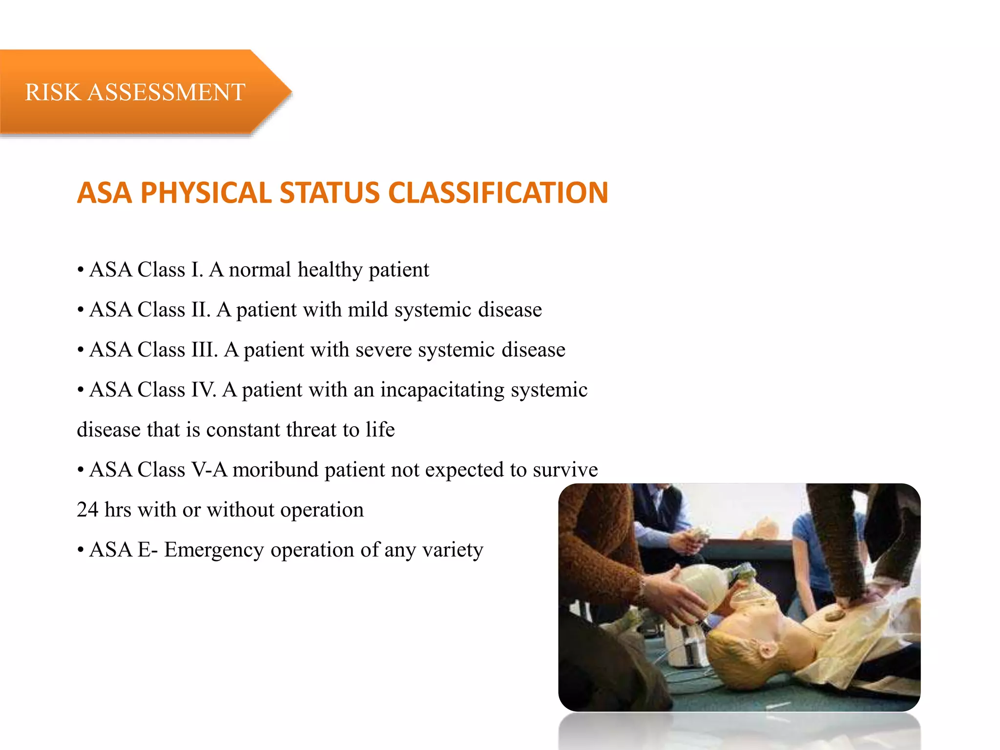
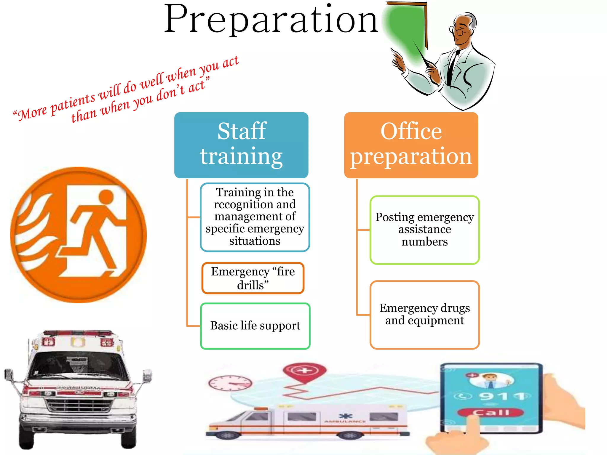
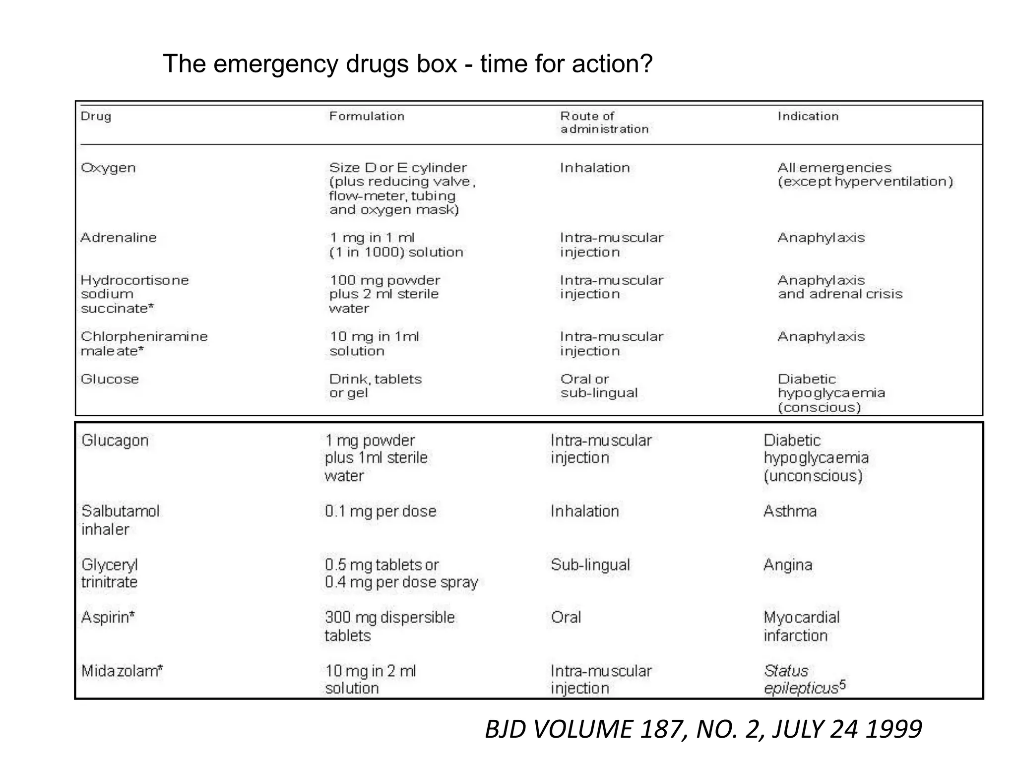
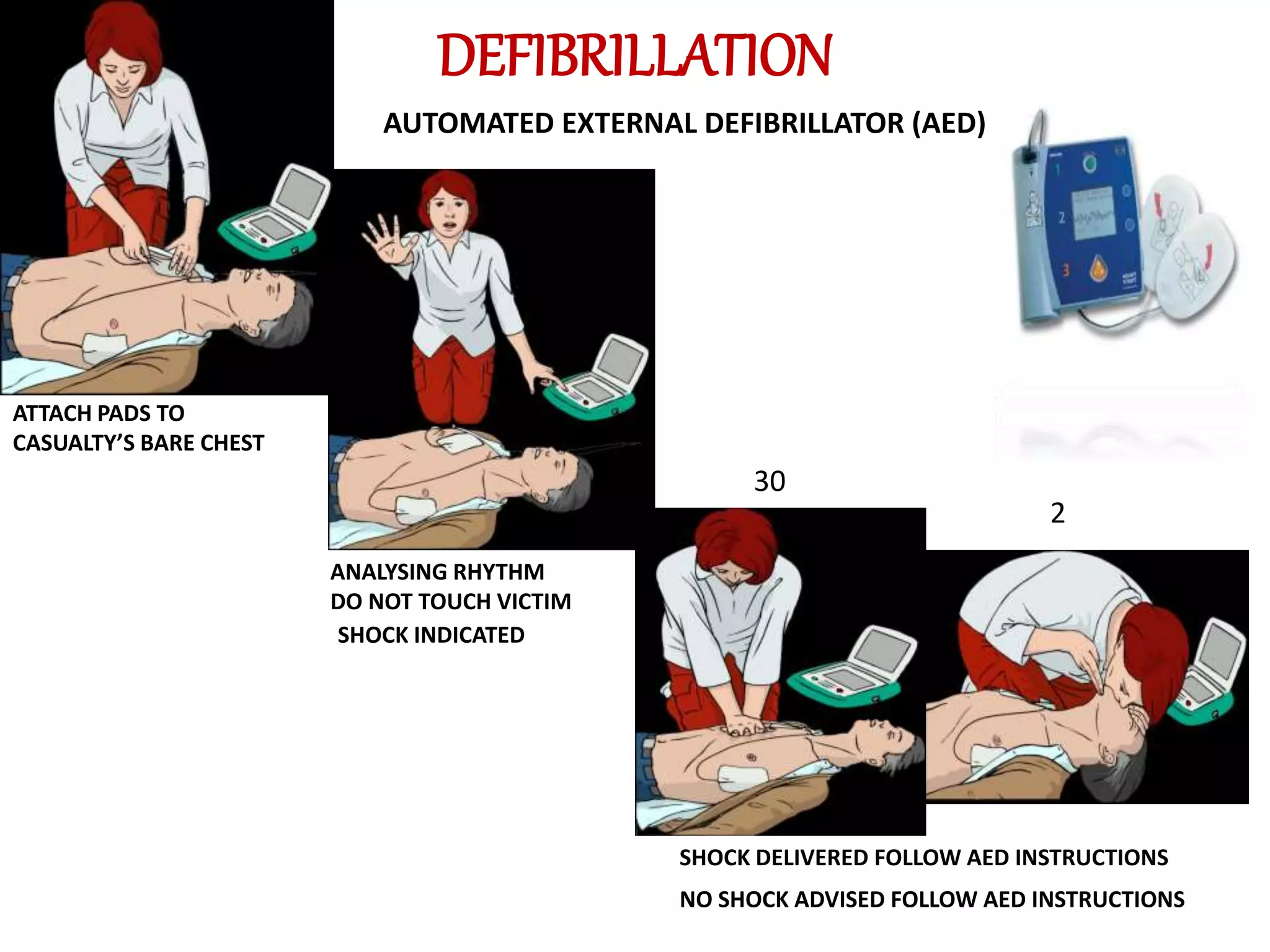
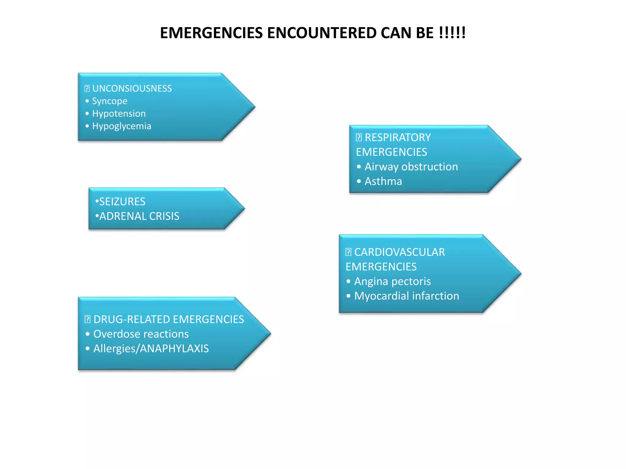
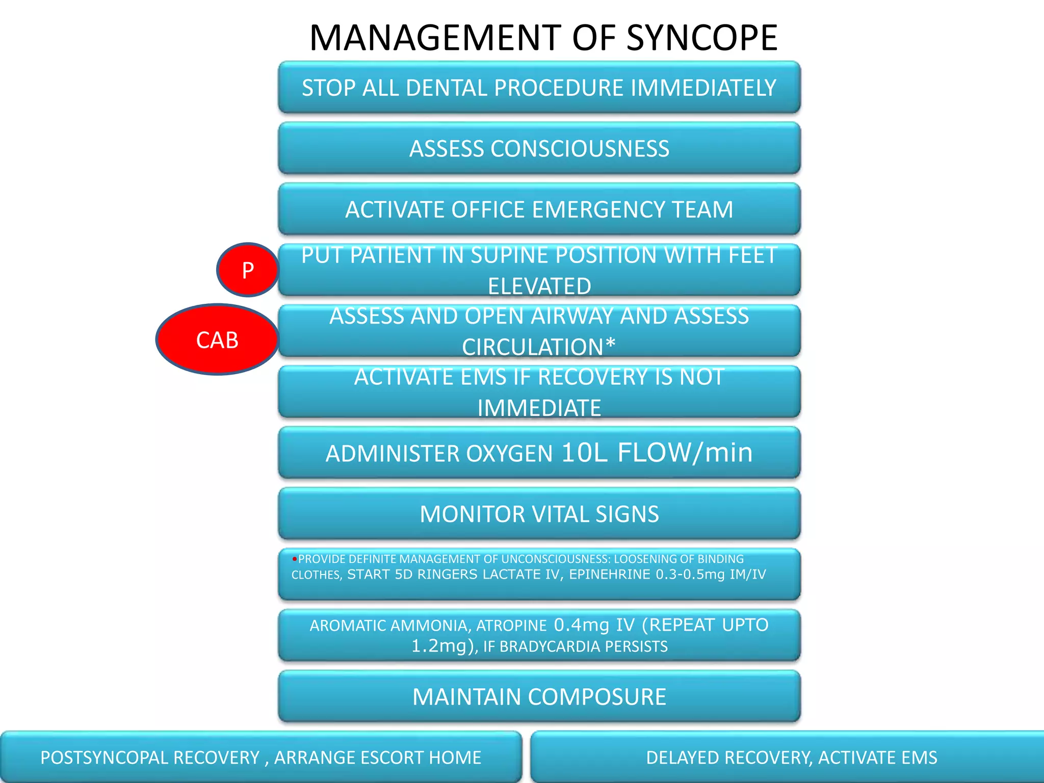
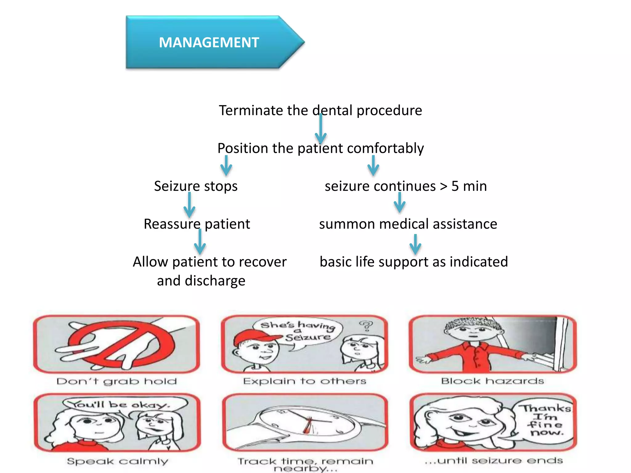
2. “When you prepare for emergencies, they cease to
exist!”
- Goldberger
What is an emergency?
3. APPROACH TO A MEDICAL EMERGENCY
Prevention
Preparation
Management
4. Prevention
“Never treat a stranger”
Medical history
Physical examination
Psychological examination
OBSERVATION
DETERMINATION OF MEDICAL RISK
5. • ASA Class I. A normal healthy patient
• ASA Class II. A patient with mild systemic disease
• ASA Class III. A patient with severe systemic disease
• ASA Class IV. A patient with an incapacitating systemic
disease that is constant threat to life
• ASA Class V-A moribund patient not expected to survive
24 hrs with or without operation
• ASA E- Emergency operation of any variety
ASA PHYSICAL STATUS CLASSIFICATION
RISK ASSESSMENT
6. Staff
training
Training in the
recognition and
management of
specific emergency
situations
Basic life support
Office
preparation
Posting emergency
assistance
numbers
Emergency drugs
and equipment
Emergency “fire
drills”
Preparation
7.
8. The emergency drugs box - time for action?
BJD VOLUME 187, NO. 2, JULY 24 1999
9. ALERT
Did you know ???
A person who receives BLS has
20%increase in survival rate than one
who
does not…so just act..
16. Airway Obstruction
◘ Re-establishment of airway:
Non-invasive procedures
Forceful coughing Back blows
Heimlich maneuverChest thrust
Finger sweeps
Invasive procedures
17. “B” BREATHING
• LOOK
• LISTEN
• FEEL
TIME FOR ACTION
METHODS OF RESCUE BREATHS
• MOUTH TO MOUTH OR NOSE
• MOUTH TO BARRIER DEVICE
•VENTILATION WITH BAG AND
MASK
•VENTILATION WITH ADVANCED
AIRWAY
18. DEFIBRILLATION
AUTOMATED EXTERNAL DEFIBRILLATOR (AED)
ATTACH PADS TO
CASUALTY’S BARE CHEST
ANALYSING RHYTHM
DO NOT TOUCH VICTIM
SHOCK INDICATED
SHOCK DELIVERED FOLLOW AED INSTRUCTIONS
NO SHOCK ADVISED FOLLOW AED INSTRUCTIONS
30
2
25. SYNCOPE
• It is defined as sudden, transient loss of consciousness that is usually secondary to period of transient
ischemia.
• Predisposing factors:
STRESS
IMPAIRED PHYSICAL CONDITION
HYPOGLYCEMIA Stress=Tachycardia=Carotidbody & sinus
stimulation
Vagal stimulation= Bradycardia,
Vasodilation=Decreased cerebral blood flow
Reflexive response to reestablish cerebral
blood flow=syncope
How to identify ?
Sings /symptoms
H.R.
UNCONSCIOUS
CONVULSION
PUPIL
DILATION
SWEATING
NAUSEA
PALLOR
ANXIETY
B.P.
Webster-Merriam’s Medical Dictionary. 12th ed.
Baltimore:Williams;2011.“syncope”;p.348
PATHOPHYSIOLOGY
26. MANAGEMENT OF SYNCOPE
STOP ALL DENTAL PROCEDURE IMMEDIATELY
ASSESS CONSCIOUSNESS
ACTIVATE OFFICE EMERGENCY TEAM
PUT PATIENT IN SUPINE POSITION WITH FEET
ELEVATED
ASSESS AND OPEN AIRWAY AND ASSESS
CIRCULATION*
ACTIVATE EMS IF RECOVERY IS NOT
IMMEDIATE
ADMINISTER OXYGEN 10L FLOW/min
MONITOR VITAL SIGNS
•PROVIDE DEFINITE MANAGEMENT OF UNCONSCIOUSNESS: LOOSENING OF BINDING
CLOTHES, START 5D RINGERS LACTATE IV, EPINEHRINE 0.3-0.5mg IM/IV
AROMATIC AMMONIA, ATROPINE 0.4mg IV (REPEAT UPTO
1.2mg), IF BRADYCARDIA PERSISTS
MAINTAIN COMPOSURE
POSTSYNCOPAL RECOVERY , ARRANGE ESCORT HOME DELAYED RECOVERY, ACTIVATE EMS
P
CAB
27. POSTURAL/ORTHOSTATIC HYPOTENSION
Drugs
Prolonged
recumbency /
convalescence
Late stage
pregnancy
Varicosities
Addison’s Disease
Severe exhaustion
Shy-Drager
Syndrome
PREDISPOSING FACTORS
PATHOPHYSIOLOGY
Pt attains upright
position
SBP falls =<60mm of Hg
due to ANS response
failure
Cerebral blood
flow<critical level
Loss of consciousness
Supination=revival
Criteria for postural
hypotension.
1.Symptomology develops on
standing
2.Increase in standing pulse
atleast 30 beats per minute
3.Decrease in standing systolic
BP atleast 25 mm of Hg
4.Decrease in standing diastolic
BP atleast 10 mm of Hg
Second leading cause of unconsciousness
How to identify ?
Sings /symptoms
•DIZZINESS
•PALLOR
•BLURRED VISION
•NAUSEA
• BP
28. MANAGEMENT OF POSTURAL HYPOTENSION
Recognize problem
Activate office emergency team
Position the patient (supine with feet slightly
elevated)
A B C &Definitive management of
unconsciousness (O2,)
Episode continuesEpisode terminates
Summon medical assistantProvide subsequent
management
Slowly reposition chair
Discharge pts
29. Acute Adrenal Insufficiency
A condition first recognized by Addison in 1844.
PREDISPOSING
FACTORS
•Addison’s disease .
•Pts on long term steroid therapy(RULE OF
TWOS)
•Stress
CLINICAL PRESENTATION
•Shock,
•Anorexia,
•Nausea,
•Vomiting,
•Abdominal Pain,
•Weakness,
•Fatigue,
•Lethargy,
•Fever,
•Confusion,or Coma
PREVENTION
Acute adrenal insufficiency is best managed by
its preventionwhich is based on
1. Medical history questionnaire.
2. Dialogue history.
30. MANAGEMENT OF ACUTE ADRENAL INSUFFICIENCY
Terminate dental therapy
Position patient comfortably if asymptomatic
Supine with feet elevated, if symptomatic
Monitor vital signs
Summon medical assistance
Administer oxygen
• Administer glucocorticoid 100 mg of
hydrocortisone sodium succinate (IV or IM) and
repeat every 6 – 8 hours
•Dexamethasone 4mg IV 6-8 HRS
• Additional management: provide Basic Life
Support as needed
• Provide oxygen as needed
• Maintain iv line
31. SEIZURES
Diagnostic clues
•Sudden onset of immobility and blank
stare
• Show blinking of eyes
• Short duration
• Rapid recovery
•It is a paroxysmal disorder of cerebral function characterized by an attack, involving changes in the state of
consciousness, motor activity or sensory phenomena.
• Usually sudden in onset and of brief duration.
32. Terminate the dental procedure
Position the patient comfortably
Seizure stops seizure continues > 5 min
Reassure patient summon medical assistance
Allow patient to recover basic life support as indicated
and discharge
MANAGEMENT
34. MANAGEMENT
CONCIOUS PATIENT UNCONCIOUS PATIENT
RECOGNISE HYPOGLYCEMIA……
TERMINATE DENTAL PROCEDURE
POSITION PATIENT ( SUPINE WITH LEGS ELEVATED)
BASIC LIFE SUPPORT ,ASCESS A-B-C
ADMINISTER ORAL CARBOHYDRATE
EPISODE TERMINATE EPISODE CONTINUES
PERMIT PATIENT RECOVERY SUMMON MEDICAL ASSIATANCE
DISCHARGE PATIENT ADMINISTER PARENTRAL CARBOHYDRATE
( GLUCAGON 1MG IM /2-3 MIN OR
50% DEXTROS IV)
MONITER VITALS
DISCHARGE
SUMMON MEDICAL ASSIATANCE( NO
RESPONSE TO BLS)
DEFINITIVE T/T
GLUCAGON 1MG IM
50% DEXTROS IV, TRANSMUCOSAL SUGAR
0.5MG EPINEPHRINE (1:1000) IM/SC,REPEAT
IN 15 MIN AS NEEDED
ALLOW PT. TO RECOVER
MONITOR VITALS
DISCHARGE
37. ANGINA PECTORIS
“A condition marked by severe pain in the chest, often also spreading to the shoulders, arms, and neck, owing
to an indequate blood supply to the heart.”
• Types:
- Stable
- Variant
- Unstable
Clinical characteristics
– Poorly localized pain
– Brief duration(2-10 minutes)
– Moderate intensity pain described as
squeezing, oppressive, burning or heavy
• Pain localized with one finger
• Lasts less than 30 seconds or longer than 30
minutes
• Pain described as sticking, jabbing,throbbing
or constantly severe
Stress reduction
Reassurance
Psychosedation
Excluded if:
PREVENTION
38.
39. MYOCARDIALINFARCTION
“A clinical syndrome caused by deficient coronary arterial blood supply resulting in
ischaemia to a region of the myocardium and causing cellular death and necrosis.”
– Atherosclerosis and coronary artery disease
– Coronary thrombosis, occlusion and spasm
– Males
– 5th and 6th decades of life
– Undue stress
Predisposing Factors: •STRESS REDUCTION PROTOCOL
•OXYGEN SUPPLEMENT
•PSYCHOSEDATION
PREVENTION
41. CARDIAC ARREST
Abrupt cessation of cardiac pump function that results in death , which may be
averted if prompt intervention is instituted.
PREDISPOSING
FACTORS
•90% Coronary artery disease
•Obesity- Male in 5th and 6th
decade
•stress
PRECAUTIONS
•Stress reduction protocol
•Oxygen supplement
•Sedation
HOW TO IDENTIFY ?
•Sudden and abrupt loss of conciousne
•Absence of respiration
•Loss of central and pheripheral pulse
•No heart sound
CLINICAL
MANIFESTATIONS
Sever retrosternal pain
radiating to lt. arm
Restlessness, distress
Pale and moist
skin
Dizziness,palpitation,
prespiration
Nausea, vomitting
Irrigular
heart rate
42. MANAGEMENT
BLS
ACLS
TERMINATE DENTAL PROCEDURE
MONITOR VITALS
DEFINITIVE TREATMENT
•NITROGLYCERINE
•ANTIPLATELET THERAPY- ASPIRIN 325MG
•MANAGE PAIN- PARENTRAL OPOIDS, N2O+ O2
DIAGNOSE- DIFFERENTIATE ANGINAL PAIN WITH NON-ANGINAL PAIN
P POSITION PATIENT COMFORTABLY
C-A-B
TRANSFER TO HOSPITAL
SUMMON MEDICAL ASSISTANCE
43.
44. AIRWAY OBSTRUCTION/FOREIGN BODY
ASPIRATION
◘ May occur due to:
o Pathology on the airway
o Dental instruments
o Tongue
PREVENTION
Rubber dam
Oral packing
Chair position
Dental assistant
Magill’s intubation forceps
45. MANAGEMENT
◘ Re-establishment of airway:
Non-invasive procedures
Forceful coughing Back blows
Heimlich maneuverChest thrust
Finger sweeps
Invasive procedures
46. FOREIGN BODY ASPIRATION
STIOP DENTAL PROCEDURE
ASK PT. TO COUGH OUT OBJECT
OBJECT EXPELLED OUT BACK BLOWS
RE-EXAMINE PT.
EVALUATE FOR RESIDUAL OBJECT/ SUCCESS
NJURY TO TRACHEOBRONCHEAL TREE UNSUCCESSFUL
( CHEST X-RAY,ENDOSCOPY)
CONCIOUS UNCONCIOUS
SUSPECT COMPLETE OBSTRUCTION
HEIMLICH MANOEUVRE HEIMLICH MANOEUVRE
SUCCESSFUL UNSUCCESSFUL
CONCIOUS UNCONCIOUS SUCCESSFUL UNSUCCESSFUL
CHECK VITALS VITALS,BLS EMERGENCYAIRWAY
MAINTAIN BREATHING AIRWAY M/M VITALS,BLS
02 /AIRWAY
RECOVERY NO RECOVERY
SHIFT TO HOSPITAL CALL EMERGENCY MEDICAL
SUCCESSFUL UNSUCCESSFUL
47. ASTHMA
A clinical state of hyper reactivity of the tracheobronchial tree, characterized by recurrent
paroxysms of dyspnea and wheezing.
Can be:
• Extrinsic Asthma
• Intrinsic Asthma
• Status Asthmaticus
Feeling of chest tightness
Dyspnea
Tachypnea
Cough
Use of Accessory/Respiratory Muscles
Agitations
Signs and symptoms
48.
49. HYPERVENTILATION
Excessive rate and depth of respiration leading to abnormal loss of carbon dioxide
from the blood primarily predisposed to stress and anxiety.
Rapid short strained breaths
Cold sweats
Palpitations
Dizziness
Chest muscle fatigue
• Characterized by:
50. MANAGEMENT
• Terminate procedure
• Rebreathing bag (exhaled air)
• Drug management – 10 to15 mg
diazepam or 3 to 5 mg Midazolam IV
•Resume procedure
• Calm patient
•Check vitals
• Correct respiratory alkalosis (breathe
in gaseous mixture of 7% CO2 and 93% O2)
• Position patient (upright)
51.
52. OVERDOSE REACTIONS
In a dental practice, commonest overdosage>>LA
• Confusion, talkativeness, blurred speech
• Muscular twitching, facial tremor
•Headache, tinnitus
•Drowsiness, disorientation
• Elevated BP, HR, RR
• If uncontrolled, seizures
CLINICAL MANIFEATATIONS
-Stop administration of LA
-Place the patient in supine position
-Monitor vital signs
-Administer oxygen
-anticonvulsants(Administer diazepam 5-10mg
slowly.)
- Perform BLS as needed
- Allow recovery to occur
- In case of continuation of symptoms,
summon EMS
MANAGEMENT
54. MANAGEMENT
Identify anaphylactic reaction
• Position patient in upright position
• Assess ABC
• Definitive management:
• Epinephrine (1:1000),0.3 mg IM or SC every 5 mins
• Oxygen by nasal hood or face mask at rate of 5 to 6
L/min
- Im histamine blocker ( chlorpheniramine maleate 10 mg )
- IV access and administration of additional histamine
blockers and corticosteroids( hydrocortisone 20 mg)
Summon Medical Assistance
CPR ,If cardiac arrest
Transfer to hospital
55. CONCLUSION
As the saying goes, “PREVENTION IS BETTER THAN CURE”.
ALWAYS BE PREPARED.
Prompt recognition and efficient management of medical emergencies by a well-prepared
dental team that can increase the likelihood of a safe & satisfactory outcome.
Basic life support training – A MUST
Editor's Notes
◘
A serious and unexpected situation requiring an
immediate action.
It is an unforeseen combination of circumstances or the
resulting state that calls for an immediate action.
Never treat a stranger”
Physical Evaluation
Medical history questionnaire
Physical examination (vital signs, visual inspection, functional tests, auscultation of heart and lungs)
Dialogue history (recognition of anxiety)
Psychological examination
Medical history questionnaire
Anxiety questionnaire
Observation
Increased BP & Heart rate
Trembling
Excessive sweating
Dilated pupils
Physical status classification system (1962, American Society of Anesthesiologists)
ASA I : A patient without systemic disease, a normal healthy patient
ASA II : A patient with mild systemic disease
ASA III : A patient with severe systemic disease that limits activity but is not incapacitating
ASA IV : A patient with incapacitating systemic disease that is a constant threat to life.
ASA V : A moribund patient not expected to survive 24 hrs with or with out surgery.
ASA VI : Clinically dead patient being maintained for harvesting organs.
ASA E : Emergency operation of any variety; E precedes the number, indicating the patients physical status( ASA E-III)
Medical consultation
Stress reduction protocol
Premedication
Appointment scheduling
Minimized waiting time
Psycosedation during therapy
Adequate pain control during therapy
Duration of dental treatment
Postoperative control of pain and anxiety
The concept of ‘‘how healthy is the patient,’’ otherwise
termed ‘‘risk assessment,’’ is key in determining the
likelihood of complications. The higher the ASA class,
the more at-risk the patient is both from a surgical and
anesthetic perspective.
• ASA Class I. A normal healthy patient
• ASA Class II. A patient with mild systemic disease
• ASA Class III. A patient with severe systemic disease
• ASA Class IV. A patient with an incapacitating systemic
disease that is constant threat to life
• ASA Class V-A moribund patient not expected to survive
24 hrs with or without operation
• ASA E- Emergency operation of any variety
Staff training should include:
Basic life support training for all members of dental office staff
Training in the recognition and management of specific emergency situations
Emergency “fire drills” A fire drill is a method of practicing how a building would be evacuated in the event of a fire or other emergencies. In most cases, the building's existing fire alarm system is activated and the building is evacuated by means of the nearest available exit as if an emergency had actually occurred.
Office preparation should include:
Posting emergency assistance numbers
Stocking emergency drugs and equipment
Suction equipment
Blood pressure monitor
Ambu bag and airways
Oxygen delivery equipment and tubing
Syringe for IM & IV injections
Tourniquet
Optional emergency equipment
Pulse oximeter
Intravenous cannulae
Nitrous oxide / oxygen delivery system
Laryngoscope
Tracheostomy kit
Criteria for selecting appropriate emergency drugs in general dental practice:
Only drugs which are essential for the first-line management of emergencies need to be kept in general dental practice
The recommended emergency drugs can be administered by simple (oral, sub-lingual, inhalation or intra-muscular) routes
All GDPs should be trained and competent to use the essential emergency drugs Criteria for selecting appropriate emergency drugs in general dental practice:
Only drugs which are essential for the first-line management of emergencies need to be kept in general dental practice
The recommended emergency drugs can be administered by simple (oral, sub-lingual, inhalation or intra-muscular) routes
All GDPs should be trained and competent to use the essential emergency drugs
Module one - basic emergency kit (critical drugs and equipment)
Module two - noncritical drugs and equipments
Module three- advanced cardiac life support
Module four - antidotal drugs
In each module
Injectable drugs
Noninjectable drugs
Module one:-Emergency equipments
Oxygen delivery system
Suction & suction tips
Tourniquets
Syringes
Magill intubation forceps
Injectable drugs
Epinephrine – 1: 1000
Antihistamine – CPM
Noninjectable drugs
Oxygen – E- Cylinder
Vasodialator – NTG, Amyl nitrite
Module two:-Injectable drugs
Anticonvulsant – Midazolam, Diazepam
Analgesic – Morphine
Vasopressor – Methoxamine, Phenylephrine.
Antihypoglycemic – 50% dextrose, Glucogon.
Corticosteroid – Hydrocortisone, Dexamethosone
Antihypertensive – Lebetalol, Propronolol
Anticholinergic – Atropine
Noninjectable drugs
Respiratory stimulant – Aromatic Ammonia
Antihypoglycemic – Sugar
Bronchodilator – Albuterol, Metaproterenol.
Emergency equipments
Device for cricothyrotomy
Artificial airways
Laryngoscope & endotracheal tubes
Module three:-Essential ACLS drugs include
Epinephrine
Oxygen
Lidocaine
Atropine
Dopamine
Morphine sulphate
Verapamil
Module four:-Narcotic antagonist – Naloxone, Nalbuphine
Benzodiazepine antagonist – Flumazenil
Antiemergence delirium drug – Physostigmine
Vasodilator – Procaine
POSITIONING THE PATIENT
SUPINE POSITION
AVOID TRENDELENBURG POSITION
FEET ELEVATED AT 10-15 DEGREES
PREGNANT WOMEN – LEFT LATERAL POSITION
Assessment of consciousness
-Importance?
-Three criteria
Lack of response to sensory stimulation
Loss of protective reflexes
An inability to maintain patent airway
EARLY DEFIBRILLATION
AED – Automatic external Defibrillator
A battery operated device
On applying to victim detects and assesses cardiac rhythm and
prompts the user for further action
AED BOX contains –
AED machine with battery and charger
Two self sticking pads with cables & connectors
one razor
AED MACHINE
On/Off switch
Plug with flashing light near it
Shock delivery button(orange)
Speaker & volume control for
voice prompt
Battery
Give ONE shock each time AED advises “SHOCK”
Resume CPR immediately- 5 cycles ( 2 min ) starting with
chest compressions
After 2 minutes, AED will automatically start analyzing again &
prompt accordingly
Non-shockable rhythm- AED prompts to check for “signs of
circulation” - Check Pulse (< 10sec)
a) No pulse : continue CPR
b) Pulse : discontinue CPR
CONTINUE RESUSCITATION
UNTIL
Qualified help arrives and takes over
Victim revives: The victim starts breathing normally
Rescuer becomes exhausted
If the victim responds, position him in the recovery
position and monitor breathing until help arrives.
Synonyms for vasodepressor Syncope:
faint, Swoon, Vasovagal Syncope,
Neurogenic Syncope
PREDISPOSING FACTORS
Psychogenic factors
• Fright
• Anxiety
• Emotional stress
• Pain
• Sight of blood or syringe
Nonpsychogenic factors
• Sitting in upright position or standing
• Hunger
• Exhaustion
• Poor physical condition
• Hot, humid environment
• Age between 16 to 35 year
• Males
Causes of Syncope
• Cardiac
• Peripheral vascular
• Cerebrovascular
• Hyperventilation
• Hypoglycemia
• Seizures
CLINICAL MANIFESTATIONS
Presyncope :
• Early :
• feeling of warmth
• ashen gray skin
• heavy perspiration
• feeling bad or faint
• nausea
• blood pressure approximately at baseline
• tachycardia
late :
• pupillary dilatation
• yawning
• hyperpnea
• coldness in hand and feet
• hypotension
• bradycardia
• visual disturbances
• dizziness
• loss of consciousness
Syncope
• irregular, gasping and jerky breathing
• or it may cease entirely (respiratory arrest/apnea)
• dilated pupils
• convulsive movements
• bradycardia
• low BP
• weak and thready pulse
• generalized muscle relaxation
• fecal incontinence
Postsyncope :
• Pallor
• Nausea
• Weakness and Sweating
• Mental confusion and disorientation which
may persist for 24 hours
Cerebral blood flow required for
maintaining consciousness is about 30ml
of blood per 100 gm of brain tissue per
minute.
• Brain weighs about 1360 gms.
• Normal value of cerebral blood flow per
minute is 50 to 55 ml per 100 gm per
minute.
• So when this decreases, syncope occurs!
Orthostatic hypotension — also called postural hypotension — is a form of low blood pressure that happens when you stand up from sitting or lying down. Orthostatic hypotension can make you feel dizzy or lightheaded, and maybe even faint. Orthostatic hypotension may be mild and last for less than a few minutes
POSTURAL HYPOTENSION
• Second leading cause of unconsciousness
• Results from failure of baroreceptor reflex
mediated increase in peripheral vascular
resistance in response to positional
changes.
• Infrequently associated with fear and
anxiety.
Predisposing factors
Administration and ingestion of drugs
• -antihypertensives esp sodium depleting diuretics, Ca
channel blockers, ganglion blocking agents
• -phenothiazines like chlorpromazine, thioridazine
• -tricyclic antidepressants like doxipen, amitryptaline,
imipramine
• -narcotics like morphine and mepiridine
• -antiparkinsonism drugs like levodopa
• -Sedatives and tranquilizers
• N2O sedation,
• Age – more chances with increasing age
• Prolonged recumbency and convalescence (as seen
in long dental appointments)
• Inadequate postural reflex
• Pregnancy
• Varicose veins in legs
• Addison’s disease
• Physical exhaustion, fatigue and starvation
• Chronic postural hypotension
Should follow the basic management steps
Step 1 to step 7
Definitive management
Usually resolve with the above steps.
If these does not elevate the BP to acceptable levels,
Establish IV line and administer rapid infusion of 500 ml of Ringer’s lactate
If the heart rate is less than 60 ATROPINE is given
If heart rate normal but BP reduced EPHEDRINE, a vasopressor which acts both on α & β adrenergic receptors is preferred
It is important that changes in position from supine to the erect be made slowly.
Recheck the BP before the patient leaves the office.
A condition first recognized by Addison in 1844.It is an uncommon, potentially life threatening and readily treatable condition
Cortisol one of the glucocorticoid a product of the adrenal cortex helps the body adapt to stress and is thereby extremely vital to survival.
Hypersecretion of cortisol leads to Cushing's syndrome characterized by “buffalo hump” on the back ,raised BP, eosinopenia, lymphopenia. Its not a life-threatening situation
Cortisol deficiency on the other hand, may lead to relatively rapid onset of clinical symptoms, quite possibly patient’s death.
Primary adrenocortical insufficiency is called Addison’s disease, an insidious and usually progressive disease.
Secondary form of the disease is usually produced by administration of exogenous glucocorticosteroids to a patient with functional adrenal glands.
In development of acute adrenal crisis, secondary adrenal insufficiency is today a much greater potential threat than is Addison's disease.
Acute adrenal insufficiency is a true medical emergency . Death is usually the result of peripheral vascular collapse (shock) and ventricular asystole (cardiac arrest).
RULE OF TWOS
PT ON 20 mg/DAY of cortisone THERAPY.
for2 weeks or longer.
Within 2 years. OF DENTAL/SURGICAL T/T
MANAGEMENT:
CONSCIOUS –
SEMIRECLINED
OXYGEN
MONITOR VITALS
HYDROCORTISONE 100mg IV OVER 30 SECS, 6-8 HRS
DEXAMETHASONE 4mg IV 6-8 HRS
EMS
TRANSFER TO HOSPITAL
UNCONSCIOUS –
SUPINE
OXYGEN
EMS
HYDROCORTISONE 100mg IV OVER 30 SECS
IV INFUSION OVER 2 HRS
EPINEPHRINE 0.5 ml
TRANSFER TO HOSPITAL
Conscious patient
Terminate dental therapy
Position the patient ( supine )
Monitor vital signs
Summon medical assistance ( patients physician )
Oxygen
Administer glucocorticosteroids
In a known adrenal insufficiency patient administer 100 mg of hydrocortisone sodium succinate (IV or IM) and repeat every 6 – 8 hours
If no prior history, dexamethasone phosphate 4 mg IV every 6 – 8 hours until diagnosis is confirmed by ACTH stimulation test.
Additional management
1 liter of normal saline infused in first hour
5 % dextrose added next to help combat hypoglycemia
If absence of IV line 1 – 2 mg of glucagon should be administered IM
SEIZURES
• EPILEPSY- “A chronic brain disorder of various
etiologies characterized by recurrent seizures due to
excessive neuronal discharge”
• SEIZURE/ICTUS- “A paroxysmal disorder of cerebral
function characterized by a short attack involving changes
in the state of consciousness, motor activity, or sensory
phenomena”
• TONUS- “Neuromuscular dysfunction characterised by
sustained contraction and tonicity of all striated muscles”
Webster-Merriam’s Medical Dictionary. 12th ed. Baltimore:Williams;2011. “Epilepsy”, “Seizure”, “Tonus”;
p166,327,428
CLONUS- “An abnormality in neuromuscular
activity characterized by rapidly alternating muscular
contraction and relaxation”
• POST-ICTAL PHASE- “A phase of centralised
neuronal depression following a clonic seizure in
which the subject demonstrates generalised
muscular relaxation observable as deep slumber”
• STATUS EPILEPTICUS- “A prolonged repetitive
seizure with no recovery between attacks leading to
a life-threatening emergency situation”
Webster-Merriam’s Medical Dictionary. 12th ed. Baltimore:Williams;2011.“Clonus”, “Post-Ictal Phase”,
“Status Epilepticus”; p98,279,369
ASA CLASSIFICATION OF EPILEPTIC
SEIZURES
• TYPE I-Absence Seizures/Petit Mal Epilepsy
• TYPE II-Myoclonic Seizures
• TYPE III-Clonic Seizures
• TYPE IV-Tonic Seizures
• TYPE V-Tonic-Clonic Seizures/Grand Mal Epilepsy
• TYPE-VI-Atonic Seizures
78%
11%
3%
4.8%
1%
2.2%
22
MANAGEMENT OF MEDICAL EMERGENCIES
IN DENTAL PRACTICE – 60
PREVENTION
• If pt is a known epileptic, make sure he/she has taken their regular dose of anti-convulsant on the day of appointment.
• Instruct him/her to alert you as the aura of the impending seizure manifests itself.
• Inhalational sedation, based onindividualised
severity levels.
• Keep life support equipment ready in case of an emergent status epilepticus.
MANAGEMENT
• Self limiting emergency
• Remove dangerous objects from the mouth and around the pt.eg. sharp instruments, needles,etc.
• Loosen any tight clothing.
• Avoid restraining the pt.
• In case the ictus fails to subside within a
maximum of 10 minutes, declare status
epilepticus and proceed with BLS + definitive
care.
Prodromal stage
Terminate the dental procedure
Ictal stage
Position the patient (supine with legs elevated slightly)
Summon medical assistance
Protect patient from injury
Basic life support as indicated
Administer oxygen
Monitor vital signs
Seizure continues > 5 min
Basic life support perform venipuncture,
until assistance arrives administer iv anticonvulsant
administer 50% dextrose iv
definitive management
( phenytoin (15mg/kg)
Phenobarbital (10 to 15 mg/kg, Neuromuscular blockade with
pancuronium)
Blood glucose concentrations below levels satisfactory to support the body's need for energy usually defined a blood glucose levels below 3.0mmol per litre. Acute hypoglycaemia may clinically occur in patients who have diabetes and who fail to eat after taking insulin.
ANGINA PECTORIS
Angina is a latin word meaning a
spasmodic, cramp like , choking feeling, or
suffocating pain. It can be
• Stable angina/chronic/classic/exhertional
angina
• Unstable angina/ crescendo angina, pre
infarction angina
• Prinzmetal angina/ vasoplastic angina /
variant angina
• Causes:
– Coronary artery atherosclerosis
– Coronary artery spasm
– Multiple other cardiac and pulmonary
etiologies:
• Aortic stenosis, cardiomyopathy,
pulmonary hypertension or infarction,
myocardial disease, pericarditis, mitral
valve prolapse, aortic dissection
• Clinical characteristics
– Poorly localized pain
• Usually retrosternal but may occur anywhere
from lower jaw to umbilicus
– Brief duration
• 2-10 minutes
– Moderate intensity pain described as
squeezing, oppressive, burning or heavy
• Clinical characteristics
– Precipitated by:
• Emotional distress
• Physical exertion
• Heavy meals
• Cold
• Walking up stairs or hills
– Exacerbated by:
• Recumbency
– Excluded if:
• Pain localized with one finger
• Lasts less than 30 seconds or longer than 30
minutes
• Pain described as sticking, jabbing, throbbing
or constantly severe
• Treatment
– Stop procedure
– Position patient to comfort
– Oxygen 2-3 L per NC or face mask
– Nitroglycerin 0.4 mg SL
• Repeat for 5 minutes
• If no response, assume MI or unstable angina
• Activate EMS and transfer to hospital
Diagnostic approach
– Nitroglycerin
• Normally relieves pain in 3 minutes or less
• Failure to relieve pain after 10 minutes
evidence against angina
• Failure to relieve pain indicates either unstable• Function of nitroglycerin
– Dilates coronary arteries to increase blood
flow and improve oxygen d•
Dental treatment
– Early morning appointments
– Short appointments
– Stress reduction protocols
– Supplemental oxygen
– Adequate pain controlelivery to
cardiac tissue
– Platelet disaggregation
angina or myocardial infarction
• LA containing vasoconstrictor can be used
with proper technique.
• Max safe dose of epinephrine for cardiac
patients is 0.04 mg which is equivalent to
1 cartridge of 1:50000 conc, 2 cartridges
of 1:100000 and 4 cartridges of 1:200000
conc.
PREVENTION
Avoid overstressing the patient
Supplemental oxygen during the treatment
Pain control during therapy (appropriate use of local anesthesia)
Psychosedation
Elective dental care is avoided until atleast 6 months after MI
IA and PSA nerve blocks should be avoided due to high risk of
hemorrhage.
Airway obstruction
◘ May occur due to:
o Pathology on the airway
o Dental instruments
o Tongue
◘ Patient demonstrates symptoms ranging from coughing,
gurgling, gagging, to choking & gasping with pain.
◘ Aspired object may pass into the trachea or oesophagus.
Is directed at rapid relief of obstruction to prevent
cardiopulmonary arrest and anoxic brain damage.
• Perform the head tilt and chin lift maneuver if
cervical spine trauma is not suspected.
• Perform a jaw thrust if cervical spine trauma is
suspected.
• Attempt to ventilate the patient with a bag-valvemask
apparatus.
Perform the Heimlich maneuver
(subdiaphragmatic abdominal thrust)
repeatedly until the object is expelled
from the airway.
• If the situation cannot be managed, the
patient should be referred to a nearby
Hospital
If the patient is unconscious:
• Place the patient in supine position.
• Open patient’s airway by using
head tilt chin lift technique.
• Place the heel of one hand against
the victims abdomen in the midline
slightly above the umbilicus & well
below the xiphoid process.
• Place one hand on top of other
hand.
• Press in to the victims abdomen
with quick inward and upward
thrust.
Discontinue the dental procedure and allow the
patient to assume a upright position.
Establish and maintain a patent airway and
administer Beta 2 agonists via inhaler or nebulizer.
Administer oxygen if possible
If no improvement is observed and symptoms are
worsening, administer epinephrine subcutaneously
(1:1,000 solution, 0.01 mg/kg of body weight to a
maximum dose of 0.3 mg).
Begin diligent basic life support.
Document in time form the beginning of the
event.
Alert emergency medical services.
Maintain a good oxygen level until the
patient stops wheezing and/or medical
assistance arrives.
Escort patient to hospital as needed
RECOGNIZE PROBLEM( RESPI.DISTRESS,WHEEZING)
DISCONTINUE DENTAL TREATMENT
ACTIVATE OFFICE EMERGENCY TEAM
POSITION PATIENT UPRIGHT OR BENDING FORWARD WITH ARMS STRAIGHT AHEAD
ASCESS A-B-C ( BASIC LIFE SUPPORT)
D-DEFINITIVE MANAGEMENT
ADMINISTER OXYGEN (5-7 L/MIN)
ADMINISTER BRONCHODILATOR – ALBUTEROL (INHALATION)
- EPINEPHRINE( ADRENALIN)0.3 MG SC OR IM (BETA 2 ADRENERGIC AGONIST)
EPISIDE TERMINATE- CONTI DENTAL TREATMENT-DISCHARGE
EPISODE CONTINUES- SUMMON EMS- ADMINISTER PARENTRAL DRUGS ;EPI 0.3 ML IV REPEAT 30 -60 MIN;IF ATTACK IS SEVER GIVE CORTICOSTEROID ( HYDROCORTISONE SODIUM SUCCINATE 100-200 MG IV) – HOSPITALIZE PATIENT
PREVENTION
Exhaled air is inhaled-in again using a
paper bag.
The point of breathing into a bag is to “rebreathe”
your exhaled CO2 to bring the
body back to a normal state.
Reduce patient’s stress and
anxiousness by any means.
The operator should stay calm and also
make the patient be relaxed
MANAGEMENT
Administration of Benzodiazepenes:
-Diazepam (2-5 mg i.m./i.v. every 3-4 hourly)
-Lorazepam (2-3 mg oral per day, BD/TD)
-Triazolam (0.25 – 0.5 mg)
-Alprazolam (0.25 – 0.5 mg oral TD)
Toxic effects – these are the result of excessive pharmacological
action of drug due to overdosage or prolonged use.
• Allergy - Allergy may be defined as a hypersensitive state acquired
through exposure to a particular allergen, re-exposure to which
produces a heightened capacity to react.
• Idiosyncrasy – it is a genetically determined abnormal reactivity to a
chemical.
• Anaphylaxis – it is a state of rapidly developing immune response to
an antigen mediated by IgE antibodies.
An overdose is when a person ingests or takes in more than normal of
recommended or prescribed amount of drug. It can be accidental or
intentional.
• In a dental practice, most common overdosage is by local anesthesia
Anaphylaxis is a severe potentially life threatening hypersensitivity reaction to AN ANTIGEN. In the dental setting anaphylaxis may follow administration of a drug or contact with substances used during care.
Anaphylaxis is defined as "an acute, potentially life-threatening hypersensitivity reaction, involving the release of mediators from mast-cells, basophils and recruited inflammatory cells. Anaphylaxis is defined by a number of signs and symptoms, alone or in combination, which occur within minutes, or up to a few hours, after exposure to a provoking agent. It can be mild, moderate to severe, or severe. Most cases are mild but any anaphylaxis has the potential to become life-threatening" (World Allergy Organization). Anaphylaxis develops rapidly, usually reaching peak severity within 5-30 min, and may, rarely, last for several days. All dental practitioners should be aware of the diagnosis and management of emergencies such as anaphylaxis that may arise from the use of local anesthetic agents in their clinical set up. Resuscitative drugs such as antihistamine, adrenaline and corticosteroids should be available at chair side for immediate use. All patients must be warned prior to local anesthetic agent administration of the possible danger that follows its use. They should be told to report back immediately to the clinic if a rash should develop. [1] Anaphylaxis may develop immediately and is usually immediately life-threatening due to respiratory embarrassment. Early symptoms and signs include a sensation of warmth, itching especially in the axilla and groin, and a feeling of anxiety and panic. These may progress into an erythematous or urticarial rash, edema of the face and neck, bronchospasm and laryngeal edema.CUSUAL PROGRESSION OF ANAPHYLAXIS
Skin
Eye , Nose , GI
Respiratory system
Cardiovascular system
AdrenalineAdrenaline (epinephrine) intramuscularly (IM) in the anterolateral aspect of the middle third of the thigh (safe, easy, and effective):Adult IM dose 0.5 mg IM (=500 μg = 0.5 mL of 1:1000) adrenaline (epinephrine).
>12 years: 500 μg IM (0.5 mL) that is, the same as the adult dose.
6-12 years: 300 μg IM (0.3 mL).
<6 years: 150 μg IM (0.15 mL).