


![CHEST
[CARDIO-VASCULAR
&
RESPIRATORY]
CENTRAL NERVOUS
SYSTEM
&
SPINE
• Clear,Vesicular Breath
Sounds
• Normal Heart Sounds
• No Added Sounds
• Within Normal
Limits](https://image.slidesharecdn.com/cccbreast-200518145117/85/MBBS-MS-DNB-Sample-EXAM-Long-Case-on-Breast-Lump-21-320.jpg)
![[MBBS/MS/DNB] Sample EXAM Long Case on Breast Lump](https://image.slidesharecdn.com/cccbreast-200518145117/85/MBBS-MS-DNB-Sample-EXAM-Long-Case-on-Breast-Lump-26-320.jpg)

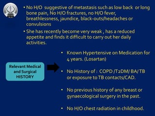
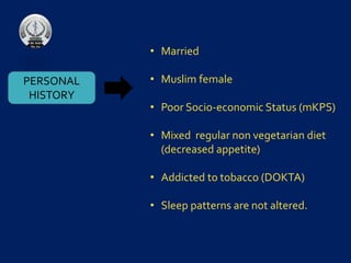
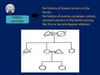
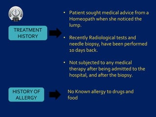
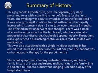
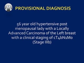
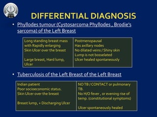
A 56-year-old hypertensive post-menopausal woman presented with a rapidly enlarging, painless lump in her left breast, which has grown from 2 cm to 6 cm over two years, accompanied by skin changes and a single axillary lump. She underwent radiological tests and biopsy but has no history of breast cancer in her family or signs of metastasis. The provisional diagnosis includes phyllodes tumor or breast tuberculosis, with a noted background of poor socio-economic status and tobacco addiction.


![BENIGN_BREAST_CONDITIONS_cases-1[1].pptx](https://cdn.slidesharecdn.com/ss_thumbnails/benignbreastconditionscases-11-230222205003-eccf02a2-thumbnail.jpg?width=640&height=640&fit=bounds)


![[MBBS/MS/DNB] Sample Long Case on Inguinal Hernia](https://cdn.slidesharecdn.com/ss_thumbnails/ccchernia-200501225130-thumbnail.jpg?width=640&height=640&fit=bounds)


![CASE_PRESENTATION_ON_subdural_hematoma(SDH)[1 FINAL PPT]-1.pptx](https://cdn.slidesharecdn.com/ss_thumbnails/casepresentationonsubduralhematomasdh1finalppt-1-260129172522-d405d375-thumbnail.jpg?width=640&height=640&fit=bounds)


