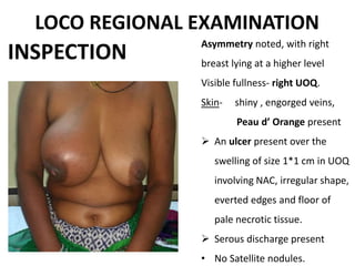
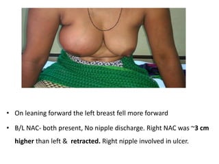
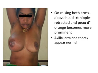
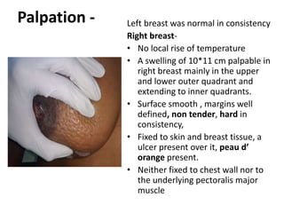
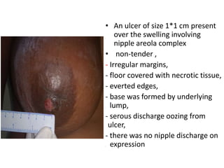
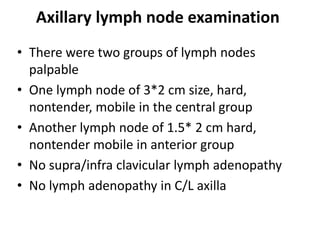
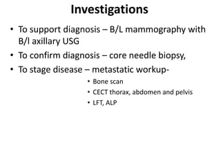
CASE PRESENTATION - LABC
Presenter – Aakansh Jain
Postgraduate student
PGIMER & Dr RML Hospital,
New delhi
SCOPE – MARCH 2017
Chief complaints
Swelling in right breast - 4months
Ulceration and pain over swelling in rt breast - 2 week
• 40 yr
• Hindu lady
• Resident of UP
• Home maker
HISTORY
History Of Present Illness
 She noticed a swelling of approx 2 cm in right breast
4 months back
 Since then it rapidly progressed to current size of
approx 10 cm, not associated with pain or fever
 2 months later another swelling appeared in right
axilla of about 1-2 cm in size, gradually increasing in
size
 No other swelling in opposite breast or axilla
HOPI cont..
 She developed an ulcer in the nipple areola complex
2 weeks back just above the swelling
 Ulcer was associated with serous discharge
 It was also associated with pain
 Pain was localised to region of ulcer, dull aching in
character, relieved on taking medication
• No h/o nipple discharge
• No h/o trauma / radiation exposure in past
• No h/o chest pain cough/ hemoptysis
• No h/o weight loss or appetite
• No h/o pain in abdomen or jaundice
• No h/o lower back ache, pain in limbs
• No h/o headaches / vomiting or weakness of any
limbs
• Not a known case of HTN/ DM / asthma
HOPI cont..
HOPI cont..
• She gave h/o multiple needle insertions and dry taps
from the swelling done by some local practitioner
Past History
• No past h/o tuberculosis
• No past h/o surgical intervention
Family history
• No h/o any breast , ovarian , prostate or GI
malignancies in 1st degree relatives or family
members
Menstrual & Obstetric history
• Menarche at the age of 12 yrs.
• Married at age of 16 yrs.
• 1st child at the age of 23 yrs.
• She has 4 children and one abortion
• All Breastfed for minimum 2 years
• Youngest child of age 5 yrs.
• No h/o intake of any birth control pills
• Has normal menstrual cycles
Personal History
• Vegetarian
• Normal sleep pattern
• Normal bowel and bladder habit
• Non smoker/ non alcoholic
• No known allergies
To summarize…..
• 40 yr premenopausal hindu lady presented
with rapidly progressing swelling in her right
breast for 4 months which is at present of
approximately 10 cm and an ulcer over the
swelling for last 2 wks associated with serous
discharge.
• She complained of another swelling in her
right axilla for last 2 months.
• Negative metastatic and family history.
General physical Examination
• Conscious, oriented to time, place and person
• Built average
• Performance status - 80 (karnofsky scale),
• Well hydrated
• Adequately Nourished with BMI – 24.1 kg/m2
• No pallor, clubbing , cyanosis, icterus, pedal edema,
generalised lymphadenopathy (axillary LNA in regional
examination)
Vitals-
• PR-80 /min
• BP -118/74 mmHg
• RR – 16 / min ,
• Temperature- 98.2° F
LOCO REGIONAL EXAMINATION
INSPECTION
Asymmetry noted, with right
breast lying at a higher level
Visible fullness- right UOQ.
Skin- shiny , engorged veins,
Peau d’ Orange present
 An ulcer present over the
swelling of size 1*1 cm in UOQ
involving NAC, irregular shape,
everted edges and floor of
pale necrotic tissue.
 Serous discharge present
• No Satellite nodules.
• On leaning forward the left breast fell more forward
• B/L NAC- both present, No nipple discharge. Right NAC was ~3 cm
higher than left & retracted. Right nipple involved in ulcer.
• On raising both arms
above head- rt nipple
retracted and peau d’
orange becomes more
prominent
• Axilla, arm and thorax
appear normal
Palpation - Left breast was normal in consistency
Right breast-
• No local rise of temperature
• A swelling of 10*11 cm palpable in
right breast mainly in the upper
and lower outer quadrant and
extending to inner quadrants.
• Surface smooth , margins well
defined, non tender, hard in
consistency,
• Fixed to skin and breast tissue, a
ulcer present over it, peau d’
orange present.
• Neither fixed to chest wall nor to
the underlying pectoralis major
muscle
• An ulcer of size 1*1 cm present
over the swelling involving
nipple areola complex
• non-tender ,
- Irregular margins,
- floor covered with necrotic tissue,
- everted edges,
- base was formed by underlying
. lump,
- serous discharge oozing from
….ulcer,
- there was no nipple discharge on
. expression
Axillary lymph node examination
• There were two groups of lymph nodes
palpable
• One lymph node of 3*2 cm size, hard,
nontender, mobile in the central group
• Another lymph node of 1.5* 2 cm hard,
nontender mobile in anterior group
• No supra/infra clavicular lymph adenopathy
• No lymph adenopathy in C/L axilla
Systemic examination
• P/A – soft , non tender, no organomegaly, no
shifting dullness
• DRE- No fissures, fistula/sinus openings
No palpable nodule
No rectal bleeding
• Per vaginal examination - NAD
• Spine examination – no spine tenderness
• Chest – B/L AE +, no added sounds
• CVS – S1S2 heard, no murmur heard
To Summarize…
 40 yr premenopausal lady presented with rapidly
progressing right breast swelling currently of size
11 cm associated with serous discharging ulcer and
peau d’ orange with I/L two mobile axillary lymph
nodes with no clinical evidence of distant metastasis.
Diagnosis
• 40 yr premenopausal lady with locally
advance carcinoma of right breast with
clinically T4b N1 M0 with stage IIIb
Investigations
• To support diagnosis – B/L mammography with
B/l axillary USG
• To confirm diagnosis – core needle biopsy,
• To stage disease – metastatic workup-
• Bone scan
• CECT thorax, abdomen and pelvis
• LFT, ALP
Management
• Neoadjuvant chemotherapy
• MRM + axillary lymph nodal dissection
• Radiotherapy to chest wall and supraclavicular
LN region + Adjuvant chemotherapy
• Hormonal therapy (as per receptor status)

Labc case presentation

  • 1.
    CASE PRESENTATION -LABC Presenter – Aakansh Jain Postgraduate student PGIMER & Dr RML Hospital, New delhi SCOPE – MARCH 2017
  • 2.
    Chief complaints Swelling inright breast - 4months Ulceration and pain over swelling in rt breast - 2 week • 40 yr • Hindu lady • Resident of UP • Home maker HISTORY
  • 3.
    History Of PresentIllness  She noticed a swelling of approx 2 cm in right breast 4 months back  Since then it rapidly progressed to current size of approx 10 cm, not associated with pain or fever  2 months later another swelling appeared in right axilla of about 1-2 cm in size, gradually increasing in size  No other swelling in opposite breast or axilla
  • 4.
    HOPI cont..  Shedeveloped an ulcer in the nipple areola complex 2 weeks back just above the swelling  Ulcer was associated with serous discharge  It was also associated with pain  Pain was localised to region of ulcer, dull aching in character, relieved on taking medication
  • 5.
    • No h/onipple discharge • No h/o trauma / radiation exposure in past • No h/o chest pain cough/ hemoptysis • No h/o weight loss or appetite • No h/o pain in abdomen or jaundice • No h/o lower back ache, pain in limbs • No h/o headaches / vomiting or weakness of any limbs • Not a known case of HTN/ DM / asthma HOPI cont..
  • 6.
    HOPI cont.. • Shegave h/o multiple needle insertions and dry taps from the swelling done by some local practitioner Past History • No past h/o tuberculosis • No past h/o surgical intervention
  • 7.
    Family history • Noh/o any breast , ovarian , prostate or GI malignancies in 1st degree relatives or family members
  • 8.
    Menstrual & Obstetrichistory • Menarche at the age of 12 yrs. • Married at age of 16 yrs. • 1st child at the age of 23 yrs. • She has 4 children and one abortion • All Breastfed for minimum 2 years • Youngest child of age 5 yrs. • No h/o intake of any birth control pills • Has normal menstrual cycles
  • 9.
    Personal History • Vegetarian •Normal sleep pattern • Normal bowel and bladder habit • Non smoker/ non alcoholic • No known allergies
  • 10.
    To summarize….. • 40yr premenopausal hindu lady presented with rapidly progressing swelling in her right breast for 4 months which is at present of approximately 10 cm and an ulcer over the swelling for last 2 wks associated with serous discharge. • She complained of another swelling in her right axilla for last 2 months. • Negative metastatic and family history.
  • 11.
    General physical Examination •Conscious, oriented to time, place and person • Built average • Performance status - 80 (karnofsky scale), • Well hydrated • Adequately Nourished with BMI – 24.1 kg/m2 • No pallor, clubbing , cyanosis, icterus, pedal edema, generalised lymphadenopathy (axillary LNA in regional examination) Vitals- • PR-80 /min • BP -118/74 mmHg • RR – 16 / min , • Temperature- 98.2° F
  • 12.
    LOCO REGIONAL EXAMINATION INSPECTION Asymmetrynoted, with right breast lying at a higher level Visible fullness- right UOQ. Skin- shiny , engorged veins, Peau d’ Orange present  An ulcer present over the swelling of size 1*1 cm in UOQ involving NAC, irregular shape, everted edges and floor of pale necrotic tissue.  Serous discharge present • No Satellite nodules.
  • 13.
    • On leaningforward the left breast fell more forward • B/L NAC- both present, No nipple discharge. Right NAC was ~3 cm higher than left & retracted. Right nipple involved in ulcer.
  • 14.
    • On raisingboth arms above head- rt nipple retracted and peau d’ orange becomes more prominent • Axilla, arm and thorax appear normal
  • 15.
    Palpation - Leftbreast was normal in consistency Right breast- • No local rise of temperature • A swelling of 10*11 cm palpable in right breast mainly in the upper and lower outer quadrant and extending to inner quadrants. • Surface smooth , margins well defined, non tender, hard in consistency, • Fixed to skin and breast tissue, a ulcer present over it, peau d’ orange present. • Neither fixed to chest wall nor to the underlying pectoralis major muscle
  • 16.
    • An ulcerof size 1*1 cm present over the swelling involving nipple areola complex • non-tender , - Irregular margins, - floor covered with necrotic tissue, - everted edges, - base was formed by underlying . lump, - serous discharge oozing from ….ulcer, - there was no nipple discharge on . expression
  • 17.
    Axillary lymph nodeexamination • There were two groups of lymph nodes palpable • One lymph node of 3*2 cm size, hard, nontender, mobile in the central group • Another lymph node of 1.5* 2 cm hard, nontender mobile in anterior group • No supra/infra clavicular lymph adenopathy • No lymph adenopathy in C/L axilla
  • 18.
    Systemic examination • P/A– soft , non tender, no organomegaly, no shifting dullness • DRE- No fissures, fistula/sinus openings No palpable nodule No rectal bleeding • Per vaginal examination - NAD • Spine examination – no spine tenderness • Chest – B/L AE +, no added sounds • CVS – S1S2 heard, no murmur heard
  • 19.
    To Summarize…  40yr premenopausal lady presented with rapidly progressing right breast swelling currently of size 11 cm associated with serous discharging ulcer and peau d’ orange with I/L two mobile axillary lymph nodes with no clinical evidence of distant metastasis.
  • 20.
    Diagnosis • 40 yrpremenopausal lady with locally advance carcinoma of right breast with clinically T4b N1 M0 with stage IIIb
  • 21.
    Investigations • To supportdiagnosis – B/L mammography with B/l axillary USG • To confirm diagnosis – core needle biopsy, • To stage disease – metastatic workup- • Bone scan • CECT thorax, abdomen and pelvis • LFT, ALP
  • 22.
    Management • Neoadjuvant chemotherapy •MRM + axillary lymph nodal dissection • Radiotherapy to chest wall and supraclavicular LN region + Adjuvant chemotherapy • Hormonal therapy (as per receptor status)

Editor's Notes

  • #5 Reddish watery; initially painless
  • #7 Radial scar appear as ductal carcinoma in situ grossely
  • #8 PTEN cowden syndrome – thyroid malignancy, GI malignancy, breast ,cerebral malignancy % of increased risk if 1* 2* 3* 4*
  • #9 LMP – 20 feb 2017
  • #10  combined oral contraceptive pill contains estrogen(estradiol) and progesterone (progestin).
  • #12 AFTER TAKING INFORMED CONSENT , IN WELL LIT ROOM, IN PRESENCE OF FEMALE ATTENDANT , Bp measured in left brachial artery ECOG- EUROPEAN COOPERATIVE ONCOLOGY GROUP Nutritional status = carbohydrates protein – mid arm circumference, fat -
  • #13 I examined the pt in sitting, sitting with hand raise above head , supine, recumbant and bending forward position Left breast -normal in size and shape Left Nipple areola complex normal
  • #17 Correction – ulcer to be called pressure ulcer as underlying lump presses to cause skin necrosis, margins to be undermined , probe test positive
  • #22 incisional biopsy from ulcer