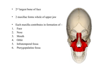
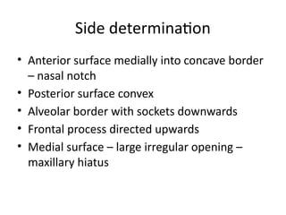
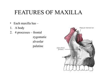
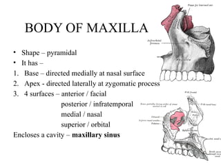
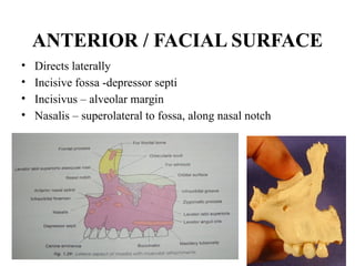
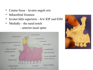
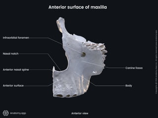
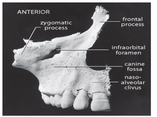
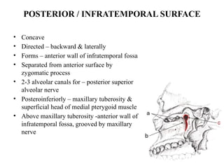
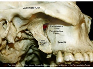
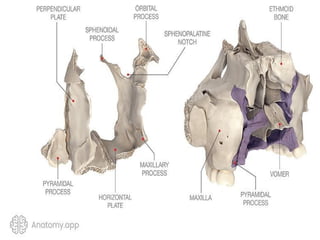
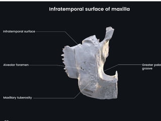
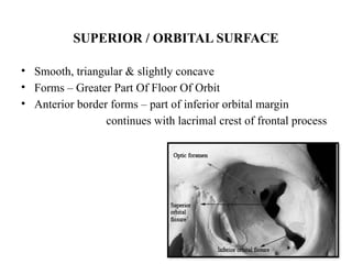
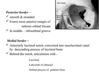
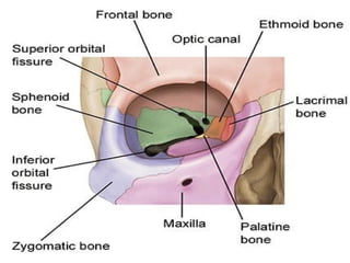
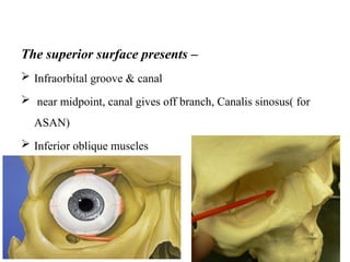
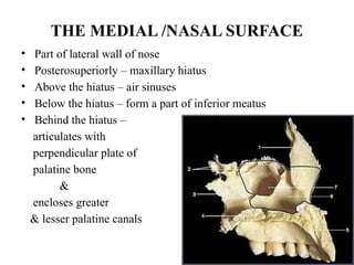
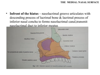
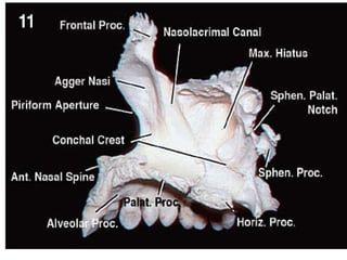
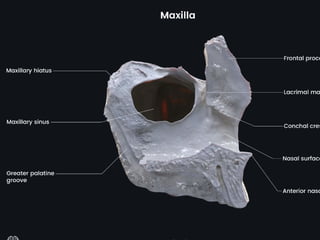
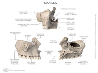
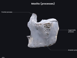
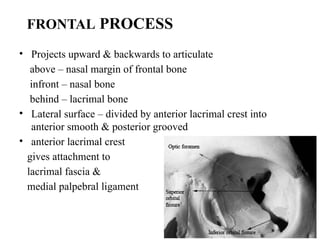
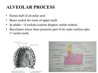
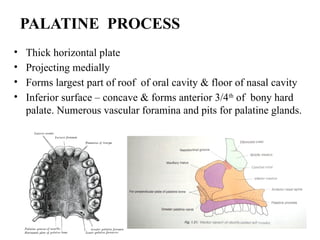
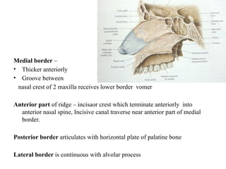
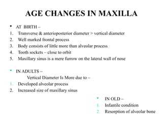
MAXILLA (PART – I)
NORMAL ANATOMY
Name of resident - Dr Niharika Oza
Name of guide - Dr Viral Prajapati
Designation - 3rd
year resident doctor
Institute - Dr M K Shah Medical College & Research Centre,
Ahmedabad
OSSIFICATION:
• MAXILLA ossifies from 3 centers in the membrane –
1. 1 center for maxilla proper – 6th
week of IUL, above the canine fossa
2. 2 centers for premaxilla
Of 2 premaxillary centers-
o Main center above the incisive fossa - 7th
week of IUL
o Second center (Paraseptal/ prevomerine)– ventral margin of nasal septum
- 10th
week of IUL
soon fuses with palatal process of maxilla.
Premaxilla begin to fuse with alveolar process almost immediately after the
ossification begins.
.
• 2nd
largest bone of face
• 2 maxillae forms whole of upper jaw
• Each maxilla contributes in formation of –
1. Face
2. Nose
3. Mouth
4. Orbit
5. Infratemporal fossa
6. Pterygopalatine fossa
Side determination
• Anterior surface medially into concave border
– nasal notch
• Posterior surface convex
• Alveolar border with sockets downwards
• Frontal process directed upwards
• Medial surface – large irregular opening –
maxillary hiatus
FEATURES OF MAXILLA
• Each maxilla has –
1. A body
2. 4 processes – frontal
zygomatic
alveolar
palatine
BODY OF MAXILLA
• Shape – pyramidal
• It has –
1. Base – directed medially at nasal surface
2. Apex - directed laterally at zygomatic process
3. 4 surfaces – anterior / facial
posterior / infratemporal
medial / nasal
superior / orbital
Encloses a cavity – maxillary sinus
ANTERIOR / FACIAL SURFACE
• Directs laterally
• Incisive fossa -depressor septi
• Incisivus – alveolar margin
• Nasalis – superolateral to fossa, along nasal notch
• Canine fossa – levator anguli oris
• Infraorbital foramen
• levator labii superioris – b/w IOF nad IOM
• Medially – the nasal notch
- anterior nasal spine
POSTERIOR / INFRATEMPORAL SURFACE
• Concave
• Directed – backward & laterally
• Forms – anterior wall of infratemporal fossa
• Separated from anterior surface by
zygomatic process
• 2-3 alveolar canals for – posterior superior
alveolar nerve
• Posteroinferiorly – maxillary tuberosity &
superficial head of medial pterygoid muscle
• Above maxillary tuberosity -anterior wall of
infratemporal fossa, grooved by maxillary
nerve
SUPERIOR / ORBITAL SURFACE
• Smooth, triangular & slightly concave
• Forms – Greater Part Of Floor Of Orbit
• Anterior border forms – part of inferior orbital margin
continues with lacrimal crest of frontal process
Posterior border –
 smooth & rounded
 Forms most anterior margin of
inferior orbital fissure
 In middle – infraorbital groove
Medial border –
 Anteriorly lacrimal notch, converted into nasolacrimal canal
by descending process of lacrimal bone
 Behind the notch, articulation with -
Lacrimal
Labyrinth of ethmoid
Orbital process of palatine bone
The superior surface presents –
 Infraorbital groove & canal
 near midpoint, canal gives off branch, Canalis sinosus( for
ASAN)
 Inferior oblique muscles
THE MEDIAL /NASAL SURFACE
• Part of lateral wall of nose
• Posterosuperiorly – maxillary hiatus
• Above the hiatus – air sinuses
• Below the hiatus – form a part of inferior meatus
• Behind the hiatus –
articulates with
perpendicular plate of
palatine bone
&
encloses greater
& lesser palatine canals
THE MEDIAL /NASAL SURFACE
• Infront of the hiatus – nasolacrimal groove articulates with
descending process of lacrimal bone & lacrimal process of
inferior nasal concha to forms nasolacrimal canal,transmit
nasolacrimal duct to inferior meatus
THE MEDIAL /NASAL SURFACE
• More anteriorly – conchal creast for articulation with inferior
nasal concha.
• Above the conchal crest – atrium of middle meatus.
FRONTAL PROCESS
• Projects upward & backwards to articulate
above – nasal margin of frontal bone
infront – nasal bone
behind – lacrimal bone
• Lateral surface – divided by anterior lacrimal crest into
anterior smooth & posterior grooved
• anterior lacrimal crest
gives attachment to
lacrimal fascia &
medial palpebral ligament
.
• Anterior smooth area gives origin to orbital
part of orbicularis oculi muscle and levator
labii superioriis alaeque nasi.
FRONTAL PROCESS
• Medial surface – forms lateral wall of nose
from above downwards –
1. Uppermost roughened area for articulation with ethmoid to close
ethmoid air sinuses
2. Ethmoidal crest – a horizontal ridge about middle of process, posterior
part of crest articulates with middle nasal concha and anterior part lies
beneath agger nasi.
3. Below the ethmoidal crest
hollowed out to form atrium of middle meatus
conchal crest articulates with inferior nasal concha
below it, inferior meatus with nasolacrimal groove ending just
behind crest
• Pyramidal lateral projection
• Anterior, posterior & superior surfaces converge here
• Superiorly – rough, to articulate with zygomatic bone
ZYGOMATIC PROCESS
• Forms half of alveolar arch
• Bears socket for roots of upper teeth
• In adults = 8 sockets (canine deepest, molar widest)
• Buccinator arises from posterior part of its outer surface upto
1st
molar tooth.
ALVEOLAR PROCESS
• Thick horizontal plate
• Projecting medially
• Forms largest part of roof of oral cavity & floor of nasal cavity
• Inferior surface – concave & forms anterior 3/4th
of bony hard
palate. Numerous vascular foramina and pits for palatine glands.
PALATINE PROCESS
• Posterolaterally –
greater & lesser
palatine foremen
• Superior surface –concave
from side to side & forms
floor of nasal cavity.
Medial border –
• Thicker anteriorly
• Groove between
nasal crest of 2 maxilla receives lower border vomer
Anterior part of ridge – incisaor crest which terminate anteriorly into
anterior nasal spine, Incisive canal traverse near anterior part of medial
border.
Posterior border articulates with horizontal plate of palatine bone
Lateral border is continuous with alvolar process
AGE CHANGES IN MAXILLA
 AT BIRTH –
1. Transverse & anterioposterior diameter > vertical diameter
2. Well marked frontal process
3. Body consists of little more than alveolar process
4. Tooth sockets – close to orbit
5. Maxillary sinus is a mere furrow on the lateral wall of nose
 IN ADULTS –
Vertical Diameter Is More due to –
1. Developed alveolar process
2. Increased size of maxillary sinus
 IN OLD –
1. Infantile condition
2. Resorption of alveolar bone
MAXILLA   anatomy is very important.pptx

MAXILLA anatomy is very important.pptx

  • 2.
    MAXILLA (PART –I) NORMAL ANATOMY Name of resident - Dr Niharika Oza Name of guide - Dr Viral Prajapati Designation - 3rd year resident doctor Institute - Dr M K Shah Medical College & Research Centre, Ahmedabad
  • 4.
    OSSIFICATION: • MAXILLA ossifiesfrom 3 centers in the membrane – 1. 1 center for maxilla proper – 6th week of IUL, above the canine fossa 2. 2 centers for premaxilla Of 2 premaxillary centers- o Main center above the incisive fossa - 7th week of IUL o Second center (Paraseptal/ prevomerine)– ventral margin of nasal septum - 10th week of IUL soon fuses with palatal process of maxilla. Premaxilla begin to fuse with alveolar process almost immediately after the ossification begins.
  • 5.
    . • 2nd largest boneof face • 2 maxillae forms whole of upper jaw • Each maxilla contributes in formation of – 1. Face 2. Nose 3. Mouth 4. Orbit 5. Infratemporal fossa 6. Pterygopalatine fossa
  • 6.
    Side determination • Anteriorsurface medially into concave border – nasal notch • Posterior surface convex • Alveolar border with sockets downwards • Frontal process directed upwards • Medial surface – large irregular opening – maxillary hiatus
  • 7.
    FEATURES OF MAXILLA •Each maxilla has – 1. A body 2. 4 processes – frontal zygomatic alveolar palatine
  • 8.
    BODY OF MAXILLA •Shape – pyramidal • It has – 1. Base – directed medially at nasal surface 2. Apex - directed laterally at zygomatic process 3. 4 surfaces – anterior / facial posterior / infratemporal medial / nasal superior / orbital Encloses a cavity – maxillary sinus
  • 9.
    ANTERIOR / FACIALSURFACE • Directs laterally • Incisive fossa -depressor septi • Incisivus – alveolar margin • Nasalis – superolateral to fossa, along nasal notch
  • 10.
    • Canine fossa– levator anguli oris • Infraorbital foramen • levator labii superioris – b/w IOF nad IOM • Medially – the nasal notch - anterior nasal spine
  • 13.
    POSTERIOR / INFRATEMPORALSURFACE • Concave • Directed – backward & laterally • Forms – anterior wall of infratemporal fossa • Separated from anterior surface by zygomatic process • 2-3 alveolar canals for – posterior superior alveolar nerve • Posteroinferiorly – maxillary tuberosity & superficial head of medial pterygoid muscle • Above maxillary tuberosity -anterior wall of infratemporal fossa, grooved by maxillary nerve
  • 17.
    SUPERIOR / ORBITALSURFACE • Smooth, triangular & slightly concave • Forms – Greater Part Of Floor Of Orbit • Anterior border forms – part of inferior orbital margin continues with lacrimal crest of frontal process
  • 18.
    Posterior border – smooth & rounded  Forms most anterior margin of inferior orbital fissure  In middle – infraorbital groove Medial border –  Anteriorly lacrimal notch, converted into nasolacrimal canal by descending process of lacrimal bone  Behind the notch, articulation with - Lacrimal Labyrinth of ethmoid Orbital process of palatine bone
  • 20.
    The superior surfacepresents –  Infraorbital groove & canal  near midpoint, canal gives off branch, Canalis sinosus( for ASAN)  Inferior oblique muscles
  • 21.
    THE MEDIAL /NASALSURFACE • Part of lateral wall of nose • Posterosuperiorly – maxillary hiatus • Above the hiatus – air sinuses • Below the hiatus – form a part of inferior meatus • Behind the hiatus – articulates with perpendicular plate of palatine bone & encloses greater & lesser palatine canals
  • 22.
    THE MEDIAL /NASALSURFACE • Infront of the hiatus – nasolacrimal groove articulates with descending process of lacrimal bone & lacrimal process of inferior nasal concha to forms nasolacrimal canal,transmit nasolacrimal duct to inferior meatus
  • 23.
    THE MEDIAL /NASALSURFACE • More anteriorly – conchal creast for articulation with inferior nasal concha. • Above the conchal crest – atrium of middle meatus.
  • 28.
    FRONTAL PROCESS • Projectsupward & backwards to articulate above – nasal margin of frontal bone infront – nasal bone behind – lacrimal bone • Lateral surface – divided by anterior lacrimal crest into anterior smooth & posterior grooved • anterior lacrimal crest gives attachment to lacrimal fascia & medial palpebral ligament
  • 29.
    . • Anterior smootharea gives origin to orbital part of orbicularis oculi muscle and levator labii superioriis alaeque nasi.
  • 31.
    FRONTAL PROCESS • Medialsurface – forms lateral wall of nose from above downwards – 1. Uppermost roughened area for articulation with ethmoid to close ethmoid air sinuses 2. Ethmoidal crest – a horizontal ridge about middle of process, posterior part of crest articulates with middle nasal concha and anterior part lies beneath agger nasi. 3. Below the ethmoidal crest hollowed out to form atrium of middle meatus conchal crest articulates with inferior nasal concha below it, inferior meatus with nasolacrimal groove ending just behind crest
  • 34.
    • Pyramidal lateralprojection • Anterior, posterior & superior surfaces converge here • Superiorly – rough, to articulate with zygomatic bone ZYGOMATIC PROCESS
  • 35.
    • Forms halfof alveolar arch • Bears socket for roots of upper teeth • In adults = 8 sockets (canine deepest, molar widest) • Buccinator arises from posterior part of its outer surface upto 1st molar tooth. ALVEOLAR PROCESS
  • 36.
    • Thick horizontalplate • Projecting medially • Forms largest part of roof of oral cavity & floor of nasal cavity • Inferior surface – concave & forms anterior 3/4th of bony hard palate. Numerous vascular foramina and pits for palatine glands. PALATINE PROCESS
  • 37.
    • Posterolaterally – greater& lesser palatine foremen • Superior surface –concave from side to side & forms floor of nasal cavity.
  • 38.
    Medial border – •Thicker anteriorly • Groove between nasal crest of 2 maxilla receives lower border vomer Anterior part of ridge – incisaor crest which terminate anteriorly into anterior nasal spine, Incisive canal traverse near anterior part of medial border. Posterior border articulates with horizontal plate of palatine bone Lateral border is continuous with alvolar process
  • 39.
    AGE CHANGES INMAXILLA  AT BIRTH – 1. Transverse & anterioposterior diameter > vertical diameter 2. Well marked frontal process 3. Body consists of little more than alveolar process 4. Tooth sockets – close to orbit 5. Maxillary sinus is a mere furrow on the lateral wall of nose  IN ADULTS – Vertical Diameter Is More due to – 1. Developed alveolar process 2. Increased size of maxillary sinus  IN OLD – 1. Infantile condition 2. Resorption of alveolar bone